


 全部商品分类
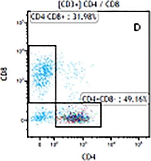


 下载产品说明书 下载SDS
 用小程序,查商品更便捷


 收藏
收藏
 对比
对比 咨询
咨询Immunocytochemistry(8-25 µg/mL)

Scientific Data
.") View Larger
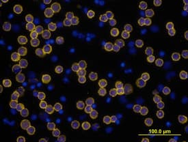
View LargerCD3 epsilon in Human PBMCs. CD3e was detected in immersion fixed human peripheral blood mononuclear cells (PBMCs) using Mouse Anti-Human CD3e Monoclonal Antibody (Catalog # MAB100) at 10 µg/mL for 3 hours at room temperature. Cells were stained using the NorthernLights™ 557-conjugated Anti-Mouse IgG Secondary Antibody (yellow; Catalog # NL007) and counterstained with DAPI (blue). View our protocol for Fluorescent ICC Staining of Non-adherent Cells.
 View Larger
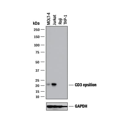
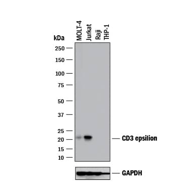
Detection of Human CD3 epsilon by Western Blot. Western blot shows lysates of MOLT‑4 human acute lymphoblastic leukemia cell line, Jurkat human acute T cell leukemia cell line, Raji human Burkitt's lymphoma cell line (negative control), and THP‑1 human acute monocytic leukemia cell line (negative control). PVDF membrane was probed with 2 µg/mL of Mouse Anti-Human CD3 epsilon Monoclonal Antibody (Catalog # MAB100) followed by HRP-conjugated Anti-Mouse IgG Secondary Antibody (HAF018). A specific band was detected for CD3 epsilon at approximately 21 kDa (as indicated). GAPDH (MAB5718) is shown as a loading control. This experiment was conducted under reducing conditions and using Western Blot Buffer Group 1.
 View Larger
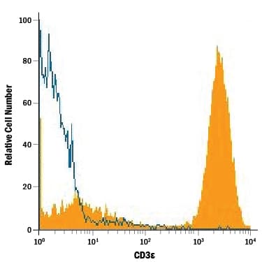
Detection of CD3 epsilon in Human Lymphocytes by Flow Cytometry. Human peripheral blood lymphocytes were stained with Mouse Anti-Human CD3e Monoclonal Antibody (Catalog # MAB100, filled histogram) or isotype control antibody (Catalog # MAB002, open histogram), followed by Phycoerythrin-conjugated Anti-Mouse IgG Secondary Antibody (Catalog # F0102B).
 View Larger
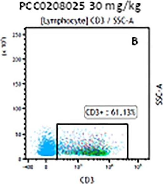
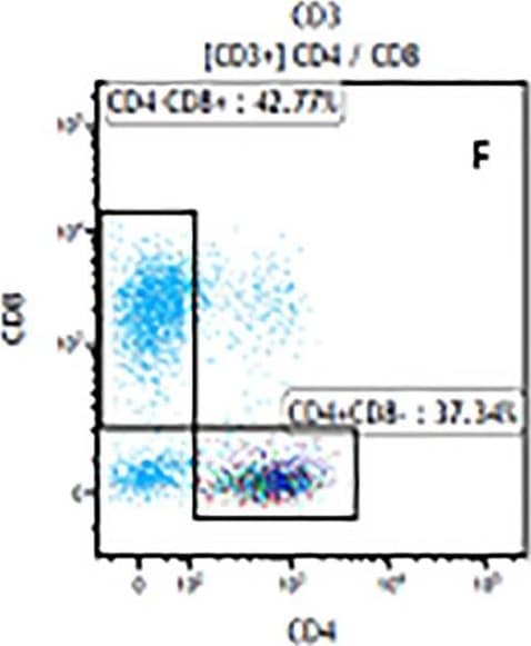
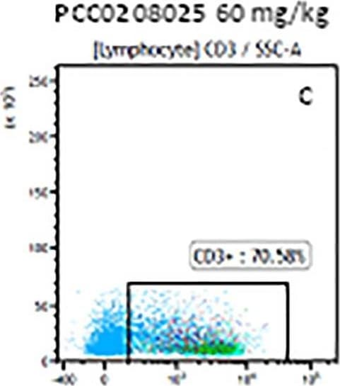
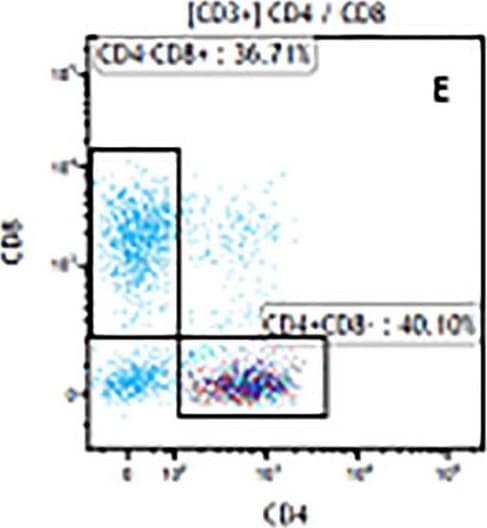
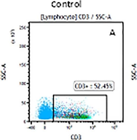
Detection of Mouse CD3 epsilon by Flow Cytometry The representative figures for T cell subsets counted by flow cytometry from tumors in B16-F10-bearing mice.The cell counts for CD3+ (A, B and C), CD3+CD4+ (D, E and F), CD3+CD8+ (D, E and F), CD4+CD25+CD127low/- (G, H and I) and CD8+IFN-gamma + (J, K and L) T lymphocytes from mouse tumor were determined by flow cytometry. Image collected and cropped by CiteAb from the following publication (https://pubmed.ncbi.nlm.nih.gov/32214351), licensed under a CC-BY license. Not internally tested by R&D Systems.
 View Larger
Detection of Mouse CD3 epsilon by Flow Cytometry The representative figures for T cell subsets counted by flow cytometry from tumors in B16-F10-bearing mice.The cell counts for CD3+ (A, B and C), CD3+CD4+ (D, E and F), CD3+CD8+ (D, E and F), CD4+CD25+CD127low/- (G, H and I) and CD8+IFN-gamma + (J, K and L) T lymphocytes from mouse tumor were determined by flow cytometry. Image collected and cropped by CiteAb from the following publication (https://pubmed.ncbi.nlm.nih.gov/32214351), licensed under a CC-BY license. Not internally tested by R&D Systems.
Human CD3 epsilon Antibody Summary
Applications
Please Note: Optimal dilutions should be determined by each laboratory for each application. General Protocols are available in the Technical Information section on our website.
Immunocytochemistry(8-25 µg/mL)
Background: CD3 epsilon
CD3 epsilon is one of at least four invariant proteins that associate with the variable antigen recognition chains of the T cell receptor and function in signal transduction.
Preparation and Storage
- 12 months from date of receipt, -20 to -70 °C as supplied.
- 1 month, 2 to 8 °C under sterile conditions after reconstitution.
- 6 months, -20 to -70 °C under sterile conditions after reconstitution.
参考图片
CD3 epsilon in Human PBMCs. CD3 epsilon was detected in immersion fixed human peripheral blood mononuclear cells (PBMCs) using Mouse Anti-Human CD3 epsilon Monoclonal Antibody (Catalog # MAB100) at 10 µg/mL for 3 hours at room temperature. Cells were stained using the NorthernLights™ 557-conjugated Anti-Mouse IgG Secondary Antibody (yellow; Catalog # NL007) and counterstained with DAPI (blue). View our protocol for Fluorescent ICC Staining of Non-adherent Cells.
Detection of CD3 epsilon in Human Lymphocytes by Flow Cytometry. Human peripheral blood lymphocytes were stained with Mouse Anti-Human CD3 epsilon Monoclonal Antibody (Catalog # MAB100, filled histogram) or isotype control antibody (Catalog # MAB002, open histogram), followed by Phycoerythrin-conjugated Anti-Mouse IgG Secondary Antibody (Catalog # F0102B).