


 全部商品分类


1/2

品牌: BD Pharmingen
 下载产品说明书 下载SDS
 用小程序,查商品更便捷


 收藏
收藏
 对比
对比 咨询
咨询反应种属:
Rat (QC Testing)
来源宿主:
Mouse BALB/c IgG2a, κ
产品介绍
产品介绍
产品信息
荧光素标记
抗原名称
CD4

宿主
Mouse BALB/c IgG2a, κ
免疫原
Rat T-cell blasts
简单描述
The OX-35 clone recognizes the CD4 antigen on most thymocytes, a subpopulation of mature T lymphocytes (i.e., MHC class II-restricted T cells, including most T helper cells), monocytes, macrophages, some dendritic cells, and microglia. CD4 is an antigen coreceptor on the T-cell surface that interacts with MHC class II molecules on antigen-presenting cells. It participates in T-cell activation through it's association with the T-cell receptor complex and protein tyrosine kinase Lck. The OX-35 clone has been reported to bind to a different epitope of CD4 than that recognized by the W3/25 and OX-38 clones.
商品描述
OX-35
The OX-35 clone recognizes the CD4 antigen on most thymocytes, a subpopulation of mature T lymphocytes (i.e., MHC class II-restricted T cells, including most T helper cells), monocytes, macrophages, some dendritic cells, and microglia. CD4 is an antigen coreceptor on the T-cell surface that interacts with MHC class II molecules on antigen-presenting cells. It participates in T-cell activation through it's association with the T-cell receptor complex and protein tyrosine kinase Lck. The OX-35 clone has been reported to bind to a different epitope of CD4 than that recognized by the W3/25 and OX-38 clones.
同种型
Mouse BALB/c IgG2a, κ
克隆号
克隆 OX-35 (RUO)
浓度
0.2 mg/ml
产品详情
PE-Cy7
PE-Cy7 dye is a part of the BD PE family of dyes. This tandem fluorochrome is comprised of a R-Phycoerythrin (PE) donor that has excitation maxima (Ex Max) of 496-nm and 566-nm and an acceptor dye, Cy™7, with an emission maximum (Em Max) at 781-nm. PE can be excited by the Blue (488-nm), Green (532-nm) and yellow-green (561-nm) lasers and detected using an optical filter centered near 781 nm (e.g., a 760/60-nm bandpass filter). The donor dye can be excited by the Blue (488-nm), Green (532-nm) and yellow-green (561-nm) lasers and the acceptor dye can be excited by the Red (627–640-nm) laser resulting in cross-laser excitation and fluorescence spillover. Please ensure that your instrument’s configurations (lasers and optical filters) are appropriate for this dye.

PE-Cy7
Yellow-Green 561 nm
496 nm, 566 nm
781 nm
应用
实验应用
Flow cytometry (Routinely Tested)
反应种属
Rat (QC Testing)
目标/特异性
CD4
背景
别名
Cd4; CD4 antigen; p55; W3/25 antigen; T-cell surface glycoprotein CD4
制备和贮存
存储溶液
Aqueous buffered solution containing ≤0.09% sodium azide.
保存方式
Aqueous buffered solution containing ≤0.09% sodium azide.
文献
文献
研发参考(7)
1. Bañuls MP, Alvarez A, Ferrero I, Zapata A, Ardavin C. Cell-surface marker analysis of rat thymic dendritic cells. Immunology. 1993; 79(2):298-304. (Biology).
2. Bierer BE, Sleckman BP, Ratnofsky SE, Burakoff SJ. The biologic roles of CD2, CD4, and CD8 in T-cell activation. Annu Rev Immunol. 1989; 7:579-599. (Biology).
3. Ford AL, Foulcher E, Goodsall AL, Sedgwick JD. Tissue digestion with dispase substantially reduces lymphocyte and macrophage cell-surface antigen expression. J Immunol Methods. 1996; 194(1):71-75. (Biology).
4. Janeway CA Jr. The T cell receptor as a multicomponent signalling machine: CD4/CD8 coreceptors and CD45 in T cell activation. Annu Rev Immunol. 1992; 10:645-674. (Biology).
5. Jefferies WA, Green JR, Williams AF. Authentic T helper CD4 (W3/25) antigen on rat peritoneal macrophages. J Exp Med. 1985; 162(1):117-127. (Immunogen: Flow cytometry, Functional assay, Immunoaffinity chromatography, Immunoprecipitation, Inhibition).
6. Liu L, Zhang M, Jenkins C, MacPherson GG. Dendritic cell heterogeneity in vivo: two functionally different dendritic cell populations in rat intestinal lymph can be distinguished by CD4 expression. J Immunol. 1998; 161(3):1146-1155. (Biology).
7. Wang CC, Wu CH, Shieh JY, Wen CY, Ling EA. Immunohistochemical study of amoeboid microglial cells in fetal rat brain. J Anat. 1996; 189(3):567-574. (Biology).
参考图片
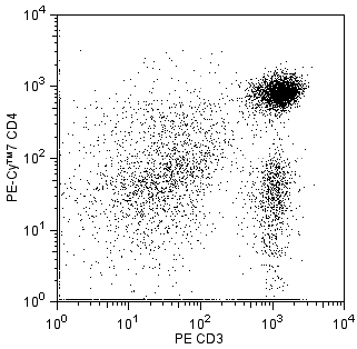
Multicolor flow cytometric analysis of CD4 expression on rat splenocytes. Splenocytes from a Lewis rat were stained with the PE-Cy™7 Mouse Anti-Rat CD4 antibody (Cat. No. 561578) in conjunction with a PE Mouse Anti-Rat CD3 antibody (Cat. No. 554833). The two-color flow cytometric dot plot shows CD3 versus CD4 expression by cells with the forward and side light-scatter characteristics of viable lymphocytes. Flow cytometry was performed on a BD LSRII™ System.
声明 :本官网所有报价均为常温或者蓝冰运输价格,如有产品需要干冰运输,需另外加收干冰运输费。