BD Horizon™ BUV496 Mouse Anti-Human CD4
1/2

BD Horizon™ BUV496 Mouse Anti-Human CD4

分享
品牌: BD Pharmingen
pdf 下载产品说明书
pdf 下载SDS
用小程序,查商品更便捷
收藏
对比对比
咨询咨询
    反应种属:
    Human (QC Testing)
    Human (QC Testing)
    来源宿主:
    Mouse BALB/c IgG1, κ
    Mouse BALB/c IgG1, κ
    展开
    产品介绍
    产品信息
    耦联标记
    BUV496
    抗原名称
    CD4
    宿主
    Mouse BALB/c IgG1, κ
    免疫原
    Human Peripheral Blood T Cells
    简单描述
    The SK3 monoclonal antibody specifically binds to CD4. The CD4 antigen is a 55 kDa type 1 transmembrane glycoprotein that belongs to the immunoglobulin superfamily. It is expressed on T-helper/inducer lymphocytes and monocytes. CD3+CD4+ T cells comprise approximately 28% to 58% of normal peripheral blood lymphocytes. CD4 is also expressed on 80% to 95% of normal thymocytes. The CD4 antigen is present in low density on the cell surface of monocytes and in the cytoplasm of monocytes and macrophages (CD3-CD4+). CD4 serves as a T cell coreceptor for MHC class II antigen recognition and as a receptor for the human immunodeficiency virus (HIV). The SK3 antibody inhibits HIV binding to CD4+ cells. The antibody was conjugated to BD Horizon BUV496 which is part of the BD Horizon Brilliant™ Ultraviolet family of dyes. This dye is a tandem fluorochrome of BD Horizon BUV395 with an Ex Max of 348-nm and an acceptor dye with an Em Max at 496-nm. BD Horizon BUV496 can be excited by the ultraviolet laser (355 nm) and detected with a 515/30 nm filter with a 450LP. Due to the excitation of the acceptor dye by other laser lines, there may be significant spillover into the channel detecting BD Horizon V500 or BV510 (eg, 525/40-nm filter). However, the spillover can be corrected through compensation as with any other dye combination.
    商品描述
    SK3 The SK3 monoclonal antibody specifically binds to CD4. The CD4 antigen is a 55 kDa type 1 transmembrane glycoprotein that belongs to the immunoglobulin superfamily. It is expressed on T-helper/inducer lymphocytes and monocytes. CD3+CD4+ T cells comprise approximately 28% to 58% of normal peripheral blood lymphocytes. CD4 is also expressed on 80% to 95% of normal thymocytes. The CD4 antigen is present in low density on the cell surface of monocytes and in the cytoplasm of monocytes and macrophages (CD3-CD4+). CD4 serves as a T cell coreceptor for MHC class II antigen recognition and as a receptor for the human immunodeficiency virus (HIV). The SK3 antibody inhibits HIV binding to CD4+ cells. The antibody was conjugated to BD Horizon BUV496 which is part of the BD Horizon Brilliant™ Ultraviolet family of dyes. This dye is a tandem fluorochrome of BD Horizon BUV395 with an Ex Max of 348-nm and an acceptor dye with an Em Max at 496-nm. BD Horizon BUV496 can be excited by the ultraviolet laser (355 nm) and detected with a 515/30 nm filter with a 450LP. Due to the excitation of the acceptor dye by other laser lines, there may be significant spillover into the channel detecting BD Horizon V500 or BV510 (eg, 525/40-nm filter). However, the spillover can be corrected through compensation as with any other dye combination.
    同种型
    Mouse BALB/c IgG1, κ
    克隆号
    克隆 SK3 (also known as Leu3a) (RUO)
    产品详情
    BUV496
    The BD Horizon Brilliant™ Ultraviolet 496 (BUV496) Dye is part of the BD Horizon Brilliant™ Ultraviolet family of dyes. This tandem fluorochrome is comprised of a BUV395 donor with an excitation maximum (Ex Max) of 350-nm and an acceptor dye with an emission maximum (Em Max) at 496-nm. BUV496, driven by BD innovation, is designed to be excited by the ultraviolet laser (355-nm) and detected using an optical filter centered near 500-nm (e.g., 515/30-nm bandpass filter). The acceptor dye can be excited by the Violet (405-nm) laser resulting in cross-laser excitation and fluorescence spillover. Please ensure that your instrument’s configurations (lasers and optical filters) are appropriate for this dye.
    BUV496
    Ultraviolet 355 nm
    350 nm
    496 nm
    应用
    实验应用
    Flow cytometry (Routinely Tested)
    推荐用量
    5 µl
    反应种属
    Human (QC Testing)
    目标/特异性
    CD4
    背景
    别名
    L3T4; Leu3a; T-cell surface antigen T4/Leu-3 ; W3/25 ; CD4 antigen (p55)
    制备和贮存
    存储溶液
    Aqueous buffered solution containing ≤0.09% sodium azide.
    保存方式
    Aqueous buffered solution containing ≤0.09% sodium azide.
    文献
    文献
    研发参考(6) 1. Bernard A, Boumsell L, Hill C. Joint report of the first international workshop on human leucocyte differentiation antigens by the investigators of the participating laboratories. In: Bernard A, Boumsell L, Dausset J, Milstein C, Schlossman SF, ed. Leucocyte Typing. New York, NY: Springer-Verlag; 1984:9-108. 2. Engleman EG, Benike CJ, Glickman E, Evans RL. Antibodies to membrane structures that distinguish suppressor/cytotoxic and helper T lymphocyte subpopulations block the mixed leukocyte reaction in man. J Exp Med. 1981; 154(1):193-198. (Clone-specific: Functional assay, Inhibition). 3. Evans RL, Wall DW, Platsoucas CD, et al. Thymus-dependent membrane antigens in man: inhibition of cell-mediated lympholysis by monoclonal antibodies to TH2 antigen. Proc Natl Acad Sci U S A. 1981; 78(1):544-548. (Immunogen: Flow cytometry, Inhibition). 4. Reichert T, DeBruyere M, Deneys V, et al. Lymphocyte subset reference ranges in adult Caucasians. Clin Immunol Immunopathol. 1991; 60(2):190-208. (Biology). 5. Sattentau QJ, Dalgleish AG, Weiss RA, Beverley PC. Epitopes of the CD4 antigen and HIV infection. Science. 1986; 234(4780):1120-1123. (Biology). 6. Wood GS, Warner NL, Warnke RA. Anti–Leu-3/T4 antibodies react with cells of monocyte/macrophage and Langerhans lineage. J Immunol. 1983; 131(1):212-216. (Biology).

    参考图片

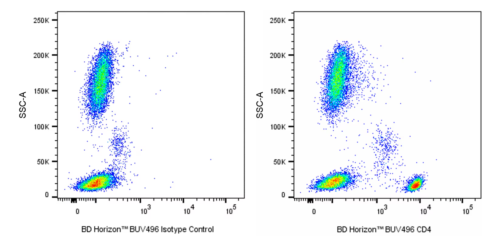
    Multiparameter flow cytometric analysis of CD4 expression on human peripheral blood leucocyte populations. Whole blood was stained with either BD Horizon™ BUV496 Mouse IgG1, κ Isotype Control (Cat. No. 612949; Left Plot) or BD Horizon BUV496 Mouse Anti-Human CD4 antibody (Cat. No. 612936/612937; Right Plot). The erythrocytes were lysed with BD FACS™ Lysing Solution (Cat. No. 349202). A two-parameter pseudocolor density plot showing the correlated expression of CD4 (or Ig Isotype control staining) versus side light-scatter (SSC-A) signals was derived from gated events with the forward and side light-scatter characteristics of intact leucocyte populations. Flow cytometry and data analysis were performed using a BD LSRFortessa™ X-20 Cell Analyzer System and FlowJo™ software.

    声明 :本官网所有报价均为常温或者蓝冰运输价格,如有产品需要干冰运输,需另外加收干冰运输费。
    货号:
    612936
    一键复制
    询价
    供应商现货
    25Tst
    100Tst
    选择数量
    当前规格1件起购 
    配送至
    预计5-6个工作日送达,快递: 免运费,若需干冰额外收费