

 全部商品分类
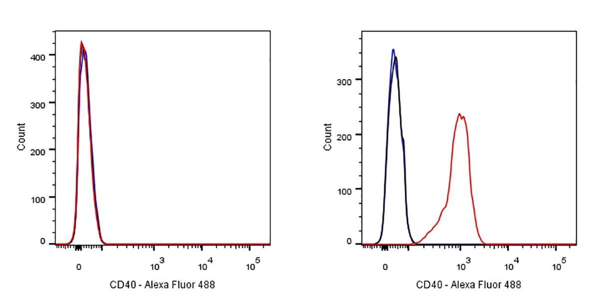


 用小程序,查商品更便捷


FCM
1:500IHC-P
1:100-500IP
1:25WB
1:1000
参考图片
Flow cytometric analysis of Jurkat (left) / Ramos (right) cells labelling CD40 antibody at 1/500 dilution (0.1ug)/ (red) compared with a Rabbit monoclonal IgG (Black) isotype control and an unlabelled control (cells without incubation with primary antibody and secondary antibody) (Blue). Goat Anti-Rabbit IgG Alexa Fluor® 488 was used as the secondary antibody. Negative control: Jurkat
IHC shows positive staining in paraffin-embedded human tonsil.
Anti-CD40 antibody was used at 1/100 dilution, followed by a Goat Anti-Rabbit IgG H&L (HRP) ready to use. Counterstained with hematoxylin.
Heat mediated antigen retrieval with Tris/EDTA buffer pH9.0 was performed before commencing with IHC staining protocol.
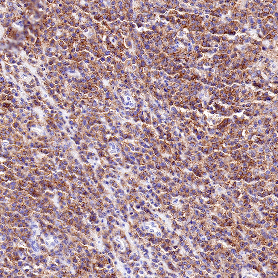
IHC shows positive staining in paraffin-embedded human spleen.
Anti-CD40 antibody was used at 1/100 dilution, followed by a Goat Anti-Rabbit IgG H&L (HRP) ready to use. Counterstained with hematoxylin.
Heat mediated antigen retrieval with Tris/EDTA buffer pH9.0 was performed before commencing with IHC staining protocol.
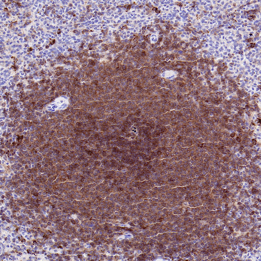
IHC shows positive staining in paraffin-embedded human diffuse large B-cell lymphoma.
Anti-CD40 antibody was used at 1/500 dilution, followed by a Goat Anti-Rabbit IgG H&L (HRP) ready to use. Counterstained with hematoxylin.
Heat mediated antigen retrieval with Tris/EDTA buffer pH9.0 was performed before commencing with IHC staining protocol.
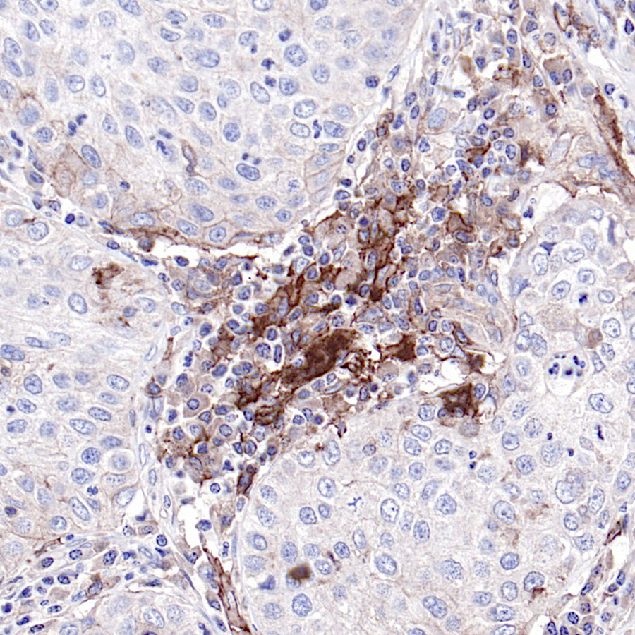
IHC shows positive staining in paraffin-embedded human lung squamous cancer.
Anti-CD40 antibody was used at 1/100 dilution, followed by a Goat Anti-Rabbit IgG H&L (HRP) ready to use. Counterstained with hematoxylin.
Heat mediated antigen retrieval with Tris/EDTA buffer pH9.0 was performed before commencing with IHC staining protocol.
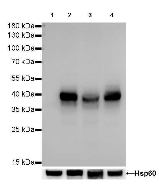
WB result of CD40 Rabbit mAb
Primary antibody:CD40 Rabbit mAb at 1/1000 dilution
Lane 1: Jurkat whole cell lysate 20 µg
Lane 2: Raji whole cell lysate 20 µg
Lane 3: Ramos whole cell lysate 20 µg
Lane 4: Daudi whole cell lysate 20 µg
Negative control: Jurkat whole cell lysate
Secondary antibody: Goat Anti-Rabbit IgG, (H+L), HRP conjugated at 1/10000 dilution
Predicted MW: 42 kDa
Observed MW: 39 kDa
Exposure time: 40s
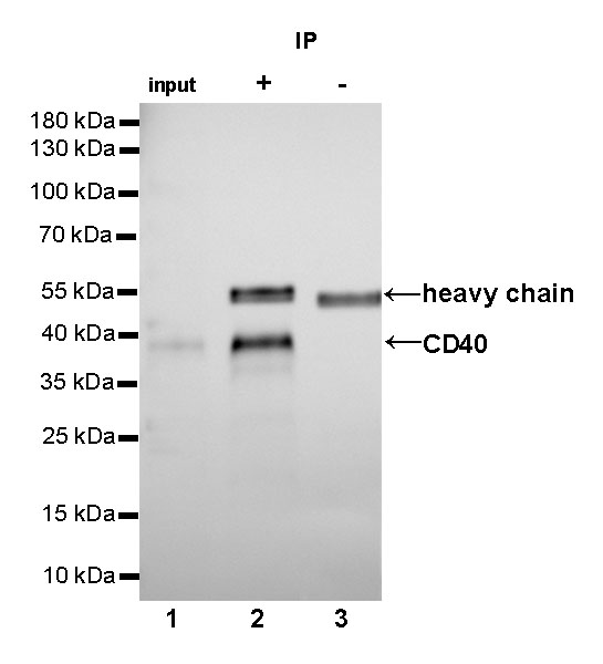
CD40 Rabbit mAb at 1/25 dilution (2µg) immunoprecipitating CD40 in 0.4mg Raji whole cell lysate.
Western blot was performed on the immunoprecipitate using CD40 Rabbit mAb at 1/1000 dilution.
Secondary antibody (HRP) for IP was used at 1/400 dilution.
Lane 1: Raji whole cell lysate 10µg (input)
Lane 2 (+): CD40 Rabbit mAb IP in Raji whole cell lysate
Lane 3 (-): Rabbit monoclonal IgG IP in Raji whole cell lysate
Predicted MW: 42 kDa
Observed MW: 39 kDa
Exposure time: 50s