


 全部商品分类


1/2
BD Pharmingen™ PE-Cy™7 Mouse Anti-Human CD68

品牌: BD Pharmingen
 下载产品说明书 下载SDS
 用小程序,查商品更便捷


 收藏
收藏
 对比
对比 咨询
咨询反应种属:
Human (QC Testing)
来源宿主:
Mouse BALB/c IgG2b, κ
产品介绍
产品使用步骤推荐 产品介绍
产品信息
荧光素标记
抗原名称
CD68

宿主
Mouse BALB/c IgG2b, κ
免疫原
PHA-stimulated Human PBMC
简单描述
The Y1/82A monoclonal antibody specifically binds to CD68 which is also known as, Scavenger receptor class D member 1 (SCARD1), Macrosialin, or GP110. CD68 is a cell surface 110 kDa-type I-transmembrane glycoprotein that is primarily expressed in cytoplasmic granules of monocytes, macrophages, dendritic cells, granulocytes, myeloid progenitor cells and, reportedly, a subset of CD34-positive hemopoietic bone marrow progenitor cells. CD68 belongs to the sialomucin family and serves as a scavenger receptor that can bind and internalize oxidized low density lipoproteins (LDL). This antibody is useful in studies of myeloid cell development and function.
商品描述
Y1/82A
The Y1/82A monoclonal antibody specifically binds to CD68 which is also known as, Scavenger receptor class D member 1 (SCARD1), Macrosialin, or GP110. CD68 is a cell surface 110 kDa-type I-transmembrane glycoprotein that is primarily expressed in cytoplasmic granules of monocytes, macrophages, dendritic cells, granulocytes, myeloid progenitor cells and, reportedly, a subset of CD34-positive hemopoietic bone marrow progenitor cells. CD68 belongs to the sialomucin family and serves as a scavenger receptor that can bind and internalize oxidized low density lipoproteins (LDL). This antibody is useful in studies of myeloid cell development and function.
同种型
Mouse BALB/c IgG2b, κ
克隆号
克隆 Y1/82A (RUO)
产品详情
PE-Cy7
PE-Cy7 dye is a part of the BD PE family of dyes. This tandem fluorochrome is comprised of a R-Phycoerythrin (PE) donor that has excitation maxima (Ex Max) of 496-nm and 566-nm and an acceptor dye, Cy™7, with an emission maximum (Em Max) at 781-nm. PE can be excited by the Blue (488-nm), Green (532-nm) and yellow-green (561-nm) lasers and detected using an optical filter centered near 781 nm (e.g., a 760/60-nm bandpass filter). The donor dye can be excited by the Blue (488-nm), Green (532-nm) and yellow-green (561-nm) lasers and the acceptor dye can be excited by the Red (627–640-nm) laser resulting in cross-laser excitation and fluorescence spillover. Please ensure that your instrument’s configurations (lasers and optical filters) are appropriate for this dye.

PE-Cy7
Yellow-Green 488 nm, 532 nm, 561 nm
496 nm, 566 nm
781 nm
应用
实验应用
Intracellular staining (flow cytometry) (Routinely Tested)
推荐用量
5 µl
反应种属
Human (QC Testing)
目标/特异性
CD68
背景
别名
GP110; Macrosialin; SCARD1; Scavenger receptor class D, member 1
制备和贮存
存储溶液
Aqueous buffered solution containing BSA and ≤0.09% sodium azide.
保存方式
Aqueous buffered solution containing BSA and ≤0.09% sodium azide.
文献
文献
研发参考(5)
1. Davey FR, Cordell JL, Erber WN, Pulford KA, Gatter KC, Mason DY. Monoclonal antibody (Y1/82A) with specificity towards peripheral blood monocytes and tissue macrophages. J Clin Pathol. 1988; 41(7):753-758. (Immunogen: Flow cytometry, Immunohistochemistry).
2. Davey FR, Erber WN, Gatter KC, Mason DY. The use of monoclonal antibody Y1/82A in the identification of acute myeloblastic and monocytic leukemias. Am J Clin Pathol. 1988; 89(1):76-80. (Clone-specific: Immunohistochemistry).
3. Kishimoto T. Tadamitsu Kishimoto .. et al., ed. Leucocyte typing VI : white cell differentiation antigens : proceedings of the sixth international workshop and conference held in Kobe, Japan, 10-14 November 1996. New York: Garland Pub.; 1997.
4. Stockinger H. Cluster report: CD68. In: Knapp W. W. Knapp .. et al., ed. Leucocyte typing IV : white cell differentiation antigens. Oxford New York: Oxford University Press; 1989:841-843.
5. Zola H. Leukocyte and stromal cell molecules : the CD markers. Hoboken, N.J.: Wiley-Liss; 2007.
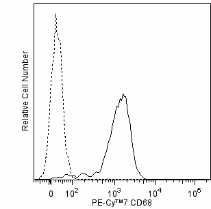
参考图片
Flow cytometric analysis of CD68 expression by human peripheral blood monocytes. Human peripheral blood mononuclear cells were fixed with BD Cytofix™ Fixation Buffer (Cat. No. 554655). After washing with BD Perm/Wash™ Buffer (Cat. No. 554723), the cells were stained with either PE-Cy™7 Mouse IgG2b, κ Isotype Control (Cat. No. 560542; dashed line histogram) or PE-Cy™7 Mouse Anti-Human CD68 antibody (Cat. No. 565595; solid line histogram). The fluorescence histogram showing CD68 expression (or Ig Isotype control staining) was derived from gated events with the forward and side light-scatter characteristics of intact monocytes. Flow cytometric analysis was performed using a BD™ LSR II Flow Cytometer System.
声明 :本官网所有报价均为常温或者蓝冰运输价格,如有产品需要干冰运输,需另外加收干冰运输费。