老带新
BD Pharmingen™ PE-Cy™7 Hamster Anti-Mouse CD69
1/2

BD Pharmingen™ PE-Cy™7 Hamster Anti-Mouse CD69

分享
品牌: BD Pharmingen
pdf 下载产品说明书
pdf 下载SDS
用小程序,查商品更便捷
收藏
对比对比
咨询咨询
反应种属:
Mouse (QC Testing)
来源宿主:
Armenian Hamster IgG1, λ3
展开
产品介绍
产品介绍
产品信息
荧光素标记
抗原名称
CD69
宿主
Armenian Hamster IgG1, λ3
免疫原
Mouse Dendritic Epidermal T Cell Line Y245
简单描述
The H1.2F3 monoclonal antibody specifically binds to CD69 (Very Early Activation antigen), an 85 kDa disulfide-linked homodimer of differentially glycosylated subunits. CD69 is a C-type lectin, most closely related to the NKR-P1 and Ly-49 NK cell-activation molecules. Its expression is rapidly induced upon activation of lymphocytes (T, B, NK, and NK-T cells), neutrophils, and macrophages. CD69 is expressed also on thymocytes that are undergoing positive selection; its role in that process is unclear. H1.2F3 mAb augments PMA-induced T-cell stimulation and IFN-γ-induced macrophage stimulation. IL-2-activated NK cells express CD69, and H1.2F3 mAb induces redirected lysis of FcR-bearing target cells by NK cells.
商品描述
H1.2F3 The H1.2F3 monoclonal antibody specifically binds to CD69 (Very Early Activation antigen), an 85 kDa disulfide-linked homodimer of differentially glycosylated subunits. CD69 is a C-type lectin, most closely related to the NKR-P1 and Ly-49 NK cell-activation molecules. Its expression is rapidly induced upon activation of lymphocytes (T, B, NK, and NK-T cells), neutrophils, and macrophages. CD69 is expressed also on thymocytes that are undergoing positive selection; its role in that process is unclear. H1.2F3 mAb augments PMA-induced T-cell stimulation and IFN-γ-induced macrophage stimulation. IL-2-activated NK cells express CD69, and H1.2F3 mAb induces redirected lysis of FcR-bearing target cells by NK cells.
同种型
Armenian Hamster IgG1, λ3
克隆号
克隆 H1.2F3 (RUO)
浓度
0.2 mg/ml
产品详情
PE-Cy7
PE-Cy7 dye is a part of the BD PE family of dyes. This tandem fluorochrome is comprised of a R-Phycoerythrin (PE) donor that has excitation maxima (Ex Max) of 496-nm and 566-nm and an acceptor dye, Cy™7, with an emission maximum (Em Max) at 781-nm. PE can be excited by the Blue (488-nm), Green (532-nm) and yellow-green (561-nm) lasers and detected using an optical filter centered near 781 nm (e.g., a 760/60-nm bandpass filter). The donor dye can be excited by the Blue (488-nm), Green (532-nm) and yellow-green (561-nm) lasers and the acceptor dye can be excited by the Red (627–640-nm) laser resulting in cross-laser excitation and fluorescence spillover. Please ensure that your instrument’s configurations (lasers and optical filters) are appropriate for this dye.
PE-Cy7
Yellow-Green 561 nm
496 nm, 566 nm
781 nm
应用
实验应用
Flow cytometry (Routinely Tested)
反应种属
Mouse (QC Testing)
目标/特异性
CD69
背景
别名
VEA; Very Early Activation Antigen; AIM; Activation Induced Molecule
制备和贮存
存储溶液
Aqueous buffered solution containing ≤0.09% sodium azide.
保存方式
Aqueous buffered solution containing ≤0.09% sodium azide.
文献
文献
研发参考(16) 1. Bendelac A, Matzinger P, Seder RA, Paul WE, Schwartz RH. Activation events during thymic selection. J Exp Med. 1992; 175(3):731-742. (Biology). 2. Brandle D, Muller S, Muller C, Hengartner H, Pircher H. Regulation of RAG-1 and CD69 expression in the thymus during positive and negative selection. Eur J Immunol. 1994; 24(1):145-151. (Biology). 3. Gabor MJ, Godfrey DI, Scollay R. Recent thymic emigrants are distinct from most medullary thymocytes. Eur J Immunol. 1997; 27(8):2010-2050. (Biology). 4. Karlhofer FM, Yokoyama WM. Stimulation of murine natural killer (NK) cells by a monoclonal antibody specific for the NK1.1 antigen. IL-2-activated NK cells possess additional specific stimulation pathways. J Immunol. 1991; 146(10):3662-3673. (Clone-specific: Cytotoxicity). 5. Keefe R, Dave V, Allman D, Wiest D, Kappes DJ. Regulation of lineage commitment distinct from positive selection. Science. 1999; 286(5442):1149-1153. (Biology). 6. Lauzurica P, Sancho D, Torres M, et al. Phenotypic and functional characteristics of hematopoietic cell lineages in CD69-deficient mice. Blood. 2000; 95(7):2312-2320. (Biology). 7. Merkenschlager M, Graf D, Lovatt M, Bommhardt U, Zamoyska R, Fisher AG. How many thymocytes audition for selection. J Exp Med. 1997; 186(7):1149-1158. (Biology). 8. Nishimura T, Kitamura H, Iwakabe K, et al. The interface between innate and acquired immunity: glycolipid antigen presentation by CD1d-expressing dendritic cells to NKT cells induces the differentiation of antigen-specific cytotoxic T lymphocytes. Int Immunol. 2000; 12(7):987-994. (Biology). 9. Punt JA, Suzuki H, Granger LG, Sharrow SO, Singer A. Lineage commitment in the thymus: only the most differentiated (TCRhibcl-2hi) subset of CD4+CD8+ thymocytes has selectively terminated CD4 or CD8 synthesis. J Exp Med. 1996; 184(6):2091-2099. (Biology). 10. Roederer M, Kantor AB, Parks DR, Herzenberg LA. Cy7PE and Cy7APC: bright new probes for immunofluorescence. Cytometry. 1996; 24(3):191-197. (Biology). 11. Sobel ES, Yokoyama WM, Shevach EM, Eisenberg RA, Cohen PL. Aberrant expression of the very early activation antigen on MRL/Mp-lpr/lpr lymphocytes. J Immunol. 1993; 150(2):673-682. (Clone-specific: (Co)-stimulation). 12. Wilkinson RW, Anderson G, Owen JJ, Jenkinson EJ. Positive selection of thymocytes involves sustained interactions with the thymic microenvironment. J Immunol. 1995; 155(11):5234-5240. (Biology). 13. Yokoyama WM, Koning F, Kehn PJ, et al. Characterization of a cell surface-expressed disulfide-linked dimer involved in murine T cell activation. J Immunol. 1988; 141(2):369-376. (Immunogen: (Co)-stimulation). 14. Yokoyama WM, Maxfield SR, Shevach EM. Very early (VEA) and very late (VLA) activation antigens have distinct functions in T lymphocyte activation. Immunol Rev. 1989; 109:153-176. (Biology). 15. Ziegler SF, Levin SD, Johnson L, et al. The mouse CD69 gene. Structure, expression, and mapping to the NK gene complex. J Immunol. 1994; 152(3):1228-1236. (Biology). 16. Ziegler SF, Ramsdell F, Alderson MR. The activation antigen CD69. Stem Cells. 1994; 12(5):456-465. (Biology).

参考图片

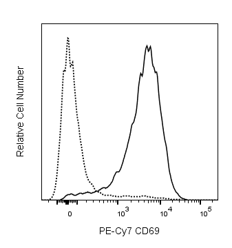
Flow cytometric analysis of CD69 expression on stimulated mouse splenocytes. Mouse splenic leucocytes were stimulated for 5 hours at 37°C with Phorbol 12-Myristate 13-Acetate (PMA, 10 ng/mL; Sigma-Aldrich, Cat. No. P-8139). The cells were then stained with either PE-Cy7 Hamster IgG1, λ1 Isotype Control (Cat. No. 557798; dashed line histogram) or PE-Cy7 Hamster Anti-Mouse CD69 antibody (Cat. No. 552879/561930; solid line histogram) at 0.25 µg/test. DAPI (4',6-Diamidino-2-Phenylindole, Dihydrochloride) Solution (Cat. No. 564907) was added to cells right before analysis. The fluorescence histogram showing CD69 expression (or Ig Isotype control staining) was derived from gated events with the forward and side light-scatter characteristics of viable (DAPI-negative) leucocytes. Flow cytometry and data analysis were performed using a BD FACSCanto™ II System and FlowJo™ software. Data shown on this Technical Data Sheet are not lot specific.

声明 :本官网所有报价均为常温或者蓝冰运输价格,如有产品需要干冰运输,需另外加收干冰运输费。
货号:
552879
一键复制
询价
优宁维现货
25ug
100ug
选择数量
当前规格1件起购 
配送至
预计3-4个工作日送达,快递: 免运费,若需干冰额外收费