The H1.2F3 monoclonal antibody specifically binds to CD69 (Very Early Activation antigen), an 85 kDa disulfide-linked homodimer of differentially glycosylated subunits. CD69 is a C-type lectin, most closely related to the NKR-P1 and Ly-49 NK cell-activation molecules. Its expression is rapidly induced upon activation of lymphocytes (T, B, NK, and NK-T cells), neutrophils, and macrophages. CD69 is expressed also on thymocytes that are undergoing positive selection; its role in that process is unclear. H1.2F3 mAb augments PMA-induced T-cell stimulation and IFN-γ-induced macrophage stimulation. IL-2-activated NK cells express CD69, and H1.2F3 mAb induces redirected lysis of FcR-bearing target cells by NK cells.
商品描述
H1.2F3
The H1.2F3 monoclonal antibody specifically binds to CD69 (Very Early Activation antigen), an 85 kDa disulfide-linked homodimer of differentially glycosylated subunits. CD69 is a C-type lectin, most closely related to the NKR-P1 and Ly-49 NK cell-activation molecules. Its expression is rapidly induced upon activation of lymphocytes (T, B, NK, and NK-T cells), neutrophils, and macrophages. CD69 is expressed also on thymocytes that are undergoing positive selection; its role in that process is unclear. H1.2F3 mAb augments PMA-induced T-cell stimulation and IFN-γ-induced macrophage stimulation. IL-2-activated NK cells express CD69, and H1.2F3 mAb induces redirected lysis of FcR-bearing target cells by NK cells.
同种型
Armenian Hamster IgG1, λ3
克隆号
克隆 H1.2F3 (RUO)
浓度
0.2 mg/ml
产品详情
PE
R-Phycoerythrin (PE), is part of the BD family of Phycobiliprotein dyes. This fluorochrome is a multimeric fluorescent phycobiliprotein with excitation maximum (Ex Max) of 496 nm and 566 nm and an emission maximum (Em Max) at 576 nm. PE is designed to be excited by the Blue (488 nm), Green (532 nm) and Yellow-Green (561 nm) lasers and detected using an optical filter centered near 575 nm (e.g., a 575/26-nm bandpass filter). As PE is excited by multiple lasers, this can result in cross-laser excitation and fluorescence spillover on instruments with various combinations of Blue, Green, and Yellow-Green lasers. Please ensure that your instrument’s configurations (lasers and optical filters) are appropriate for this dye.
PE
Yellow-Green 488 nm, 532 nm, 561 nm
496 nm, 566 nm
576 nm
应用
实验应用
Flow cytometry (Routinely Tested)
反应种属
Mouse (QC Testing)
目标/特异性
CD69
背景
别名
VEA; Very Early Activation Antigen; AIM; Activation Induced Molecule
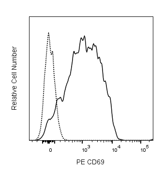
Flow cytometric analysis of CD69 expression on stimulated mouse splenocytes. Mouse splenic leucocytes were stimulated for 5 hours at 37°C with Phorbol 12-Myristate 13-Acetate (PMA, 10 ng/mL; Sigma-Aldrich, Cat. No. P-8139). The stimulated cells were preincubated with Purified Rat Anti-Mouse CD16/CD32 antibody (Mouse BD Fc Block™) (Cat. No. 553141/553142). The cells were then stained with either PE Hamster IgG1, λ1 Isotype Control (Cat. No. 553954; dashed line histogram) or PE Hamster Anti-Mouse CD69 antibody (Cat. No. 561932/553237; solid line histogram) at 0.06 µg/test. DAPI (4',6-Diamidino-2-Phenylindole, Dihydrochloride) Solution (Cat. No. 564907) was added to cells right before analysis. The fluorescence histogram showing CD69 expression (or Ig Isotype control staining) was derived from gated events with the forward and side light-scatter characteristics of viable (DAPI-negative) stimulated leucocytes. Flow cytometry and data analysis was performed using a BD FACSCelesta™ Flow Cytometer System and FloJo™ software.


 全部商品分类


 下载产品说明书
 用小程序,查商品更便捷


 收藏
收藏
 对比
对比 咨询
咨询
