The FN50 monoclonal antibody specifically binds to human CD69. CD69 is also known as activation-induced molecule (AIM), early activation antigen (EA-1), very early activation antigen (VEA), C-type lectin domain family 2 member C (CLEC2C), MLR-3, GP32/28 and Leu-23. CD69 is a transmembrane type II homodimer receptor. CD69 is comprised of disulfide-linked, differentially glycosylated core protein subunits that are approximately 28 and 34 kDa in size. Each subunit contains a C-type lectin domain. CD69 is expressed on activated T, B, and natural killer (NK) lymphocytes, thymocytes, neutrophils, eosinophils and platelets. In normal peripheral blood, a small and variable percentage of lymphocytes typically express detectable membrane CD69 antigen. Upon activation, CD69 antigen expression increases on lymphocytes. Peak CD69 expression generally occurs within 18 hours of activation, preceding the appearance of HLA-DR, IL-2Rα (CD25) and transferrin receptor (CD71). CD69 is highly expressed on the bright CD3+ subset of thymocytes. FN50 monoclonal antibody labels NK cells and most lymphocytes of the follicular mantle and perifollicular/interfollicular zone as well as germinal center T cells of lymph nodes and tonsils. Studies indicate that CD69 serves as a signaling receptor in the activation of a variety of cell types.
Clone FN50 reacts with the human form of the 28/34 kDa dimeric glycoprotein expressed early during activation of lymphocytes, monocytes, and platelets. It also cross-reacts with a subset of peripheral blood mononuclear cells (lymphocytes and monocytes) of rhesus and cynomolgus macaque monkeys. The distribution on lymphocytes is similar to that observed with human peripheral blood lymphocytes with the majority of the cells demonstrating an increase in FN50 positivity following overnight incubation with phorbol myristrate acetate (PMA).
商品描述
FN50
The FN50 monoclonal antibody specifically binds to human CD69. CD69 is also known as activation-induced molecule (AIM), early activation antigen (EA-1), very early activation antigen (VEA), C-type lectin domain family 2 member C (CLEC2C), MLR-3, GP32/28 and Leu-23. CD69 is a transmembrane type II homodimer receptor. CD69 is comprised of disulfide-linked, differentially glycosylated core protein subunits that are approximately 28 and 34 kDa in size. Each subunit contains a C-type lectin domain. CD69 is expressed on activated T, B, and natural killer (NK) lymphocytes, thymocytes, neutrophils, eosinophils and platelets. In normal peripheral blood, a small and variable percentage of lymphocytes typically express detectable membrane CD69 antigen. Upon activation, CD69 antigen expression increases on lymphocytes. Peak CD69 expression generally occurs within 18 hours of activation, preceding the appearance of HLA-DR, IL-2Rα (CD25) and transferrin receptor (CD71). CD69 is highly expressed on the bright CD3+ subset of thymocytes. FN50 monoclonal antibody labels NK cells and most lymphocytes of the follicular mantle and perifollicular/interfollicular zone as well as germinal center T cells of lymph nodes and tonsils. Studies indicate that CD69 serves as a signaling receptor in the activation of a variety of cell types.
Clone FN50 reacts with the human form of the 28/34 kDa dimeric glycoprotein expressed early during activation of lymphocytes, monocytes, and platelets. It also cross-reacts with a subset of peripheral blood mononuclear cells (lymphocytes and monocytes) of rhesus and cynomolgus macaque monkeys. The distribution on lymphocytes is similar to that observed with human peripheral blood lymphocytes with the majority of the cells demonstrating an increase in FN50 positivity following overnight incubation with phorbol myristrate acetate (PMA).
同种型
Mouse IgG1, κ
克隆号
克隆 FN50 (also known as FN 50) (RUO)
产品详情
PE-Cy7
PE-Cy7 dye is a part of the BD PE family of dyes. This tandem fluorochrome is comprised of a R-Phycoerythrin (PE) donor that has excitation maxima (Ex Max) of 496-nm and 566-nm and an acceptor dye, Cy™7, with an emission maximum (Em Max) at 781-nm. PE can be excited by the Blue (488-nm), Green (532-nm) and yellow-green (561-nm) lasers and detected using an optical filter centered near 781 nm (e.g., a 760/60-nm bandpass filter). The donor dye can be excited by the Blue (488-nm), Green (532-nm) and yellow-green (561-nm) lasers and the acceptor dye can be excited by the Red (627–640-nm) laser resulting in cross-laser excitation and fluorescence spillover. Please ensure that your instrument’s configurations (lasers and optical filters) are appropriate for this dye.
PE-Cy7
Yellow-Green 561 nm
496 nm, 566 nm
781 nm
应用
实验应用
Flow cytometry (Routinely Tested)
推荐用量
5 µl
反应种属
Human (QC Testing), Rhesus,Cynomolgus,Baboon (Tested in Development)
Aqueous buffered solution containing BSA and ≤0.09% sodium azide.
保存方式
Aqueous buffered solution containing BSA and ≤0.09% sodium azide.
文献
文献
研发参考(3)
1. Knapp W. W. Knapp .. et al., ed. Leucocyte typing IV : white cell differentiation antigens. Oxford New York: Oxford University Press; 1989:1-1182.
2. Roederer M, Kantor AB, Parks DR, Herzenberg LA. Cy7PE and Cy7APC: bright new probes for immunofluorescence. Cytometry. 1996; 24(3):191-197. (Biology).
3. Schlossman SF. Stuart F. Schlossman .. et al., ed. Leucocyte typing V : white cell differentiation antigens : proceedings of the fifth international workshop and conference held in Boston, USA, 3-7 November, 1993. Oxford: Oxford University Press; 1995.
参考图片
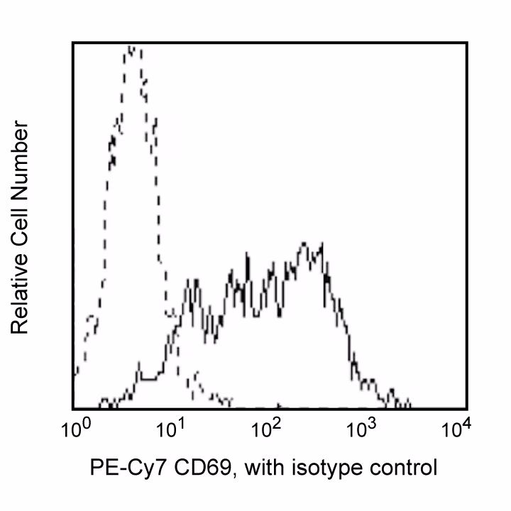
Flow cytometric analysis of CD69 expressed on peripheral blood mononuclear cells. Human PBMC were stimulated for 24 hours with phytohemagglutinin for 1 day. Cells were then stained with either a PE-Cy™7 Mouse IgG1, κ Isotype control (Cat. No. 557872; dashed line histogram) or with the PE-Cy™7 Mouse Anti-Human CD69 antibody (Cat. No. 557745/561928/560712; solid line histogram). The fluorescence histograms were derived from gated events with the forward and side light-scatter characteristics of viable lymphocytes.
Flow cytometric analysis of CD69 expressed on peripheral blood mononuclear cells. Human PBMC were stimulated for 24 hours with phytohemagglutinin for 1 day. Cells were then stained with either a PE-Cy™7 Mouse IgG1, κ Isotype control (Cat. No. 557872; dashed line histogram) or with the PE-Cy™7 Mouse Anti-Human CD69 antibody (Cat. No. 557745/561928/560712; solid line histogram). The fluorescence histograms were derived from gated events with the forward and side light-scatter characteristics of viable lymphocytes.


 全部商品分类


 下载产品说明书
 用小程序,查商品更便捷


 收藏
收藏
 对比
对比 咨询
咨询
