




 全部商品分类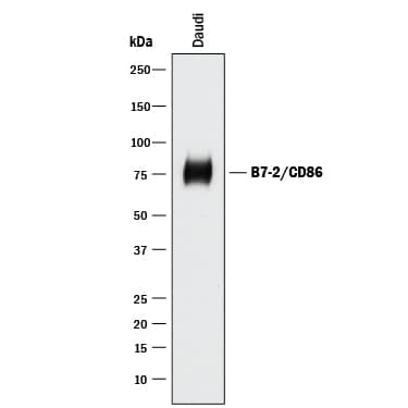




Human B7-2/CD86 Affinity Purified Polyclonal Ab (25 ug)


 用小程序,查商品更便捷



 收藏
收藏
 对比
对比 咨询
咨询
Ala23-His244
Accession # P42081
Human B7-2/CD86 Antibody Summary
Ala23-His244
Accession # P42081
Applications
Please Note: Optimal dilutions should be determined by each laboratory for each application. General Protocols are available in the Technical Information section on our website.
Scientific Data
 View Larger
Detection of Human B7‑2/CD86 by Western Blot. Western blot shows lysates of Daudi human Burkitt's lymphoma cell line. PVDF membrane was probed with 0.5 µg/mL of Goat Anti-Human B7-2/CD86 Antigen Affinity-purified Polyclonal Antibody (Catalog # AF-141-NA) followed by HRP-conjugated Anti-Goat IgG Secondary Antibody (Catalog # HAF017). A specific band was detected for B7-2/CD86 at approximately 75 kDa (as indicated). This experiment was conducted under reducing conditions and using Immunoblot Buffer Group 1.
 View Larger
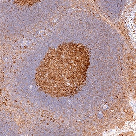
B7‑2/CD86 in Human Tonsil. B7-2/CD86 was detected in immersion fixed paraffin-embedded sections of human tonsil using Goat Anti-Human B7-2/CD86 Antigen Affinity-purified Polyclonal Antibody (Catalog # AF-141-NA) at 15 µg/mL overnight at 4 °C. Tissue was stained using the Anti-Goat HRP-DAB Cell & Tissue Staining Kit (brown; Catalog # CTS008) and counterstained with hematoxylin (blue). View our protocol for Chromogenic IHC Staining of immersion fixed paraffin-embedded Tissue Sections.
 View Larger
B7‑2/CD86 in Human Tonsil. B7-2/CD86 was detected in immersion fixed paraffin-embedded sections of human tonsil using Goat Anti-Human B7-2/CD86 Antigen Affinity-purified Polyclonal Antibody (Catalog # AF-141-NA) at 3 µg/mL for 1 hour at room temperature followed by incubation with the Anti-Goat IgG VisUCyte™ HRP Polymer Antibody (Catalog # VC004). Tissue was stained using DAB (brown) and counterstained with hematoxylin (blue). Specific staining was localized to lymphocytes. View our protocol for IHC Staining with VisUCyte HRP Polymer Detection Reagents.
 View Larger
IL‑2 secretion Induced by B7‑2/CD86 and Neutralization by Human B7‑2/CD86 Antibody. Recombinant Human B7-2/CD86 Fc Chimera (Catalog # 141-B2) co-stimulates IL-2 secretion in the Jurkat human acute T cell leukemia cell line in the presence of PHA in a dose-dependent manner (orange line), as measured by the Human IL-2 Quantikine ELISA Kit (Catalog # D2050). IL-2 secretion elicited by Recombinant Human B7-2/CD86 Fc Chimera (2 µg/mL) and PHA (10 µg/mL) is neutralized (green line) by increasing concentrations of Goat Anti-Human B7-2/CD86 Antigen Affinity-purified Polyclonal Antibody (Catalog # AF-141-NA). The ND50 is typically 0.25-1.25 µg/mL.
 View Larger
Western Blot Shows Human B7‑2/CD86 Specificity by Using Knockout Cell Line. Western blot shows lysates of Ramos human Burkitt's lymphoma parental cell line and B7-2/CD86 knockout Ramos cell line (KO). PVDF membrane was probed with 0.5 µg/mL of Goat Anti-Human B7-2/CD86 Antigen Affinity-purified Polyclonal Antibody (Catalog # AF-141-NA) followed by HRP-conjugated Anti-Goat IgG Secondary Antibody (Catalog # HAF017). A specific band was detected for B7-2/CD86 at approximately 74 kDa (as indicated) in the parental Ramos cell line, but is not detectable in knockout Ramos cell line. GAPDH (Catalog # AF5718) is shown as a loading control. This experiment was conducted under reducing conditions and using Immunoblot Buffer Group 1.
Reconstitution Calculator
Preparation and Storage
- 12 months from date of receipt, -20 to -70 °C as supplied.
- 1 month, 2 to 8 °C under sterile conditions after reconstitution.
- 6 months, -20 to -70 °C under sterile conditions after reconstitution.
Background: B7-2/CD86
B7-1 and B7-2, together with their receptors CD28 and CTLA-4, constitute one of the dominant costimulatory pathways that regulate T- and B-cell responses. Although both CTLA-4 and CD28 can bind to the same ligands, CTLA-4 binds to B7-1 and B7-2 with a 20‑100 fold higher affinity than CD28 and is involved in the down‑regulation of the immune response. B7-1 is expressed on activated B cells, activated T cells, and macrophages. B7-2 is constitutively expressed on interdigitating dendritic cells, Langerhans cells, peripheral blood dendritic cells, memory B cells, and germinal center B cells. Additionally, B7-2 is expressed at low levels on monocytes and can be up-regulated through interferon gamma. B7-1 and B7-2 are both members of the immunoglobulin superfamily. Human B7-2 is a 329 amino acid (aa) protein containing a putative 23 aa signal peptide, a 224 aa extracellular domain, a 21 aa transmembrane domain, and a 61 aa cytoplasmic domain. Human B7-2 and B7-1 share 26% amino acid identity. Human and mouse B7-2 share 50% amino acid identity. However, it has been observed that both human and mouse B7‑1 and
B7‑2 can bind to either human or mouse CD28 and CTLA-4, suggesting that there are conserved amino acids which form the B7-1/B7-2/CD28/CTLA-4 critical binding sites.
- Azuma, M. et al. (1993) Nature 366:76.
- Freeman, G.J. et al. (1993) Science 262:909.
- Freeman, G. et al. (1991) J. Exp. Med. 174:625.
- Selvakumar, A. et al. (1993) Immunogenetics 38:292.
- Chen, C. et al. (1994) J. Immunol. 152:4929.
- Freeman, G.J. et al. (1993) J. Exp. Med. 178:2185.
Product Datasheets
Background: B7-2/CD86
B7-1 and B7-2, together with their receptors CD28 and CTLA-4, constitute one of the dominant costimulatory pathways that regulate T- and B-cell responses. Although both CTLA-4 and CD28 can bind to the same ligands, CTLA-4 binds to B7-1 and B7-2 with a 20‑100 fold higher affinity than CD28 and is involved in the down‑regulation of the immune response. B7-1 is expressed on activated B cells, activated T cells, and macrophages. B7-2 is constitutively expressed on interdigitating dendritic cells, Langerhans cells, peripheral blood dendritic cells, memory B cells, and germinal center B cells. Additionally, B7-2 is expressed at low levels on monocytes and can be up-regulated through interferon gamma. B7-1 and B7-2 are both members of the immunoglobulin superfamily. Human B7-2 is a 329 amino acid (aa) protein containing a putative 23 aa signal peptide, a 224 aa extracellular domain, a 21 aa transmembrane domain, and a 61 aa cytoplasmic domain. Human B7-2 and B7-1 share 26% amino acid identity. Human and mouse B7-2 share 50% amino acid identity. However, it has been observed that both human and mouse B7‑1 and
B7‑2 can bind to either human or mouse CD28 and CTLA-4, suggesting that there are conserved amino acids which form the B7-1/B7-2/CD28/CTLA-4 critical binding sites.
- Azuma, M. et al. (1993) Nature 366:76.
- Freeman, G.J. et al. (1993) Science 262:909.
- Freeman, G. et al. (1991) J. Exp. Med. 174:625.
- Selvakumar, A. et al. (1993) Immunogenetics 38:292.
- Chen, C. et al. (1994) J. Immunol. 152:4929.
- Freeman, G.J. et al. (1993) J. Exp. Med. 178:2185.
Preparation and Storage
- 12 months from date of receipt, -20 to -70 °C as supplied.
- 1 month, 2 to 8 °C under sterile conditions after reconstitution.
- 6 months, -20 to -70 °C under sterile conditions after reconstitution.