


 全部商品分类


CDK Antibody Sampler Kit

 下载产品说明书 下载SDS
 用小程序,查商品更便捷


 收藏
收藏
 对比
对比 咨询
咨询
The CDK Antbody Sampler Kit provides and economical means of evaluating Cdk proteins. The kit contains enough primary and secondary antibodies to perform two western blot experiments.
参考图片
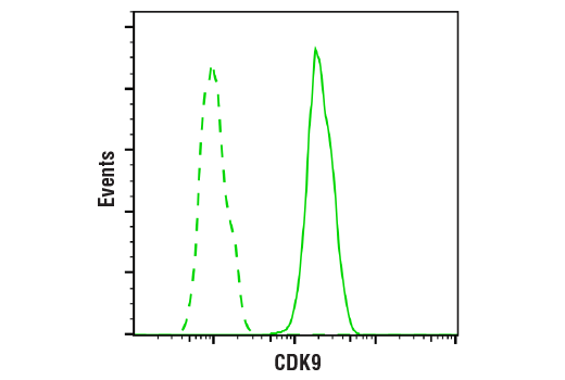
Flow cytometric analysis of HeLa cells using CDK9 (C12F7) Rabbit mAb (solid line) compared to concentration-matched Rabbit (DA1E) mAb IgG XP® Isotype Control #3900 (dashed line). Anti-rabbit IgG (H+L), F(ab')2 Fragment (Alexa Fluor® 488 Conjugate) #4412 was used as a secondary antibody.
Western blot analysis of extracts from various cell lines using CDK4 (D9G3E) Rabbit mAb.
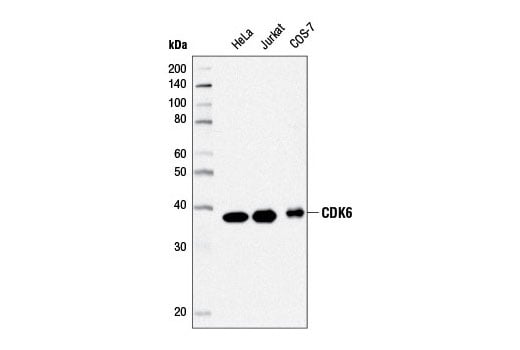
Western blot analysis of extracts from HeLa, Jurkat, and COS-7 cells using CDK6 (D4S8S) Rabbit mAb.
Simple Western™ analysis of lysates (0.1 mg/mL) from COS-7 cells using CDK6 (D4S8S) Rabbit mAb #13331. The virtual lane view (left) shows the target band (as indicated) at 1:10 and 1:50 dilutions of primary antibody. The corresponding electropherogram view (right) plots chemiluminescence by molecular weight along the capillary at 1:10 (blue line) and 1:50 (green line) dilutions of primary antibody. This experiment was performed under reducing conditions on the Jess™ Simple Western instrument from ProteinSimple, a BioTechne brand, using the 12-230 kDa separation module.
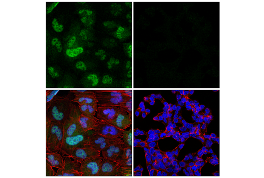
Confocal immunofluorescent analysis of T98G cells (left, positive) or VCaP cells (right, negative) using CDK6 (D4S8S) Rabbit mAb (green), DyLight™ 650 Phalloidin #12956 (red), and DAPI #4083 (blue).
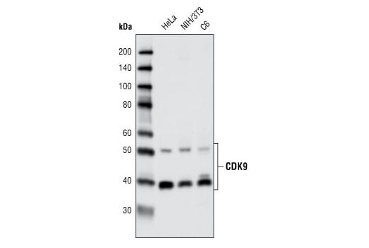
Western blot analysis of extracts from various cell types using CDK9 (C12F7) Rabbit mAb.
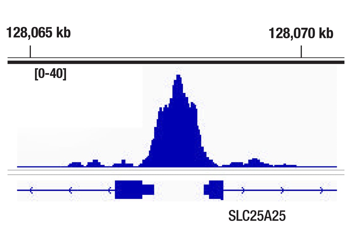
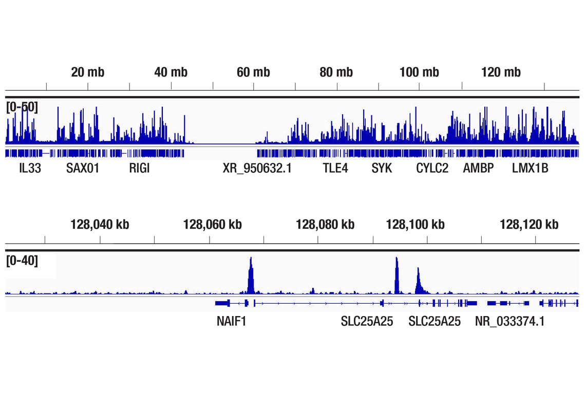
CUT&RUN was performed with HeLa cells and CDK9 (C12F7) Rabbit mAb, using CUT&RUN Assay Kit #86652. DNA Library was prepared using DNA Library Prep Kit for Illumina® (ChIP-seq, CUT&RUN) #56795. The figure shows binding across the SLC25A25 gene.
Enhanced cross-linking and immunoprecipitation (eCLIP) was performed with RNA from Hep G2 cells and CDK9 (C12F7) Rabbit mAb using a protocol based on the RBP-eCLIP method from Eclipsebio. The figure shows binding across the PUM2 transcript. Data is kindly provided by the laboratory of Dr. Gene Yeo and used with permission.
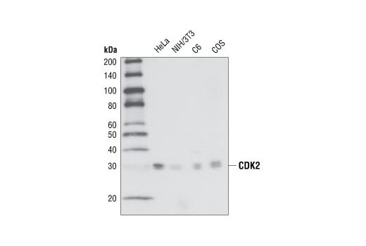
Western blot analysis of extracts from HeLa, NIH/3T3, C6 and COS cells, using CDK2 (78B2) Rabbit mAb.
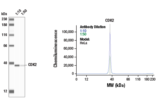
Simple Western™ analysis of lysates (1.0 mg/mL) from HeLa cells using CDK2 (78B2) Rabbit mAb #2546. The virtual lane view (left) shows a single target band (as indicated) at 1:10 and 1:50 dilutions of primary antibody. The corresponding electropherogram view (right) plots chemiluminescence by molecular weight along the capillary at 1:10 (blue line) and 1:50 (green line) dilutions of primary antibody. This experiment was performed under reducing conditions on the Jess™ Simple Western instrument from ProteinSimple, a BioTechne brand, using the 12-230 kDa separation module.
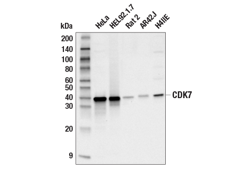
Western blot analysis of extracts from various cell lines using CDK7 (MO1) Mouse mAb.
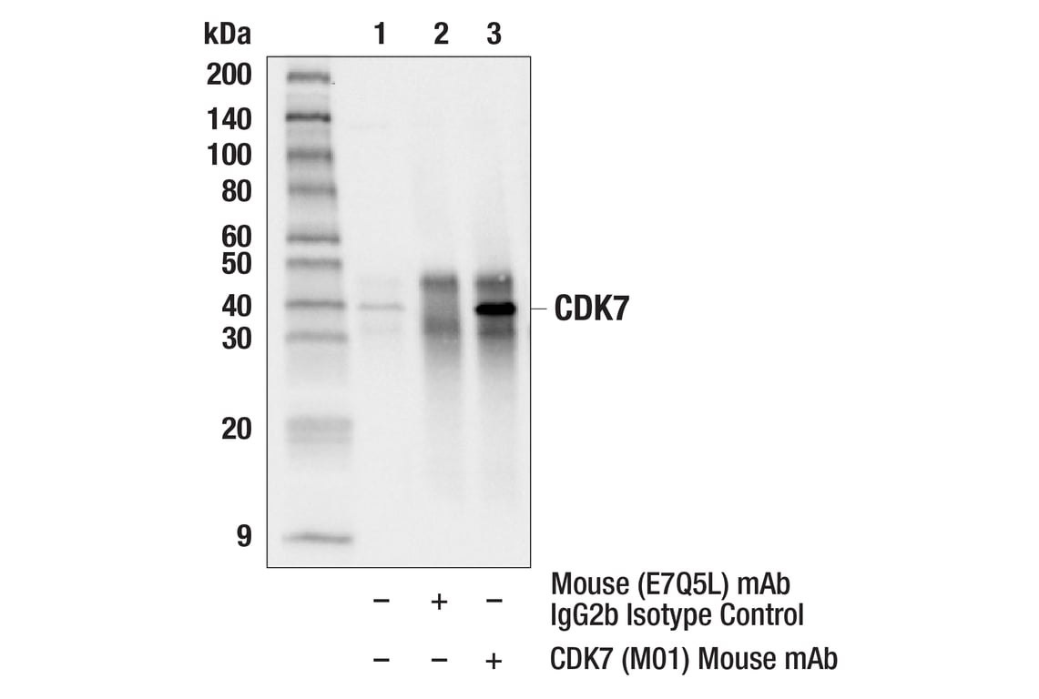
Immunoprecipitation of CDK7 protein from HeLa cell extracts. Lane 1 is 10% input, lane 2 is Mouse (E7Q5L) mAb IgG2b Isotype Control #53484, and lane 3 is CDK7 (MO1) Mouse mAb. Western blot analysis was performed using CDK7 (E4K4W) Rabbit mAb #42863.
After the primary antibody is bound to the target protein, a complex with HRP-linked secondary antibody is formed. The LumiGLO® is added and emits light during enzyme catalyzed decomposition.
After the primary antibody is bound to the target protein, a complex with HRP-linked secondary antibody is formed. The LumiGLO* is added and emits light during enzyme catalyzed decomposition.
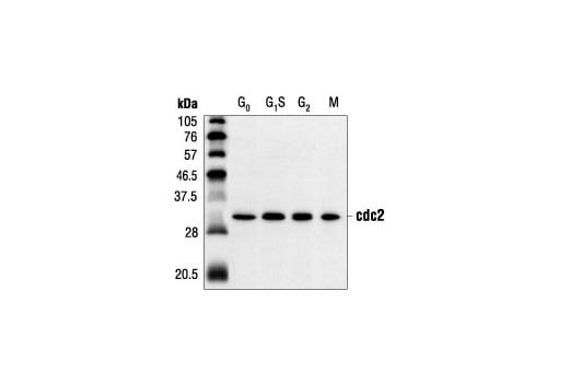
Western blot analysis of extracts from HeLa cells synchronized at various stages of the cell cycle, using cdc2 (POH1) Mouse mAb.
Immunohistochemical analysis of paraffin-embedded human breast carcinoma using CDK4 (D9G3E) Rabbit mAb.
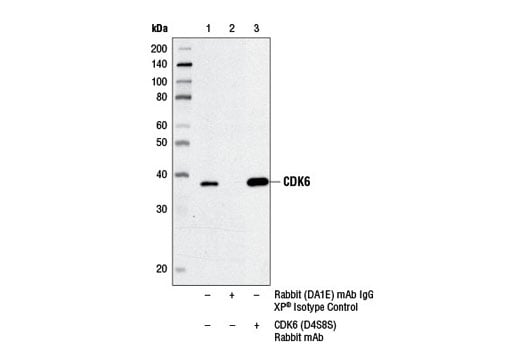
Immunoprecipitation of CDK6 from HeLa cell extracts using Rabbit (DA1E) mAb IgG XP® Isotype Control #3900 (lane 2) or CDK6 (D4S8S) Rabbit mAb (lane 3). Lane 1 is 10% input. Western blot was performed using CDK6 (DCS83) Mouse mAb #3136.
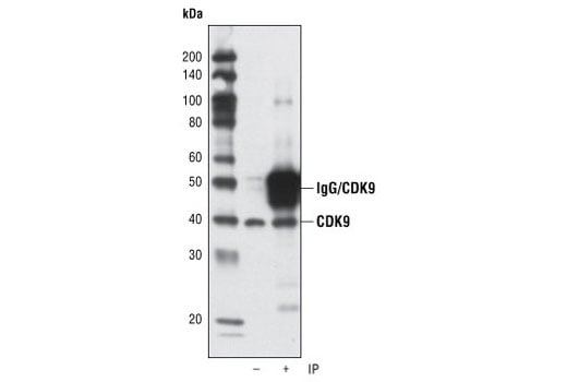
Immunoprecipitation of CDK9 from HeLa cells using CDK9 (C12F7) Rabbit mAb. Western blot detection was performed using the same antibody. Lane 1 is 5% input.
CUT&RUN was performed with HeLa cells and CDK9 (C12F7) Rabbit mAb, using CUT&RUN Assay Kit #86652. DNA Library was prepared using DNA Library Prep Kit for Illumina® (ChIP-seq, CUT&RUN) #56795. The figures show binding across chromosome 9 (upper), including the SLC25A25 gene (lower).
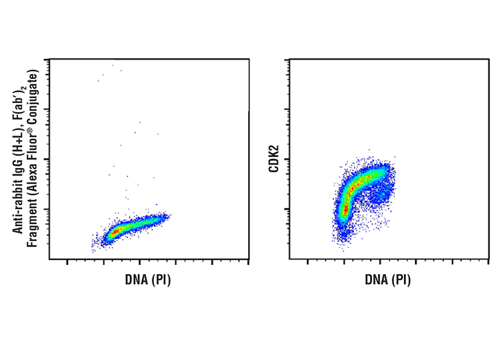
Flow cytometric analysis of Jurkat cells using CDK2 (78B2) Rabbit mAb (right) and Propidium Iodide (PI)/RNase Staining Solution #4087, compared to a secondary only sample (left). Anti-rabbit IgG (H+L), F(ab')2 Fragment (Alexa Fluor® 488 Conjugate) #4412 was used as a secondary antibody.
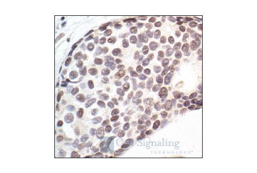
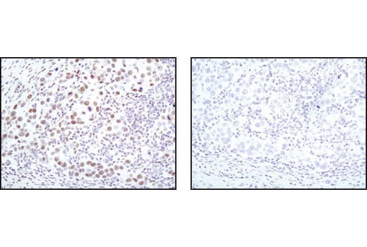
Immunohistochemical analysis of paraffin-embedded human breast carcinoma, showing nuclear localization, using CDK7 (MO1) Mouse mAb.
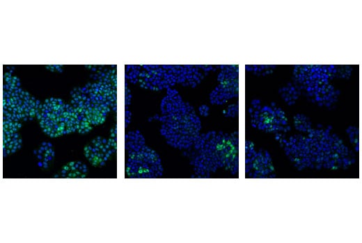
Confocal immunofluorescent analysis of HT-29 cells, transfected with SignalSilence® Control siRNA (Unconjugated) #6568 (left), SignalSilence® cdc2 siRNA I #3500 (center) or SignalSilence® cdc2 siRNA II #3600 (right), using cdc2 (POH1) Mouse mAb (green). Blue pseudocolor = DRAQ5® #4084 (fluorescent DNA dye).
Immunohistochemical analysis of paraffin-embedded human lung carcinoma using CDK4 (D9G3E) Rabbit mAb in the presence of control peptide (left) or antigen-specific peptide (right).
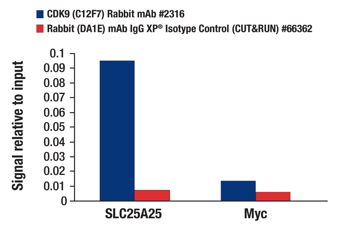
CUT&RUN was performed with HeLa cells and either CDK9 (C12F7) Rabbit mAb or Rabbit (DA1E) mAb IgG XP® Isotype Control (CUT&RUN) #66362, using CUT&RUN Assay Kit #86652. The enriched DNA was quantified by real-time PCR using human SLC25A25 promoter primers and Myc intron 1 primers. The amount of immunoprecipitated DNA in each sample is represented as signal relative to the total amount of input chromatin, which is equivalent to one.
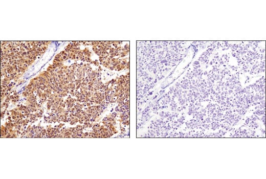
Immunohistochemical analysis of paraffin-embedded human breast carcinoma using CDK9 (C12F7) Rabbit mAb in the presence of control peptide (left) or antigen specific peptide (right).
Confocal immunofluorescent analysis of MCF7 cells using CDK4 (D9G3E) Rabbit mAb (green), p21 Waf1/Cip1 (12D1) Rabbit mAb (Alexa Fluor® 555 Conjugate) #8493 (red), and Phospho-Histone H3 (Ser10) (D2C8) XP® Rabbit mAb (Alexa Fluor® 647 Conjugate) #3458 (blue pseudocolor).
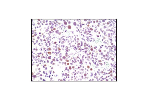
Immunohistochemical analysis of paraffin-embedded K7M2 mouse syngeneic tumor using CDK9 (C12F7) Rabbit mAb.
Flow cytometric analysis of Jurkat cells using CDK4 (D9G3E) Rabbit mAb (right) and Propidium Iodide (PI)/RNase Staining Solution #4087, compared to concentration-matched Rabbit (DA1E) mAb IgG XP® Isotype Control #3900 (left). Anti-rabbit IgG (H+L), F(ab')2 Fragment (Alexa Fluor® 488 Conjugate) #4412 was used as a secondary antibody.
Confocal immunofluorescent analysis of HeLa cells using CDK9 (C12F7) Rabbit mAb (green). Actin filaments have been labeled with DY-555 phalloidin (red).