


 全部商品分类
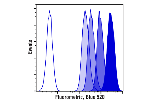


Cell Proliferation Tracer Kit, (Fluorometric, Blue 520)

 下载产品说明书 下载SDS
 用小程序,查商品更便捷


 收藏
收藏
 对比
对比 咨询
咨询The Cell Proliferation Tracer Kit (Fluorometric, Blue 520) contains Cell Proliferation Tracer Dye, Blue 520 that diffuses passively into live cells and is used for long-term cell labeling. This dye is initially a non-fluorescent ester, but is converted to a fluorescent dye by intracellular esterases. The dye then covalently reacts with amine groups on proteins, forming fluorescent conjugates that are retained in the cell. Immediately after staining, a single, bright fluorescent population will be detected by flow cytometry. Each cell division that occurs after labeling results in the appearance of a dimmer fluorescent peak on a flow cytometry histogram. The Cell Proliferation Tracer Kit (Fluorometric, Blue 520) can be used to track cell divisions in vivo or in vitro. Staining can withstand fixation and permeabilization for subsequent immunostaining.

参考图片
Cell division tracking in live Jurkat cells over the course of 4 days (d0-d3). Cells were labeled with Cell Proliferation Tracer Kit (Fluorometric, Blue 520) on day 0, and analyzed by flow cytometry each day. Each successively dimmer peak represents one cell division. Unstained cells are represented by the unshaded peak.
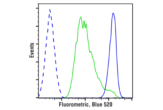
Live human peripheral blood mononuclear cells were labeled with Cell Proliferation Tracer Kit (Fluorometric, Blue 520). Cells cultured in medium containing Human Interleukin-2 (hIL-2) #8907 (50 ng/mL) were then left untreated (blue solid line) or stimulated with anti-CD3 (10 μg/mL) and anti-CD28 (5 μg/ml) (green solid line). Cells were analyzed by flow cytometry after 72 hr. Unstained cells were also acquired for comparison (blue dashed line).