


 全部商品分类


BD Horizon™ CFSE

 下载产品说明书 下载SDS
 用小程序,查商品更便捷


 收藏
收藏
 对比
对比 咨询
咨询
参考图片
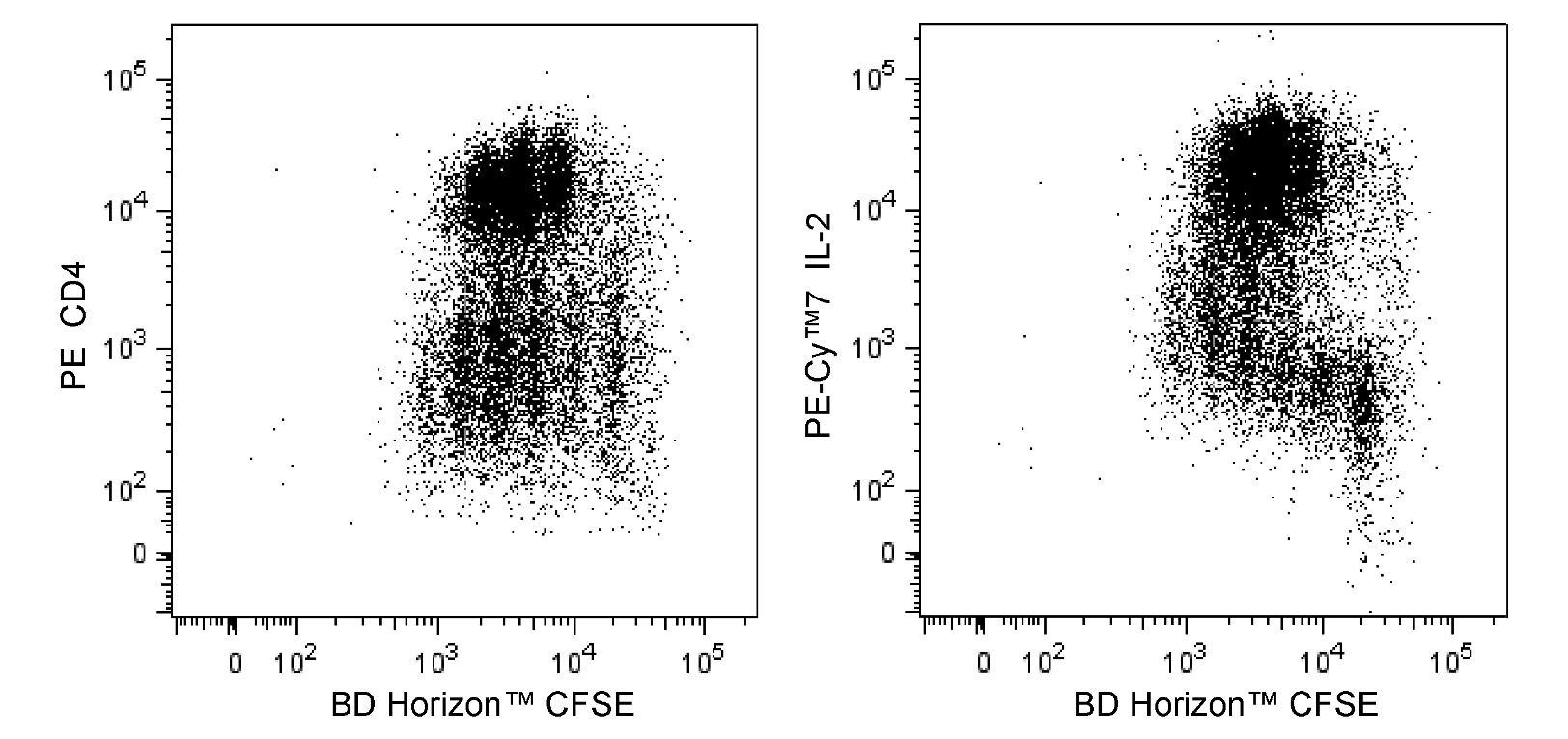
Two-color flow cytometric analysis of proliferating mouse splenocytes stained with BD Horizon™ CFSE. BALB/c mouse spleen cells were treated with BD Pharm Lyse™ Lysing Buffer (Cat. No. 555899) to lyse erythrocytes. The splenic leucocytes were washed and labeled with BD Horizon™ CFSE (1 μM) for 10 minutes at 37°C. The cells were washed twice and then cultured with Purified NA/LE Hamster Anti-Mouse CD3e (Cat. No. 553057) and Purified NA/LE Hamster Anti-Mouse CD28 (Cat. No. 553294) antibodies. After 3 days, the cells were harvested, washed, and restimulated (4 hrs) with Phorbol 12-Myristate 13-Acetate and Ionomycin in the presence of BD GolgiStop™ Protein Transport Inhibitor (containing Monensin) (Cat. No. 554724). Cells were harvested, stained with BD Horizon™ Fixable Viability Stain 660 (Cat. No. 564405; to exclude nonviable cells), fixed, permeabilized using a BD Cytofix/Cytoperm™ Fixation/Permeabilization Solution Kit (Cat. No. 554714), and then stained with PE Anti-Mouse CD4 (Cat. No. 553048/553049/561837) and PE-Cy™7 Anti-Mouse IL-2 (Cat. No. 560538) antibodies. Two-color flow cytometric dot plots showing the correlated expression patterns of CFSE versus CD4 (Left Panel) or IL-2 (Right Panel) were derived from gated events with the forward and side light-scatter characteristics of viable lymphocytes. Flow cytometric analysis was performed using a BD LSRFortessa™ Cell Analyzer System.
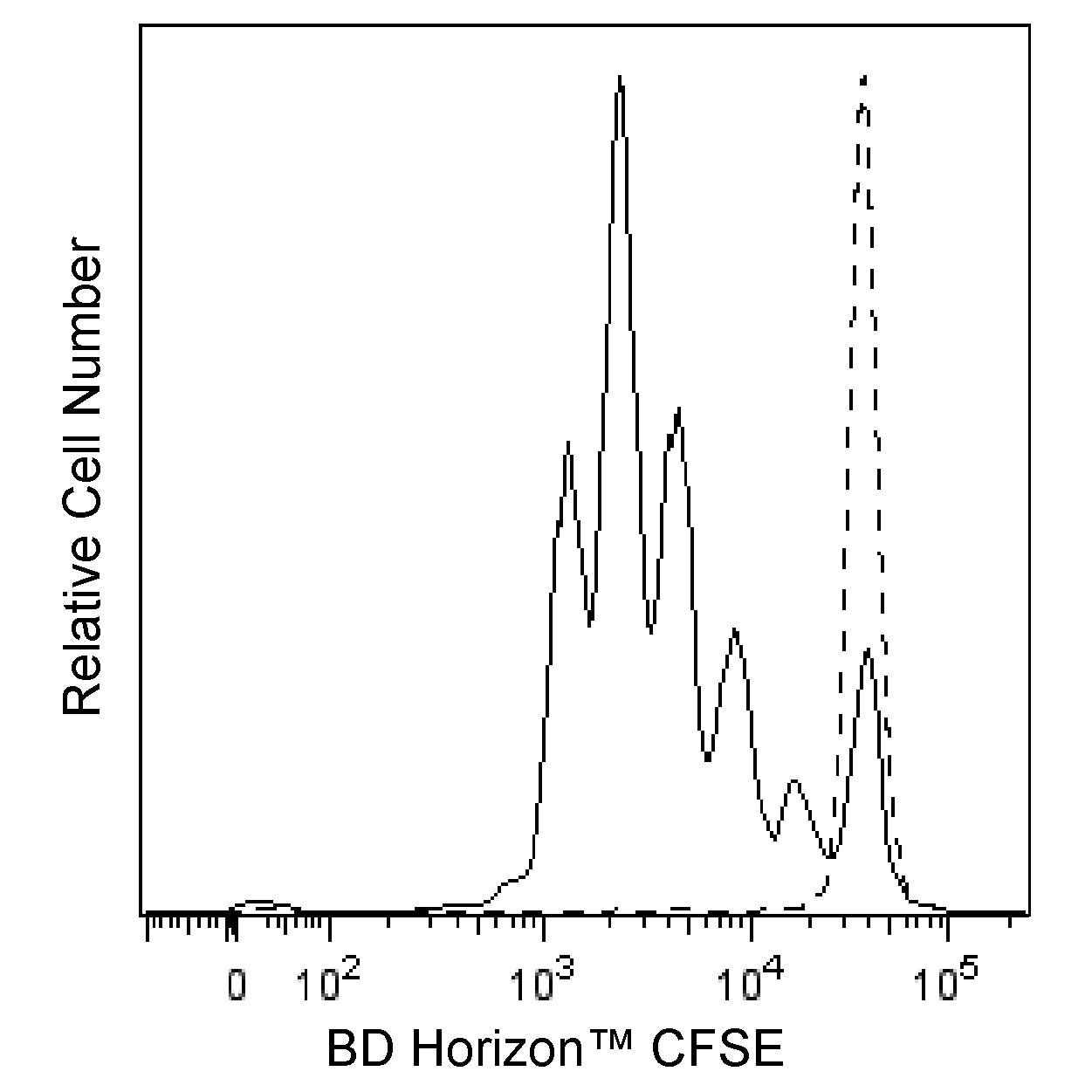
Flow cytometric analysis of proliferating peripheral blood lymphocytes stained with BD Horizon™ CFSE. Human PBMC were labeled (10 min, 37°C) with BD Horizon™ CFSE (Cat. No. 565082; 2.5 μM). Cells were washed twice and cultured (4 days) alone (dashed line histogram) or with (solid line histogram) phytohemagglutinin. The cells were stained with 0.1 μg/mL DAPI (Cat. No. 564907) to exclude nonviable cells. The histogram shows CFSE fluorescence peaks of gated events with the light-scatter characteristics of viable lymphocytes (DAPI-negative). Unstimulated cells show a single, bright CFSE fluorescence peak, indicating no proliferation. Stimulated cells show multiple CFSE fluorescence peaks, indicating multiple generations of proliferating cells. Analysis was performed using a BD LSRFortessa™ Cell Analyzer System.
Two-color flow cytometric analysis of proliferating mouse splenocytes stained with BD Horizon™ CFSE. BALB/c mouse spleen cells were treated with BD Pharm Lyse™ Lysing Buffer (Cat. No. 555899) to lyse erythrocytes. The splenic leucocytes were washed and labeled with BD Horizon™ CFSE (1 μM) for 10 minutes at 37°C. The cells were washed twice and then cultured with Purified NA/LE Hamster Anti-Mouse CD3e (Cat. No. 553057) and Purified NA/LE Hamster Anti-Mouse CD28 (Cat. No. 553294) antibodies. After 3 days, the cells were harvested, washed, and restimulated (4 hrs) with Phorbol 12-Myristate 13-Acetate and Ionomycin in the presence of BD GolgiStop™ Protein Transport Inhibitor (containing Monensin) (Cat. No. 554724). Cells were harvested, stained with BD Horizon™ Fixable Viability Stain 660 (Cat. No. 564405; to exclude nonviable cells), fixed, permeabilized using a BD Cytofix/Cytoperm™ Fixation/Permeabilization Solution Kit (Cat. No. 554714), and then stained with PE Anti-Mouse CD4 (Cat. No. 553048/553049/561837) and PE-Cy™7 Anti-Mouse IL-2 (Cat. No. 560538) antibodies. Two-color flow cytometric dot plots showing the correlated expression patterns of CFSE versus CD4 (Left Panel) or IL-2 (Right Panel) were derived from gated events with the forward and side light-scatter characteristics of viable lymphocytes. Flow cytometric analysis was performed using a BD LSRFortessa™ Cell Analyzer System.