


 全部商品分类
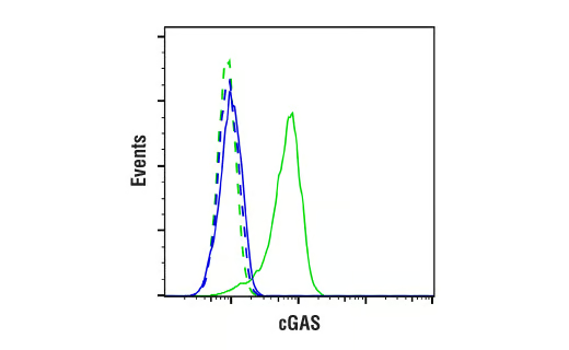


 用小程序,查商品更便捷


Monoclonal antibody is produced by immunizing animals with a synthetic peptide corresponding to residues surrounding Asn489 of human cGAS protein.
Product Usage Information
| Application | Dilution |
|---|---|
| Western Blotting | 1:1000 |
| Simple Western™ | 1:10 - 1:50 |
| Immunohistochemistry (Paraffin) | 1:50 - 1:200 |
| Immunofluorescence (Immunocytochemistry) | 1:100 - 1:200 |
| Flow Cytometry (Fixed/Permeabilized) | 1:50 - 1:200 |
Specificity/Sensitivity
Species Reactivity:
Human
Supplied in 10 mM sodium HEPES (pH 7.5), 150 mM NaCl, 100 µg/ml BSA, 50% glycerol and less than 0.02% sodium azide. Store at –20°C. Do not aliquot the antibody.
For a carrier-free (BSA and azide free) version of this product see product #86793.
参考图片
Flow cytometric analysis of SNB-19 cells (blue) and NK-92 cells (green) using cGAS (E5V3W) Rabbit mAb (solid lines) or a concentration-matched Rabbit (DA1E) mAb IgG XP® Isotype Control #3900 (dashed lines). Anti-rabbit IgG (H+L), F(ab')2 Fragment (Alexa Fluor® 488 Conjugate) #4412 was used as a secondary antibody.
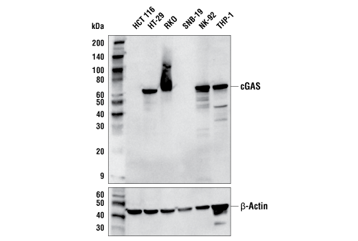
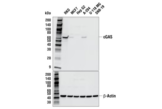
Western blot analysis of extracts from various cell lines using cGAS (E5V3W) Rabbit mAb (upper) or β-Actin (D6A8) Rabbit mAb #8457 (lower).
Western blot analysis of extracts from various cell lines using cGAS (E5V3W) Rabbit mAb (upper) or β-Actin (D6A8) Rabbit mAb #8457 (lower).
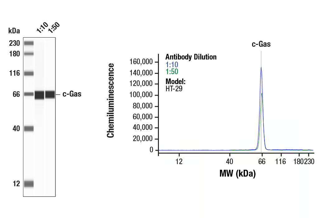
Simple Western™ analysis of HT-29 lysates (0.1 mg/mL) using cGAS (E5V3W) Rabbit mAb #79978. The virtual lane view (left) shows the target band (as indicated) at 1:10 and 1:50 dilutions of primary antibody. The corresponding electropherogram view (right) plots chemiluminescence by molecular weight along the capillary at 1:10 (blue line) and 1:50 (green line) dilutions of primary antibody. This experiment was performed under reducing conditions on the Jess™ Simple Western instrument from ProteinSimple, a BioTechne brand, using the 12-230 kDa separation module.
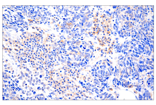
Immunohistochemical analysis of paraffin-embedded human neuroendocrine lung carcinoma using cGAS (E5V3W) Rabbit mAb.
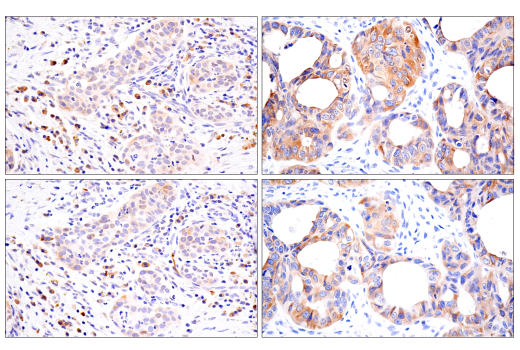
Immunohistochemical analysis of paraffin-embedded human mucoepidermoid carcinoma of the larynx (left) or endometrioid adenocarcinoma (right) using cGAS (E5V3W) Rabbit mAb (top) or cGAS Rabbit mAb (bottom). These two antibodies detect independent, unique epitopes on human cGAS. The similar staining patterns obtained with both antibodies help to confirm the specificity of the staining.
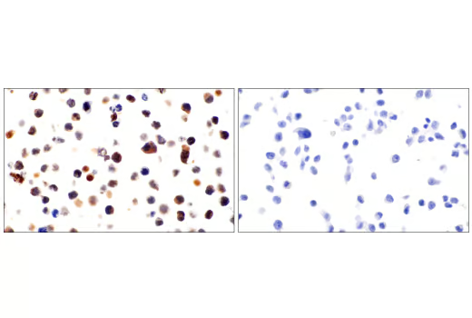
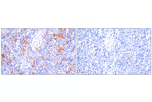
Immunohistochemical analysis of paraffin-embedded NK-92 cell pellet (left, positive) or HCT 116 cell pellet (right, negative) using cGAS (E5V3W) Rabbit mAb.
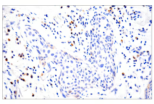
Immunohistochemical analysis of paraffin-embedded human squamous cell carcinoma of the tongue using cGAS (E5V3W) Rabbit mAb.
Immunohistochemical analysis of paraffin-embedded human tonsil using cGAS (E5V3W) Rabbit mAb (left) compared to concentration-matched Rabbit (DA1E) mAb IgG XP® Isotype Control #3900 (right).
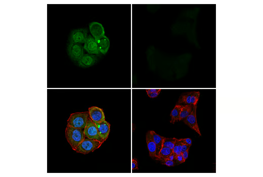
Confocal immunofluorescent analysis of HT-29 cells (left, positive) and Hep G2 cells (right, negative) using cGAS (E5V3W) Rabbit mAb (green). Actin filaments were labeled with Alexa Fluor® 647 Phalloidin #8940 (red). Cells were mounted in ProLong® Gold Antifade Reagent with DAPI #8961 (blue).