


 全部商品分类


Cleaved-IL-1beta (Asp116) (D3A3Z) Rabbit mAb

 下载产品说明书 下载COA 下载SDS
 用小程序,查商品更便捷


 收藏
收藏
 对比
对比 咨询
咨询Monoclonal antibody is produced by immunizing animals with a synthetic peptide corresponding to residues surrounding Asp116 of human IL-1β protein.

Product Usage Information
| Application | Dilution |
|---|---|
| Western Blotting | 1:1000 |
| Immunofluorescence (Immunocytochemistry) | 1:800 - 1:3200 |
Specificity/Sensitivity
Species Reactivity:
Human
Supplied in 10 mM sodium HEPES (pH 7.5), 150 mM NaCl, 100 µg/ml BSA, 50% glycerol and less than 0.02% sodium azide. Store at –20°C. Do not aliquot the antibody.
参考图片
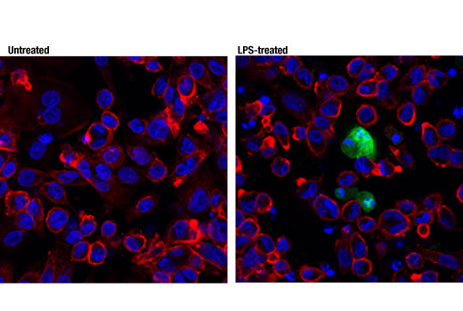
Confocal immunofluorescent analysis of THP-1 cells, differentiated with TPA #4174 (80 nM, 24 hr) and subsequently treated with (right) or without (left) Lipopolysaccharides (LPS) #14011 (1 μg/ml, 6 hr), using Cleaved-IL-1β (Asp116) (D3A3Z) Rabbit mAb (green). Actin filaments were labeled with DyLight™ 554 Phalloidin #13054 (red). Blue pseudocolor = DRAQ5® #4084 (fluorescent DNA dye).
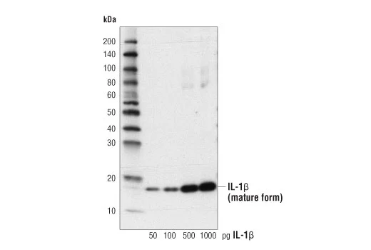
Western blot analysis of recombinant Human Interleukin-1β (hIL-1β) #8900 using Cleaved-IL-1β (Asp116) (D3A3Z) Rabbit mAb.
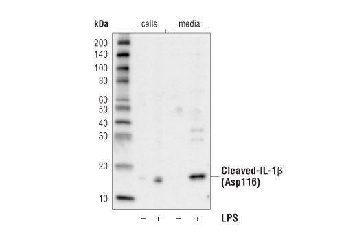
Western blot analysis of extracts from cells or media collected from THP-1 cells, differentiated with TPA #4147 (80 nM, overnight) and subsequently treated with (+) or without (-) Lipopolysaccharides (LPS) #14011 (1 μg/ml, 6 hr), using Cleaved-IL-1β (Asp116) (D3A3Z) Rabbit mAb.