


 全部商品分类


COL1A1 (E8F4L) XP ® Rabbit mAb

 下载产品说明书 下载COA 下载SDS
 用小程序,查商品更便捷


 收藏
收藏
 对比
对比 咨询
咨询
Monoclonal antibody is produced by immunizing animals with a synthetic peptide corresponding to residues surrounding Phe1196 of human COL1A1 protein.
Product Usage Information
| Application | Dilution |
|---|---|
| Western Blotting | 1:1000 |
| IHC Leica Bond | 1:100 - 1:400 |
| Immunohistochemistry (Paraffin) | 1:50 - 1:200 |
| Immunofluorescence (Frozen) | 1:100 - 1:400 |
| Immunofluorescence (Immunocytochemistry) | 1:100 - 1:400 |
Specificity/Sensitivity
Species Reactivity:
Human, Mouse, Rat, Monkey
Supplied in 10 mM sodium HEPES (pH 7.5), 150 mM NaCl, 100 µg/ml BSA, 50% glycerol and less than 0.02% sodium azide. Store at –20°C. Do not aliquot the antibody.
For a carrier free (BSA and azide free) version of this product see product #81375.
参考图片
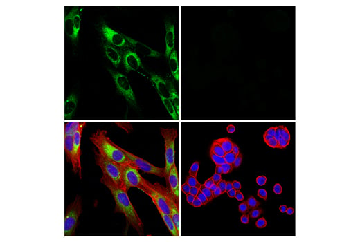
Confocal immunofluorescent analysis of U-118 MG cells (left, positive) and HT-29 cells (right, negative) using COL1A1 (E8F4L) XP® Rabbit mAb (green). Actin filaments were labeled with DyLight™ 554 Phalloidin #13054 (red). Samples were mounted in ProLong® Gold Antifade Reagent with DAPI #8961 (blue).
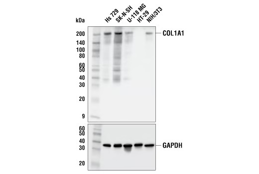
Western blot analysis of extracts from various cell lines using COL1A1 (E8F4L) XP® Rabbit mAb (upper) and GAPDH (D16H11) XP® Rabbit mAb #5174 (lower).
Immunohistochemical analysis of paraffin-embedded human endometrioid adenocarcinoma using COL1A1 (E8F4L) XP® Rabbit mAb performed on the Leica® BOND™ Rx.
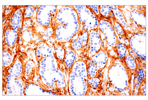
Immunohistochemical analysis of paraffin-embedded human prostate carcinoma using COL1A1 (E8F4L) XP® Rabbit mAb performed on the Leica® BOND™ Rx.
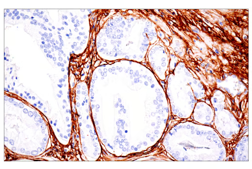
Immunohistochemical analysis of paraffin-embedded human prostate carcinoma using COL1A1 (E8F4L) XP® Rabbit mAb.
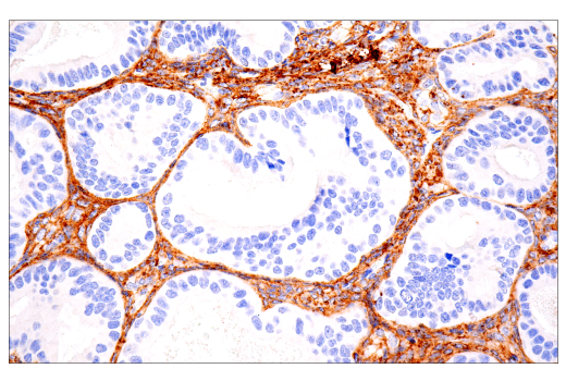
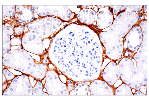
Immunohistochemical analysis of paraffin-embedded normal human kidney using COL1A1 (E8F4L) XP® Rabbit mAb.
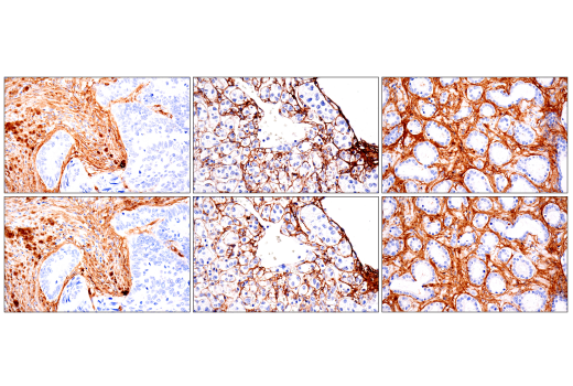
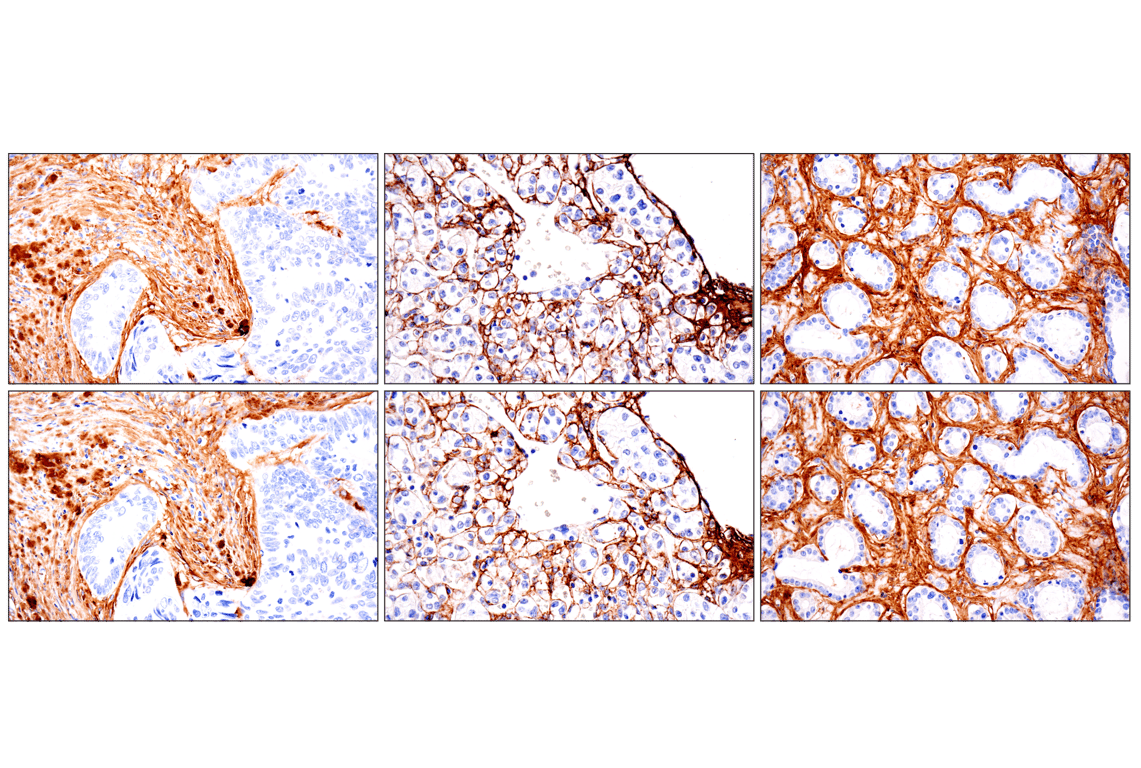
Immunohistochemical analysis of paraffin-embedded human colon carcinoma (left), renal cell carcinoma (middle) or prostate carcinoma (right) using COL1A1 (E8F4L) XP® Rabbit mAb (top) or COL1A1 (E3E1X) Mouse mAb #66948 (bottom). These two antibodies detect independent, unique epitopes on human COL1A1. The similar staining patterns obtained with both antibodies help to confirm the specificity of the staining.
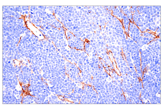
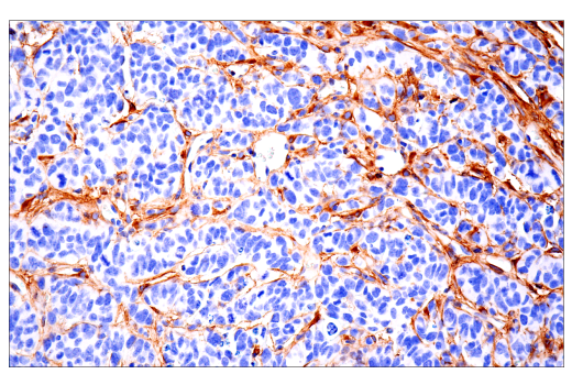
Immunohistochemical analysis of paraffin-embedded A20 syngeneic tumor using COL1A1 (E8F4L) XP® Rabbit mAb.
Immunohistochemical analysis of paraffin-embedded GL261 syngeneic tumor using COL1A1 (E8F4L) XP® Rabbit mAb.
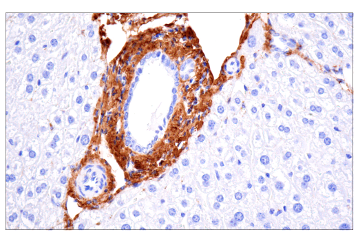
Immunohistochemical analysis of paraffin-embedded mouse liver using COL1A1 (E8F4L) XP® Rabbit mAb.
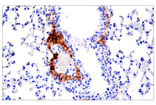
Immunohistochemical analysis of paraffin-embedded mouse lung using COL1A1 (E8F4L) XP® Rabbit mAb.
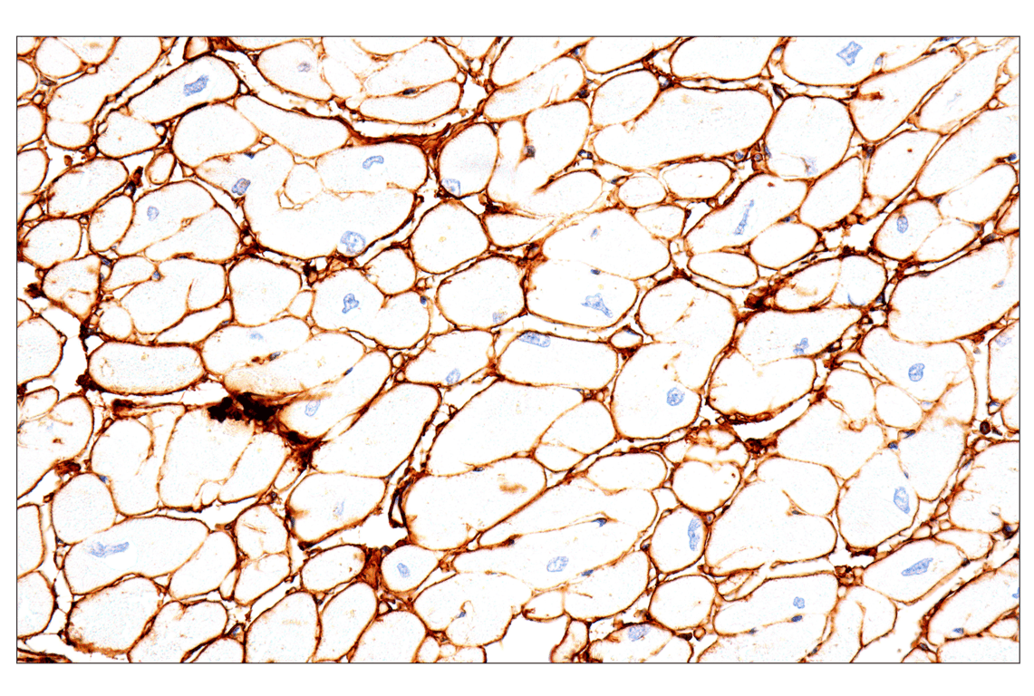
Immunohistochemical analysis of paraffin-embedded rhesus monkey heart using COL1A1 (E8F4L) XP® Rabbit mAb.
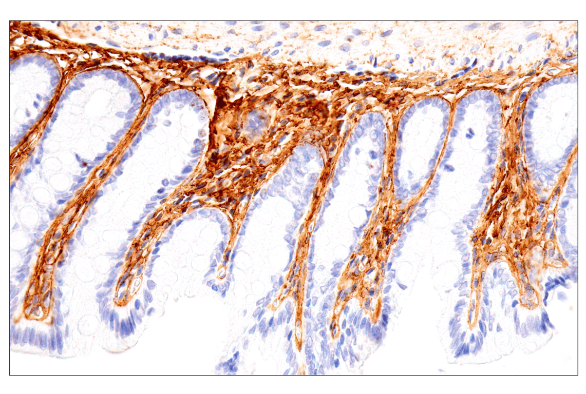
Immunohistochemical analysis of paraffin-embedded rat colon using COL1A1 (E8F4L) XP® Rabbit mAb.
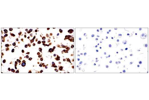
Immunohistochemical analysis of paraffin-embedded human esophageal adenocarcinoma using COL1A1 (E8F4L) XP® Rabbit mAb (left) compared to concentration-matched Rabbit (DA1E) mAb IgG XP® Isotype Control #3900 (right).
Immunohistochemical analysis of paraffin-embedded human colon carcinoma (left), renal cell carcinoma (middle) or prostate carcinoma (right) using COL1A1 (E8F4L) XP® Rabbit mAb (top) or COL1A1 (E3E1X) Mouse mAb #66948 (bottom). These two antibodies detect independent, unique epitopes on human COL1A1. The similar staining patterns obtained with both antibodies help to confirm the specificity of the staining.
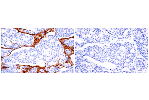
Immunohistochemical analysis of paraffin-embedded U-118 MG cell pellet (left, positive) or HT-29 cell pellet (right, negative) using COL1A1 (E8F4L) XP® Rabbit mAb.
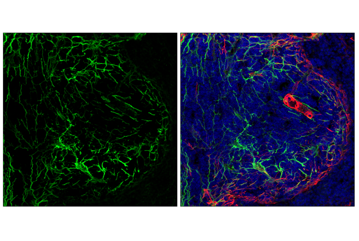
Confocal immunofluorescent analysis of fixed frozen mouse spleen labeled with COL1A1 (E8F4L) XP® Rabbit mAb (green), α-Smooth Muscle Actin (D4K9N) XP® Rabbit mAb (Alexa Fluor® 647 Conjugate) #76113 (red), and ProLong Gold Antifade Reagent with DAPI #8961 (blue).
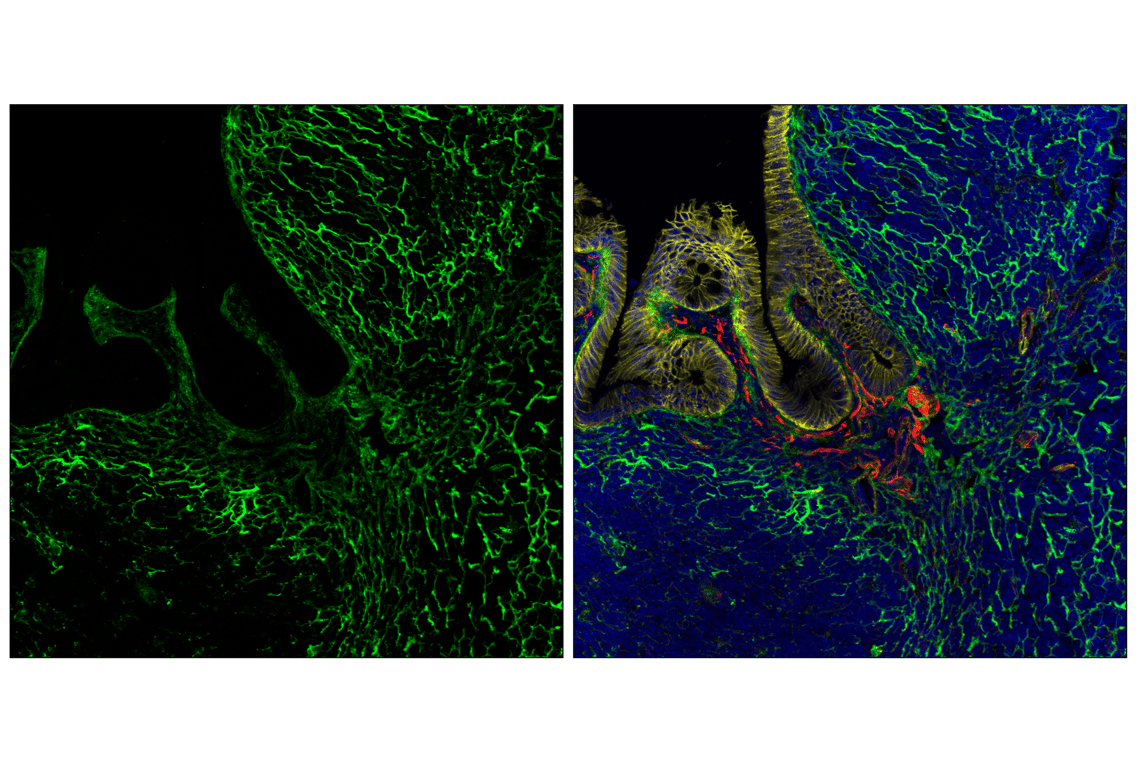
Confocal immunofluorescent analysis of fixed frozen mouse colon labeled with COL1A1 (E8F4L) XP® Rabbit mAb (green), β-Catenin (D10A8) XP® Rabbit mAb (Alexa Fluor® 555 Conjugate) #83539 (yellow), α-Smooth Muscle Actin (D4K9N) XP® Rabbit mAb (Alexa Fluor® 647 Conjugate) #76113 (red), and ProLong Gold Antifade Reagent with DAPI #8961 (blue).
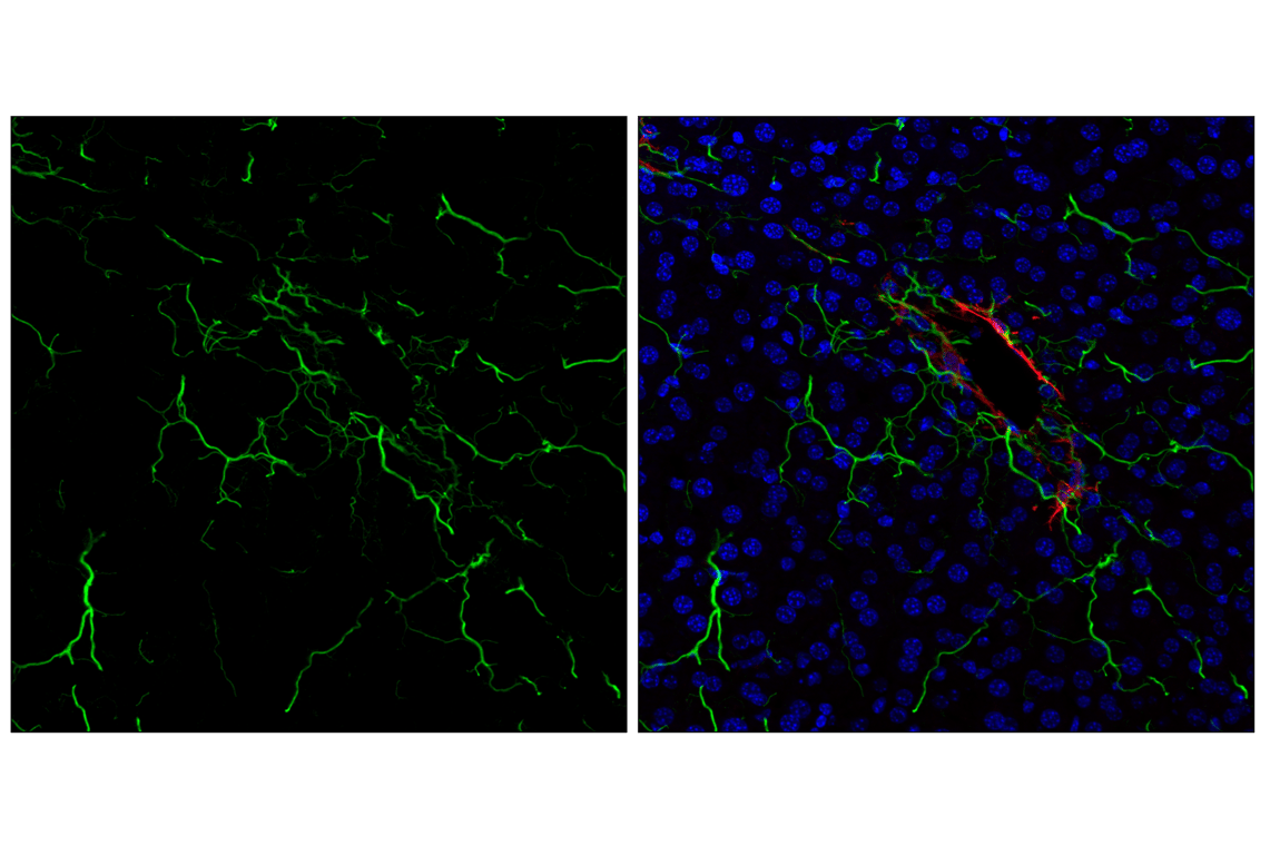
Confocal immunofluorescent analysis of fixed frozen mouse liver labeled with COL1A1 (E8F4L) XP® Rabbit mAb (green), α-Smooth Muscle Actin (D4K9N) XP® Rabbit mAb (Alexa Fluor® 647 Conjugate) #76113 (red) and ProLong Gold Antifade Reagent with DAPI #8961 (blue).