


 全部商品分类
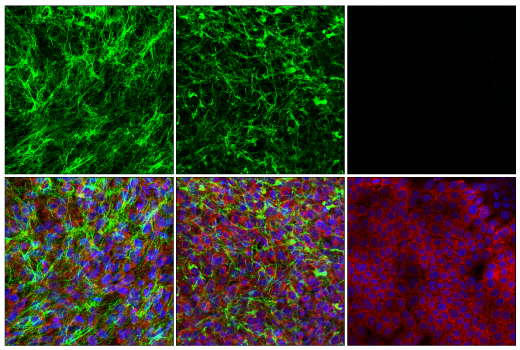


 用小程序,查商品更便捷


Monoclonal antibody is produced by immunizing animals with a synthetic peptide corresponding to residues surrounding Pro364 of human COL5A1 protein.

Product Usage Information
| Application | Dilution |
|---|---|
| Western Blotting | 1:1000 |
| Immunoprecipitation | 1:100 |
| IHC Leica Bond | 1:50 - 1:200 |
| Immunohistochemistry (Paraffin) | 1:50 - 1:200 |
| Immunofluorescence (Immunocytochemistry) | 1:200 - 1:800 |
Specificity/Sensitivity
Species Reactivity:
Human
Supplied in 10 mM sodium HEPES (pH 7.5), 150 mM NaCl, 100 µg/ml BSA, 50% glycerol and less than 0.02% sodium azide. Store at –20°C. Do not aliquot the antibody.
For a carrier free (BSA and azide free) version of this product see product #80085.
参考图片
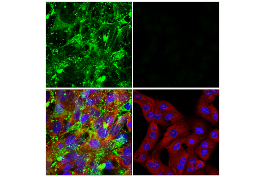
Confocal immunofluorescent analysis of SF539 cells (left, positive) or ACHN cells (right, negative) using COL5A1 (E6U9W) Rabbit mAb (green), S6 Ribosomal Protein (54D2) Mouse mAb (Alexa Fluor® 647 Conjugate) #5548 (red), and DAPI #4083 (blue).
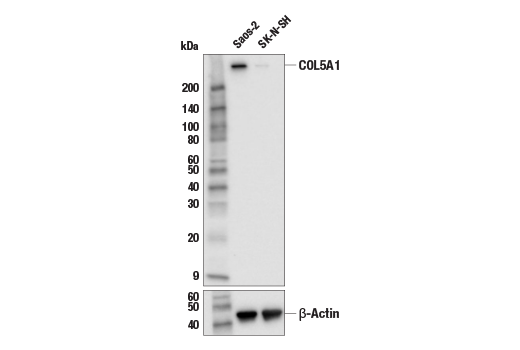
Western blot analysis of extracts from Saos-2 and SK-N-SH cells using COL5A1 (E6U9W) Rabbit mAb (upper) and β-Actin (D6A8) Rabbit mAb #8457 (lower). The differential in COL5A1 expression between Saos-2 and SK-N-SH cells is consistent with their reported molecular expression profiles (www.biogps.org), confirming specificity of the antibody for COL5A1.
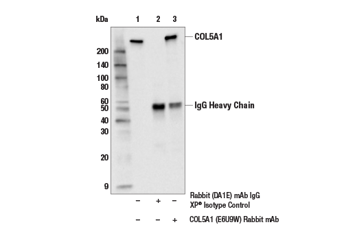
Immunoprecipitation of COL5A1 protein from SF-539 cell extracts. Lane 1 is 10% input, lane 2 is Rabbit (DA1E) mAb IgG XP® Isotype Control #3900, and lane 3 is COL5A1 (E6U9W) Rabbit mAb. Western blot analysis was performed using COL5A1 (E6U9W) Rabbit mAb. Anti-rabbit IgG, HRP-linked Antibody #7074 was used as the secondary antibody.
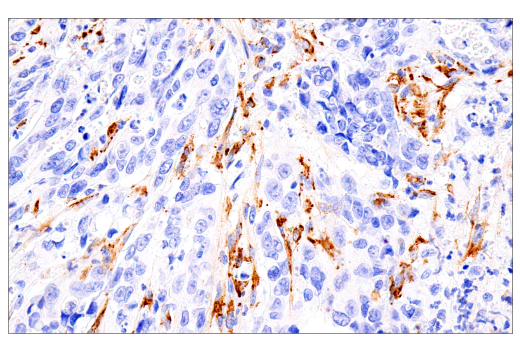
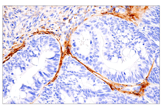
Immunohistochemical analysis of paraffin-embedded human esophageal carcinoma using COL5A1 (E6U9W) Rabbit mAb performed on the Leica® BOND™ Rx.
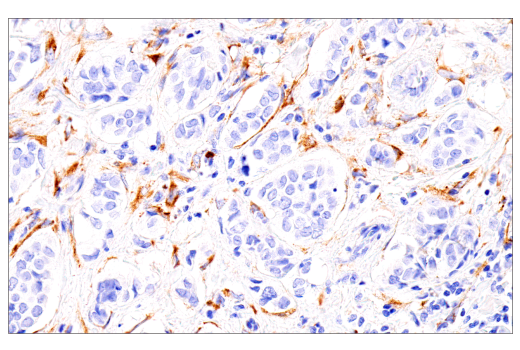
Immunohistochemical analysis of paraffin-embedded human breast carcinoma using COL5A1 (E6U9W) Rabbit mAb performed on the Leica® BOND™ Rx.
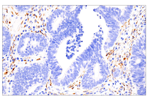
Immunohistochemical analysis of paraffin-embedded human colon carcinoma using COL5A1 (E6U9W) Rabbit mAb performed on the Leica® BOND™ Rx.
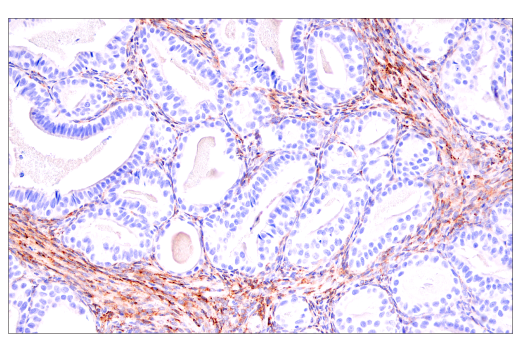
Immunohistochemical analysis of paraffin-embedded human ovarian adenocarcinoma using COL5A1 (E6U9W) Rabbit mAb performed on the Leica® BOND™ Rx.
Immunohistochemical analysis of paraffin-embedded human endometrioid adenocarcinoma using COL5A1 (E6U9W) Rabbit mAb.
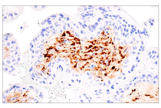
Immunohistochemical analysis of paraffin-embedded normal human placenta using COL5A1 (E6U9W) Rabbit mAb.
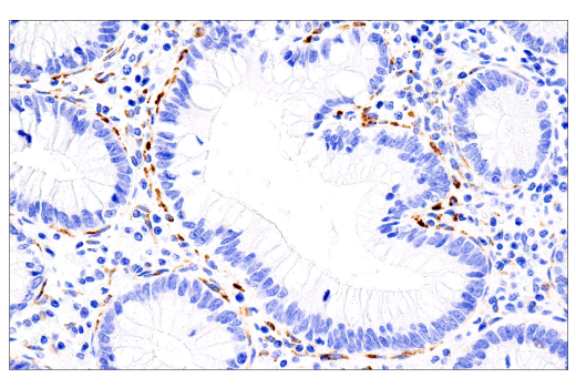
Immunohistochemical analysis of paraffin-embedded normal human colon using COL5A1 (E6U9W) Rabbit mAb.
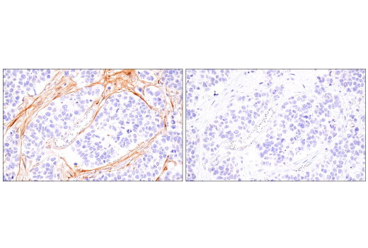
Immunohistochemical analysis of paraffin-embedded human neuroendocrine lung carcinoma using COL5A1 (E6U9W) Rabbit mAb (left) compared to concentration-matched Rabbit (DA1E) mAb IgG XP® Isotype Control #3900 (right).
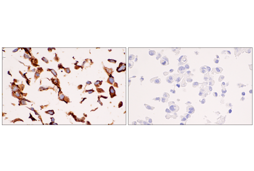
Immunohistochemical analysis of paraffin-embedded SF-539 cell pellet (left, positive) or ACHN cell pellet (right, negative) using COL5A1 (E6U9W) Rabbit mAb.
Confocal immunofluorescent analysis of U-118 MG cells (left, positive), RD cells (center, positive) or OVCAR-5 cells (right, negative) using COL5A1 (E6U9W) Rabbit mAb (green), S6 Ribosomal Protein (54D2) Mouse mAb (Alexa Fluor® 647 Conjugate) #5548 (red), and DAPI #4083 (blue).