


 全部商品分类


 用小程序,查商品更便捷


Monoclonal antibody is produced by immunizing animals with a synthetic peptide corresponding to residues surrounding Lys170 of human COL1A1 protein.

Product Usage Information
| Application | Dilution |
|---|---|
| Western Blotting | 1:1000 |
| IHC Leica Bond | 1:200 - 1:800 |
| Immunohistochemistry (Paraffin) | 1:50 - 1:200 |
| Immunofluorescence (Immunocytochemistry) | 1:50 - 1:200 |
Specificity/Sensitivity
Species Reactivity:
Human
Supplied in 10 mM sodium HEPES (pH 7.5), 150 mM NaCl, 100 µg/ml BSA, 50% glycerol and less than 0.02% sodium azide. Store at –20°C. Do not aliquot the antibody.
For a carrier free (BSA and azide free) version of this product see product #66154.
参考图片
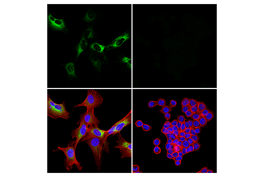
Confocal immunofluorescent analysis of U-118 MG cells (left, positive) and HT-29 cells (right, negative) using COL1A1 (E3E1X) Mouse mAb (green). Actin filaments were labeled with DyLight™ 554 Phalloidin #13054 (red). Samples were mounted in ProLong® Gold Antifade Reagent with DAPI #8961 (blue).
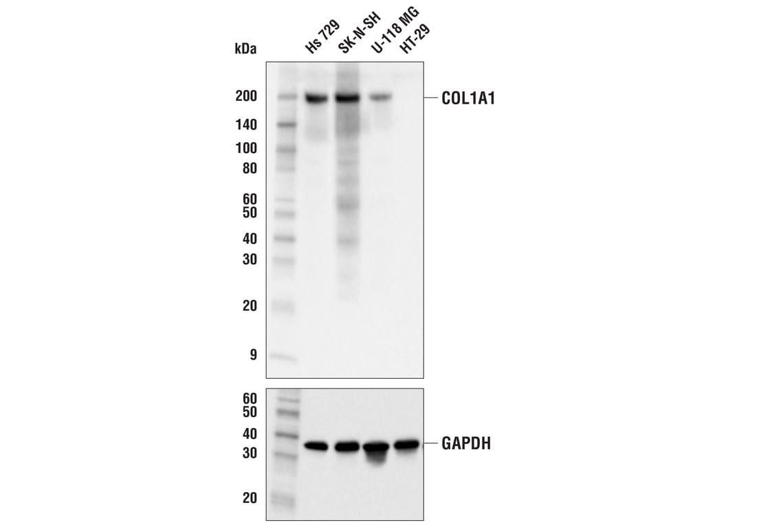
Western blot analysis of extracts from various cell lines using COL1A1 (E3E1X) Mouse mAb (upper) and GAPDH (D16H11) XP® Rabbit mAb #5174 (lower).
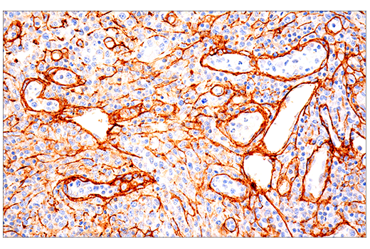
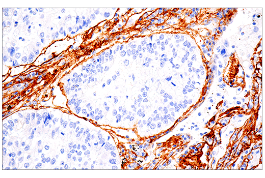
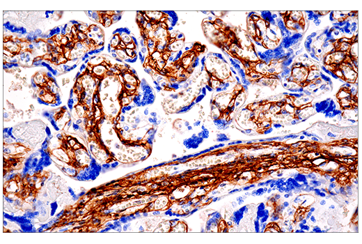
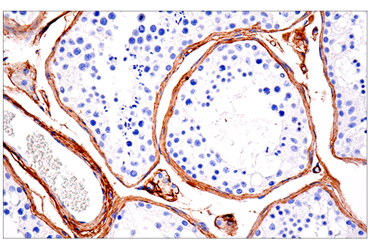
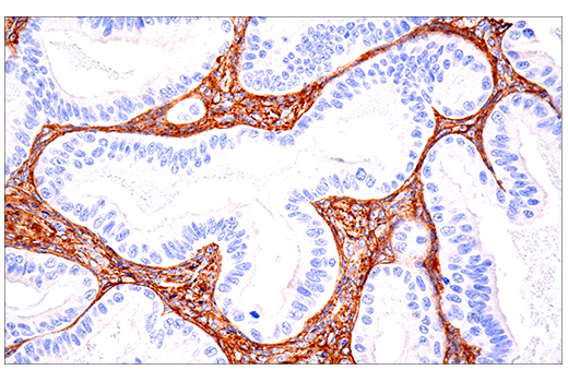
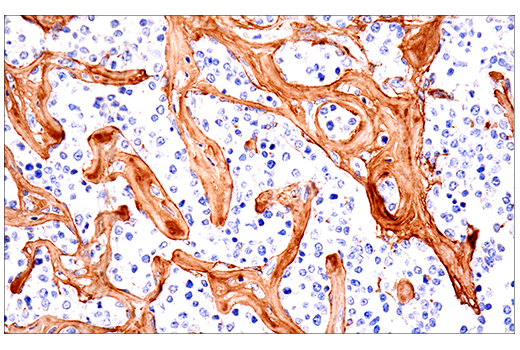
Immunohistochemical analysis of paraffin-embedded human T cell lymphoma using COL1A1 (E3E1X) Mouse mAb performed on the Leica® BOND™ Rx.
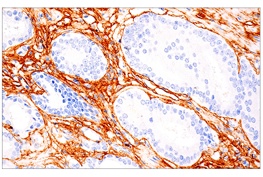
Immunohistochemical analysis of paraffin-embedded human prostate carcinoma using COL1A1 (E3E1X) Mouse mAb performed on the Leica® BOND™ Rx.
Immunohistochemical analysis of paraffin-embedded human non-small cell lung carcinoma using COL1A1 (E3E1X) Mouse mAb performed on the Leica® BOND™ Rx.
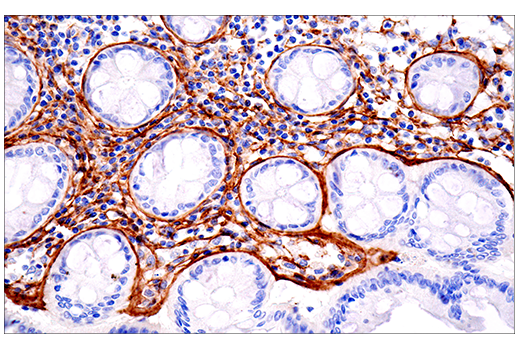
Immunohistochemical analysis of paraffin-embedded normal human placenta using COL1A1 (E3E1X) Mouse mAb.
Immunohistochemical analysis of paraffin-embedded normal human testis using COL1A1 (E3E1X) Mouse mAb.
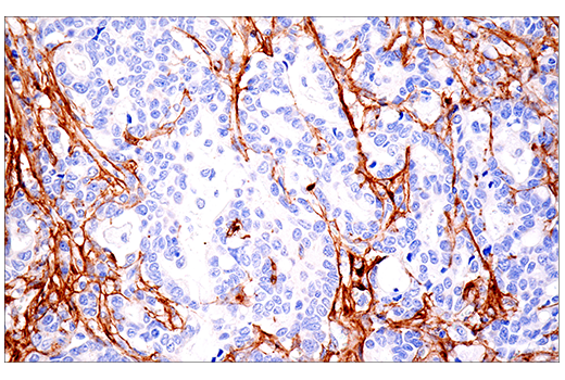
Immunohistochemical analysis of paraffin-embedded human endometrioid adenocarcinoma using COL1A1 (E3E1X) Mouse mAb.
Immunohistochemical analysis of paraffin-embedded human B cell non-Hodgkin's lymphoma with fibrosis using COL1A1 (E3E1X) Mouse mAb.
Immunohistochemical analysis of paraffin-embedded normal human colon using COL1A1 (E3E1X) Mouse mAb.
Immunohistochemical analysis of paraffin-embedded human esophageal adenocarcinoma using COL1A1 (E3E1X) Mouse mAb.
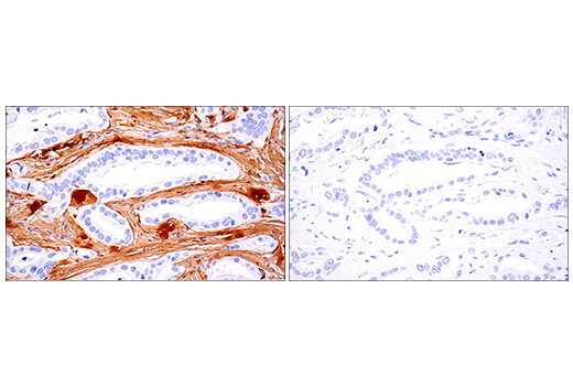
Immunohistochemical analysis of paraffin-embedded human ductal breast carcinoma using COL1A1 (E3E1X) Mouse mAb (left) compared to concentration-matched Mouse (E5Y6Q) mAb IgG2a Isotype Control #61656 (right).
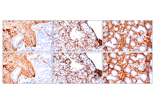
Immunohistochemical analysis of paraffin-embedded human colon carcinoma (left), renal cell carcinoma (middle), or prostate carcinoma (right) using COL1A1 (E3E1X) Mouse mAb (top) or COL1A1 (E8F4L) XP® Rabbit mAb #72026 (bottom). These two antibodies detect independent, unique epitopes on human COL1A1. The similar staining patterns obtained with both antibodies help to confirm the specificity of the staining.
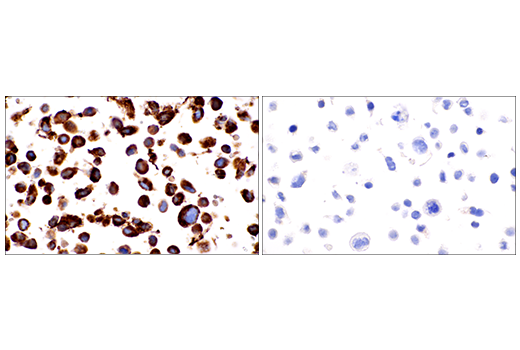
Immunohistochemical analysis of paraffin-embedded U-118 MG cell pellet (left, positive) or HT-29 cell pellet (right, negative) using COL1A1 (E3E1X) Mouse mAb.