


 全部商品分类


COX IV (3E11) Rabbit mAb

 下载产品说明书 下载COA 下载SDS
 用小程序,查商品更便捷


 收藏
收藏
 对比
对比 咨询
咨询
Monoclonal antibody is produced by immunizing animals with a synthetic peptide corresponding to residues surrounding Lys29 of human COX IV. Antibodies were purified by protein A and peptide affinity chromatography.
Product Usage Information
| Application | Dilution |
|---|---|
| Western Blotting | 1:1000 |
| Simple Western™ | 1:10 - 1:50 |
| Immunoprecipitation | 1:100 |
| Immunohistochemistry (Paraffin) | 1:500 - 1:2000 |
| Immunofluorescence (Immunocytochemistry) | 1:200 - 1:800 |
| Flow Cytometry (Fixed/Permeabilized) | 1:400 - 1:1600 |
Specificity/Sensitivity
Species Reactivity:
Human, Rat, Monkey, Zebrafish, Bovine, Pig
Supplied in 10 mM sodium HEPES (pH 7.5), 150 mM NaCl, 100 µg/ml BSA, 50% glycerol and less than 0.02% sodium azide. Store at –20°C. Do not aliquot the antibody.
For a carrier free (BSA and azide free) version of this product see product #97783.
参考图片
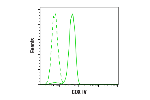
Flow cytometric analysis of HeLa cells using COX IV (3E11) Rabbit mAb (solid line) compared to concentration-matched Rabbit (DA1E) mAb IgG XP® Isotype Control #3900 (dashed line). Anti-rabbit IgG (H+L), F(ab')₂ Fragment (Alexa Fluor® 488 Conjugate) #4412 was used as a secondary antibody.
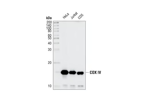
Western blot analysis of extracts from HeLa, Jurkat and COS cell lines, using COX IV (3E11) Rabbit mAb.
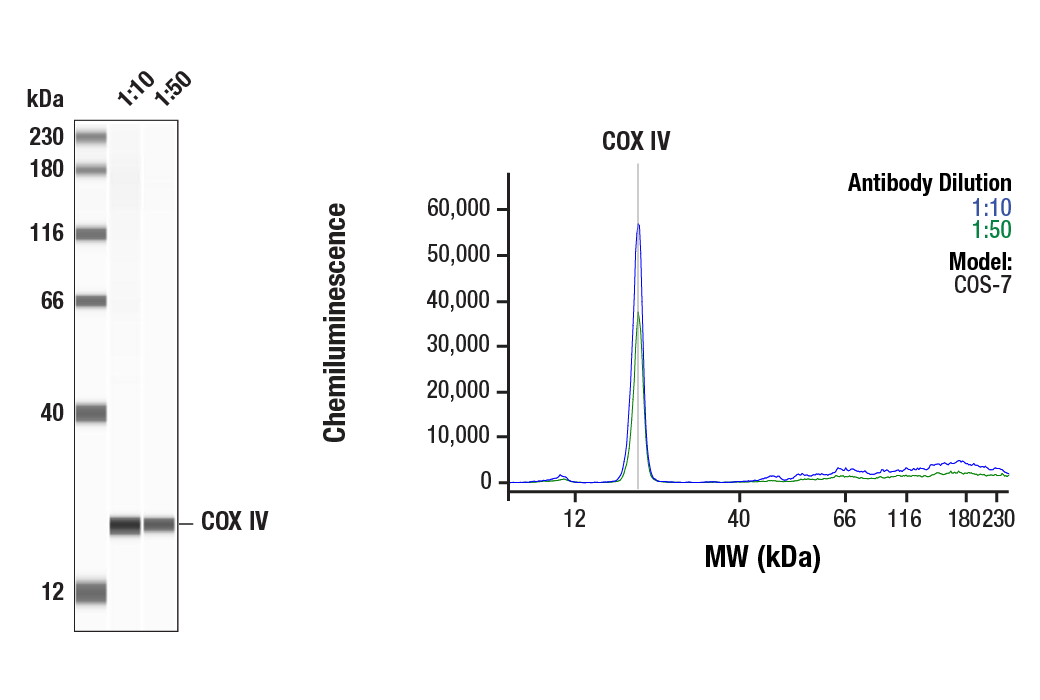
Simple Western™ analysis of lysates (1.0 mg/mL) from COS-7 cells using COX IV (3E11) Rabbit mAb #4850. The virtual lane view (left) shows the target bands (as indicated) at 1:10 and 1:50 dilutions of primary antibody. The corresponding electropherogram view (right) plots chemiluminescence by molecular weight along the capillary at 1:10 (blue line) and 1:50 (green line) dilutions of primary antibody. This experiment was performed under reducing conditions on the Jess™ Simple Western instrument from ProteinSimple, a BioTechne brand, using the 12-230 kDa separation module.
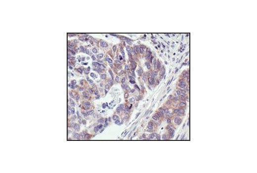
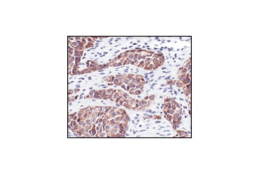
Immunohistochemical analysis of paraffin-embedded human colon carcinoma, showing staining of the mitochondria, using COX IV (3E11) Rabbit mAb.
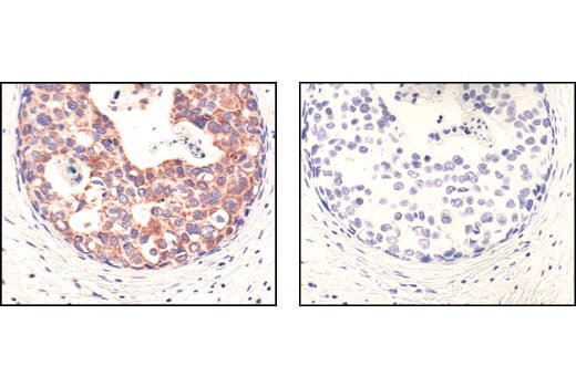
Immunohistochemical analysis of paraffin-embedded human breast carcinoma, using COX IV (3E11) Rabbit mAb in the presence of control peptide (left) or Cox IV Blocking Peptide #1034 (right).
Immunohistochemical analysis of paraffin-embedded H1650 xenograft, using COX IV Rabbit mAb. Note specific staining of human cancer cells.
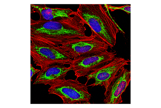
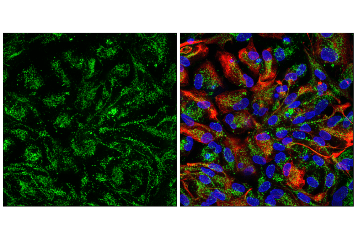
Confocal immunofluorescent analysis of HeLa cells labeled with COX IV (3E11) Rabbit mAb (green) and β-Actin (8H10D10) Mouse mAb #3700 (red). Samples were mounted in ProLong® Gold Antifade Reagent with DAPI #8961 (blue).
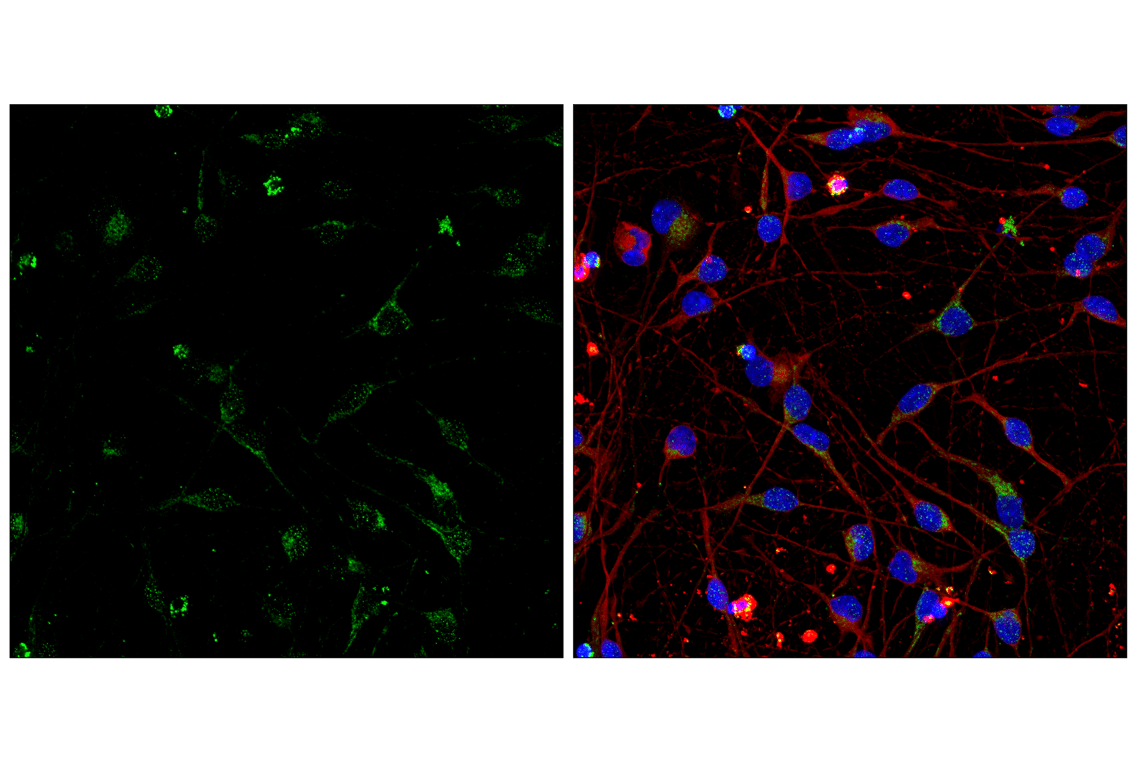
Confocal immunofluorescent analysis of human iPSC-derived cortical glutamatergic neurons at 7 days in vitro using COX IV (3E11) Rabbit mAb (green), β3-Tubulin (E9F3E) Mouse mAb #45058 (red), and DAPI #4083 (blue). iCell GlutaNeurons were kindly provided by FUJIFILM Cellular Dynamics, Inc.
Confocal immunofluorescent analysis of human iPSC-derived astrocyte cells at 14 days in vitro using COX IV (3E11) Rabbit mAb (green), GFAP (GA5) Mouse mAb #3670 (red), and DAPI #4083 (blue). iCell Astrocytes 2.0 were kindly provided by FUJIFILM Cellular Dynamics, Inc.