


 全部商品分类
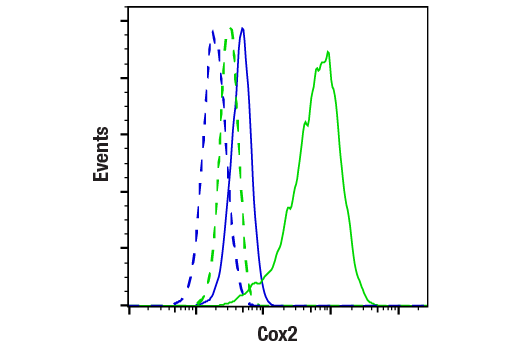


Cox2 (D5H5) XP® Rabbit mAb


 下载产品说明书 下载COA 下载SDS
 用小程序,查商品更便捷


 收藏
收藏
 对比
对比 咨询
咨询
Monoclonal antibody is produced by immunizing animals with a synthetic peptide corresponding to residues surrounding His108 of human Cox2 protein.
Product Usage Information
| Application | Dilution |
|---|---|
| Western Blotting | 1:1000 |
| Simple Western™ | 1:10 - 1:50 |
| Immunohistochemistry (Paraffin) | 1:300 - 1:1200 |
| Immunofluorescence (Immunocytochemistry) | 1:400 - 1:800 |
| Flow Cytometry (Fixed/Permeabilized) | 1:100 - 1:400 |
Specificity/Sensitivity
Species Reactivity:
Human, Mouse, Rat
Supplied in 10 mM sodium HEPES (pH 7.5), 150 mM NaCl, 100 µg/ml BSA, 50% glycerol and less than 0.02% sodium azide. Store at –20°C. Do not aliquot the antibody.
For a carrier free (BSA and azide free) version of this product see product #73315.
参考图片
Flow cytometric analysis of Raw264.7 cells, untreated (blue) or treated with LPS #14011 (1 µg/ml, 18 hr; green) using Cox2 (D5H5) XP® Rabbit mAb (solid lines) or concentration matched Rabbit (DA1E) mAb IgG XP® Isotype Control #3900 (dashed lines). Anti-rabbit IgG (H+L), F(ab')2 Fragment (Alexa Fluor® 488 Conjugate) #4412 was used as a secondary antibody.
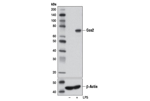
Western blot analysis of extracts from Raw 264.7 cells, untreated (-) or LPS-treated (1 μg/ml, 24 hr; +), using Cox2 (D5H5) XP® Rabbit mAb or β-Actin (D6A8) Rabbit mAb #8457.
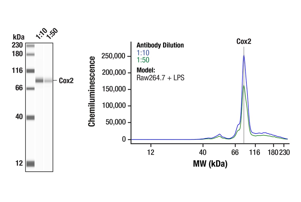
Simple Western™ analysis of lysates (0.05 mg/mL) from Raw264,7 cells treated with LPS (1ug/mL, 6 hr) using Cox2 (D5H5) XP® Rabbit mAb #12282. The virtual lane view (left) shows the target band (as indicated) at 1:10 and 1:50 dilutions of primary antibody. The corresponding electropherogram view (right) plots chemiluminescence by molecular weight along the capillary at 1:10 (blue line) and 1:50 (green line) dilutions of primary antibody. This experiment was performed under reducing conditions on the Jess™ Simple Western instrument from ProteinSimple, a BioTechne brand, using the 12-230 kDa separation module.
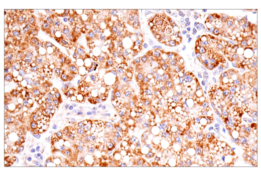
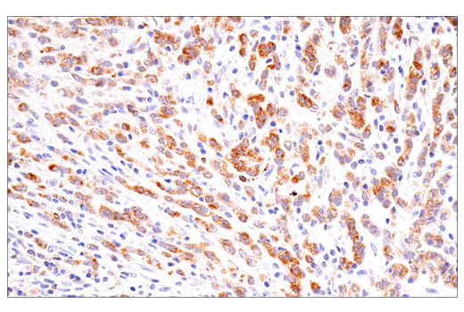
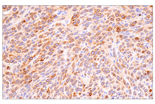
Immunohistochemical analysis of paraffin-embedded human hepatocellular carcinoma using Cox2 (D5H5) XP® Rabbit mAb.
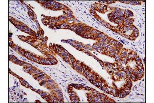
Immunohistochemical analysis of paraffin-embedded human colon carcinoma using Cox2 (D5H5) XP® Rabbit mAb.
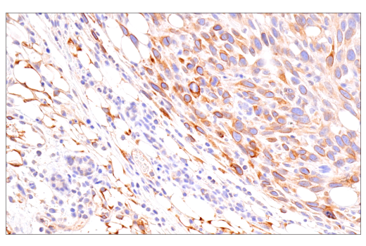
Immunohistochemical analysis of paraffin-embedded human renal cell carcinoma using Cox2 (D5H5) XP® Rabbit mAb.
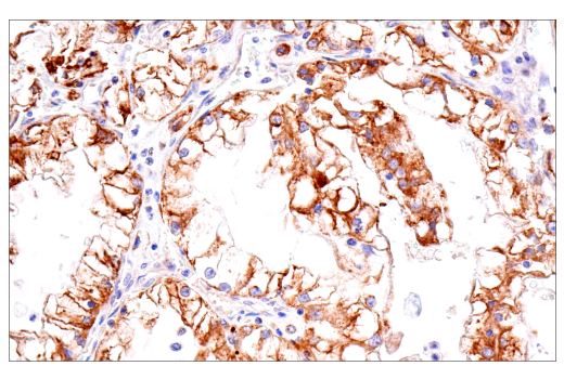
Immunohistochemical analysis of paraffin-embedded human esophageal adenocarcinoma using Cox2 (D5H5) XP® Rabbit mAb.
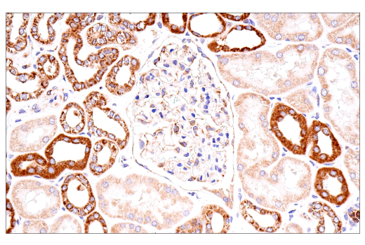
Immunohistochemical analysis of paraffin-embedded normal human kidney using Cox2 (D5H5) XP® Rabbit mAb.
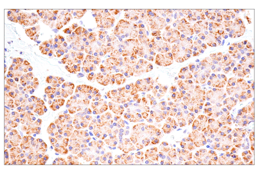
Immunohistochemical analysis of paraffin-embedded normal human pancreas using Cox2 (D5H5) XP® Rabbit mAb.
Immunohistochemical analysis of paraffin-embedded 4T1 syngeneic mammary tumor using Cox2 (D5H5) XP® Rabbit mAb.
Immunohistochemical analysis of paraffin-embedded CT26.WT syngeneic tumor using Cox2 (D5H5) XP® Rabbit mAb.
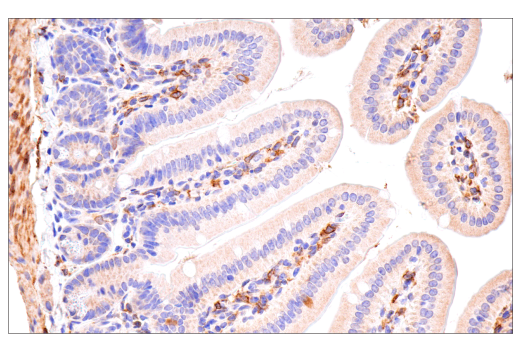
Immunohistochemical analysis of paraffin-embedded mouse small intestine using Cox2 (D5H5) XP® Rabbit mAb.
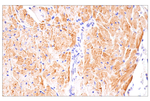
Immunohistochemical analysis of paraffin-embedded mouse heart using Cox2 (D5H5) XP® Rabbit mAb.
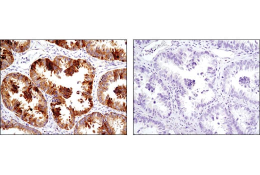
Immunohistochemical analysis of paraffin-embedded human ovarian carcinoma using Cox2 (D5H5) XP® Rabbit mAb in the presence of control peptide (left) or antigen-specific peptide (right).
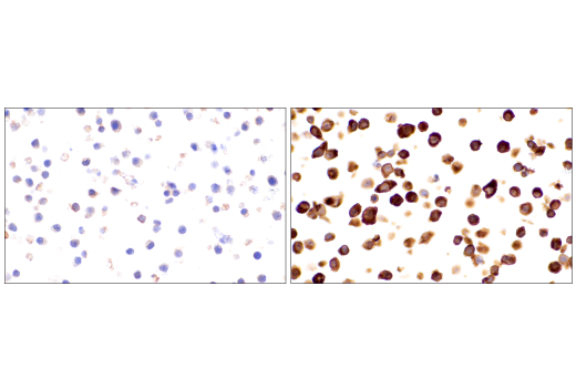
Immunohistochemical analysis of paraffin-embedded RAW 264.7 cell pellets, untreated (left) or treated with Lipopolysaccharides (LPS) #14011 (1 μg/ml, 16 hr, right), using Cox2 (D5H5) XP® Rabbit mAb.
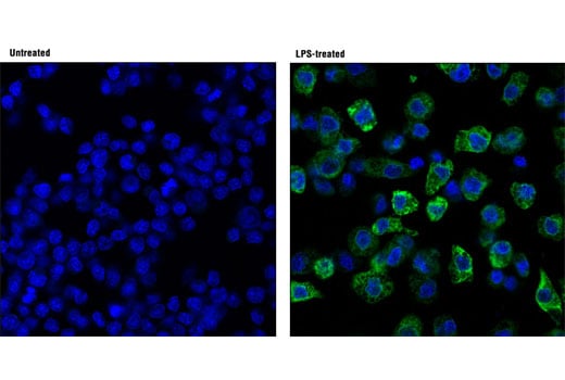
Confocal immunofluorescent analysis of Raw 264.7 cells, untreated (left; -) or LPS-treated (right; 1ug/ml, 24 hrs; +), using Cox2 (D5H5) XP® Rabbit mAb (green). Blue pseudocolor = DRAQ5® #4084 (fluorescent DNA dye).