


 全部商品分类
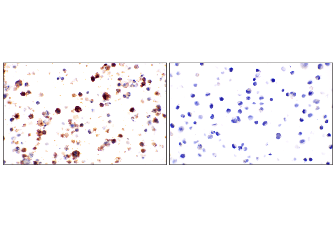


 用小程序,查商品更便捷


Monoclonal antibody is produced by immunizing animals with recombinant protein specific to the extracellular domain of human CR1/CD35 protein.

Product Usage Information
| Application | Dilution |
|---|---|
| Western Blotting | 1:1000 |
| Immunohistochemistry (Paraffin) | 1:800 - 1:3200 |
Specificity/Sensitivity
Species Reactivity:
Human
Supplied in 10 mM sodium HEPES (pH 7.5), 150 mM NaCl, 100 µg/mL BSA, 50% glycerol, and less than 0.02% sodium azide. Store at –20°C. Do not aliquot the antibody.
参考图片
Immunohistochemical analysis of paraffin-embedded TF-1 cell pellet (left, positive) or K-562 cell pellet (right, negative) using CR1/CD35 (E8B1Z) Rabbit mAb.
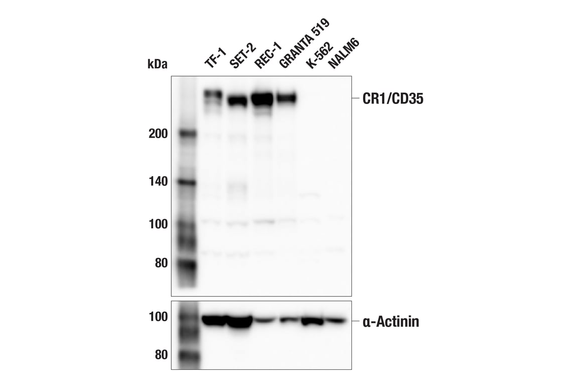
Western blot analysis of extracts from various cell lines using CR1/CD35 (E8B1Z) Rabbit mAb (upper) or α-Actinin (D6F6) XP® Rabbit mAb #6487 (lower). Negative expression of CR1/CD35 protein in K-562 and NALM6 cells is consistent with the predicted expression pattern.
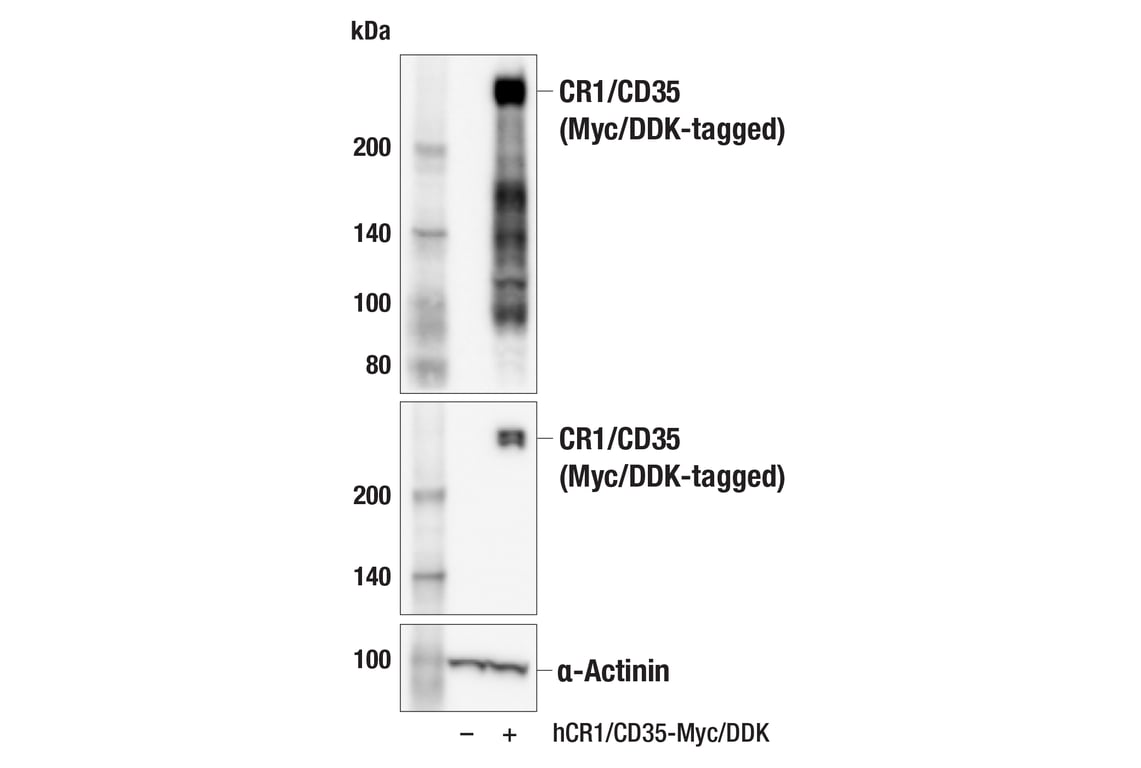
Western blot analysis of extracts from 293T cells, mock transfected (-) or transfected with a construct expressing Myc/DDK-tagged full-length human CR1/CD35 (hCR1/CD35-Myc/DDK; +), using CR1/CD35 (E8B1Z) Rabbit mAb (upper), DYKDDDDK Tag (D6W5B) Rabbit mAb #14793 (middle), or α-Actinin (D6F6) XP® Rabbit mAb #6487 (lower).
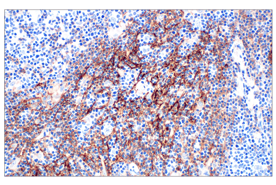
Immunohistochemical analysis of paraffin-embedded human Hodgkin lymphoma using CR1/CD35 (E8B1Z) Rabbit mAb.
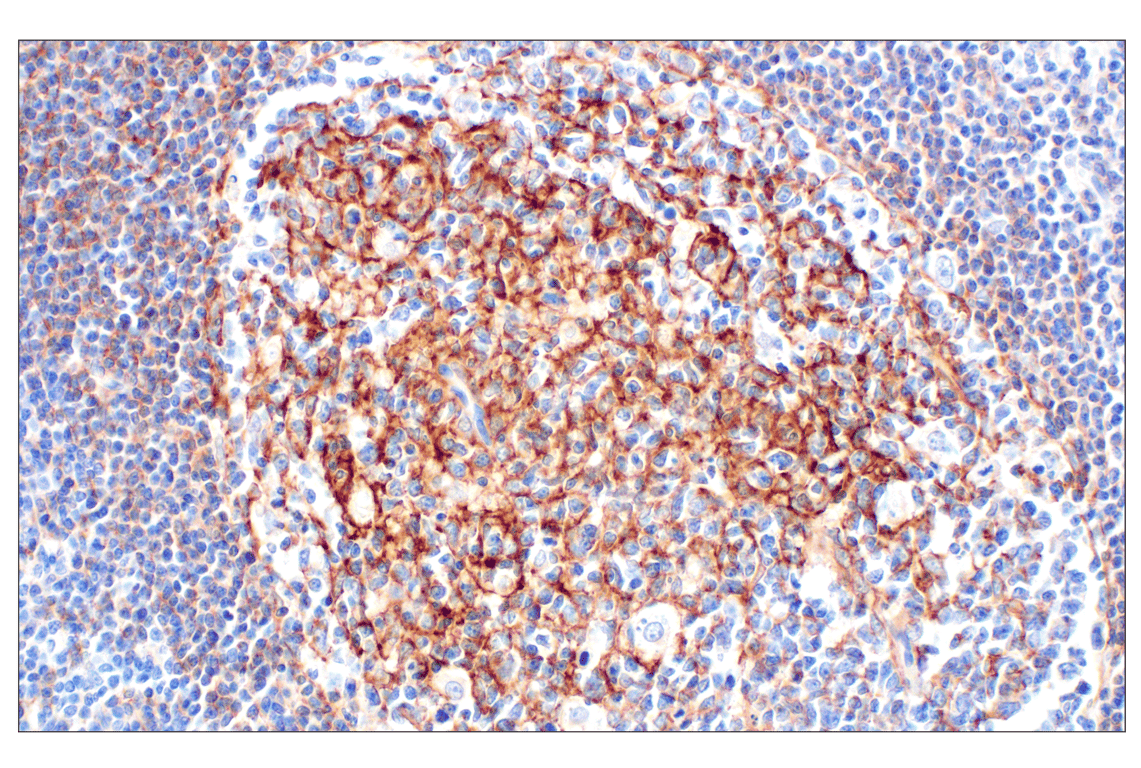
Immunohistochemical analysis of paraffin-embedded human tonsil using CR1/CD35 (E8B1Z) Rabbit mAb.
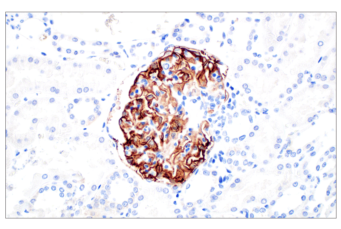
Immunohistochemical analysis of paraffin-embedded normal human kidney using CR1/CD35 (E8B1Z) Rabbit mAb.
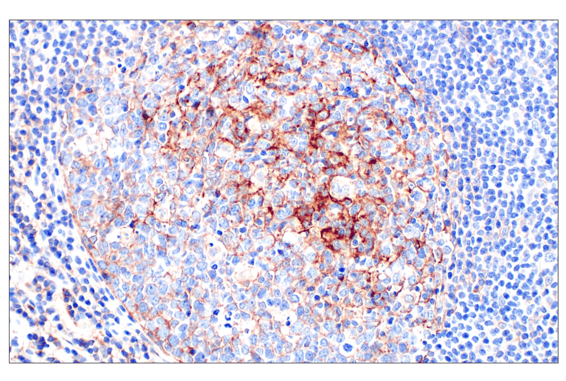
Immunohistochemical analysis of paraffin-embedded Peyer's patch within normal human small intestine using CR1/CD35 (E8B1Z) Rabbit mAb.
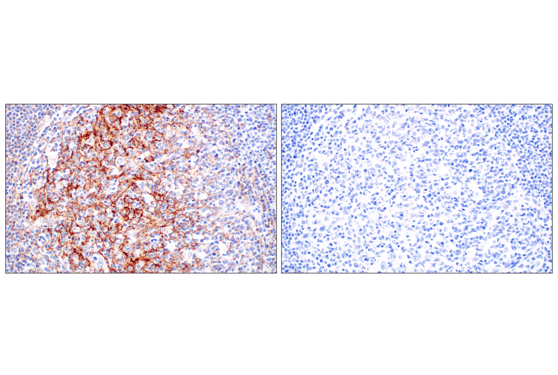
Immunohistochemical analysis of paraffin-embedded human tonsil using CR1/CD35 (E8B1Z) Rabbit mAb (left) compared to concentration-matched Rabbit (DA1E) mAb IgG XP® Isotype Control #3900 (right).