


1/2

品牌: BD Pharmingen
 下载产品说明书 下载SDS
 用小程序,查商品更便捷


 收藏
收藏
 对比
对比 咨询
咨询反应种属:
Human (QC Testing), Rhesus,Cynomolgus,Baboon (Tested in Development)
Human (QC Testing), Rhesus,Cynomolgus,Baboon (Tested in Development)
来源宿主:
Mouse BALB/c IgG2a, κ
Mouse BALB/c IgG2a, κ
产品介绍
产品信息
耦联标记
PE

抗原名称
CD152
宿主
Mouse BALB/c IgG2a, κ
免疫原
Human CTLA4 Recombinant Protein
简单描述
The BNI3 monoclonal antibody specifically binds to the human cytolytic T lymphocyte-associated antigen (CTLA-4), also known as CD152. CTLA-4 is transiently expressed on activated CD28+ T cells and binds to CD80 and CD86 present on antigen presenting cells (APC) with high avidity. This interaction appears to deliver a negative regulatory signal to the T cell. Recent reports indicate that CTLA-4 is also expressed on B cells when cultured with activated T cells, suggesting a role for CTLA-4 in the regulation of B-cell response. Immobilized BNI3 antibody enhances T-cell proliferation induced by antibody-mediated crosslinking of CD3 and CD28. Recent studies have shown that CD152 can be expressed by regulatory T (Treg) cells. After cellular fixation and permeabilization, the BNI3 antibody can stain intracellular CD152 expressed in T cells including Treg cells. Clone BNI3 was studied in the VI Leukocyte Typing Workshop.
商品描述
BNI3
The BNI3 monoclonal antibody specifically binds to the human cytolytic T lymphocyte-associated antigen (CTLA-4), also known as CD152. CTLA-4 is transiently expressed on activated CD28+ T cells and binds to CD80 and CD86 present on antigen presenting cells (APC) with high avidity. This interaction appears to deliver a negative regulatory signal to the T cell. Recent reports indicate that CTLA-4 is also expressed on B cells when cultured with activated T cells, suggesting a role for CTLA-4 in the regulation of B-cell response. Immobilized BNI3 antibody enhances T-cell proliferation induced by antibody-mediated crosslinking of CD3 and CD28. Recent studies have shown that CD152 can be expressed by regulatory T (Treg) cells. After cellular fixation and permeabilization, the BNI3 antibody can stain intracellular CD152 expressed in T cells including Treg cells. Clone BNI3 was studied in the VI Leukocyte Typing Workshop.
同种型
Mouse BALB/c IgG2a, κ
克隆号
克隆 BNI3 (RUO)
产品详情
PE
R-Phycoerythrin (PE), is part of the BD family of Phycobiliprotein dyes. This fluorochrome is a multimeric fluorescent phycobiliprotein with excitation maximum (Ex Max) of 496 nm and 566 nm and an emission maximum (Em Max) at 576 nm. PE is designed to be excited by the Blue (488 nm), Green (532 nm) and Yellow-Green (561 nm) lasers and detected using an optical filter centered near 575 nm (e.g., a 575/26-nm bandpass filter). As PE is excited by multiple lasers, this can result in cross-laser excitation and fluorescence spillover on instruments with various combinations of Blue, Green, and Yellow-Green lasers. Please ensure that your instrument’s configurations (lasers and optical filters) are appropriate for this dye.

PE
Yellow-Green 488 nm, 532 nm, 561 nm
496 nm, 566 nm
576 nm
应用
实验应用
Flow cytometry (Routinely Tested), Intracellular staining (flow cytometry) (Tested During Development)
推荐用量
20 µl
反应种属
Human (QC Testing), Rhesus,Cynomolgus,Baboon (Tested in Development)
目标/特异性
CD152 (CTLA-4)
背景
别名
CTLA-4; AILIM; Cytotoxic T-lymphocyte protein 4
制备和贮存
存储溶液
Aqueous buffered solution containing BSA and ≤0.09% sodium azide.
保存方式
Aqueous buffered solution containing BSA and ≤0.09% sodium azide.
文献
文献
研发参考(10)
1. Cabezon R, Sintes J, Llinas L, Benitez-Ribas D. Analysis of HLDA9 mAbs on plasmacytoid dendritic cell. Immunol Lett. 2011; 134(2):167-173. (Clone-specific: Flow cytometry).
2. Castan J, Klauenberg U, Kalmar P, Fleischer B, Broker BM. Expression of CTLA-4 (CD152) on human medullary CD4+ thymocytes. Med Microbiol Immunol (Berl). 1998; 187(1):49-52. (Immunogen: Fluorescence microscopy, Immunocytochemistry, Immunofluorescence, Immunohistochemistry).
3. Castan J, Tenner-Racz K, Racz P, Fleischer B, Broker BM. Accumulation of CTLA-4 expressing T lymphocytes in the germinal centres of human lymphoid tissues. Immunology. 1997; 90(2):265-271. (Immunogen: ELISA, Fluorescence microscopy, Immunofluorescence, Immunohistochemistry).
4. Healy ZR, Murdoch DM. OMIP-036: Co-inhibitory receptor (immune checkpoint) expression analysis in human T cell subsets.. Cytometry A. 2016; 89(10):889-892. (Clone-specific: Intracellular Staining/Flow Cytometry).
5. Kuiper HM, Brouwer M, Linsley PS, van Lier RA. Activated T cells can induce high levels of CTLA-4 expression on B cells. J Immunol. 1995; 155(4):1776-1783. (Biology).
6. Lindsten T, Lee KP, Harris ES, et al. Characterization of CTLA-4 structure and expression on human T cells. J Immunol. 1993; 151(7):3489-3499. (Biology).
7. Morton PA, Fu XT, Stewart JA, et al. Differential effects of CTLA-4 substitutions on the binding of human CD80 (B7-1) and CD86 (B7-2). J Immunol. 1996; 156(3):1047-1054. (Biology).
8. Rabe H, Lundell AC, Andersson K, Adlerberth I, Wold AE, Rudin A. Higher proportions of circulating FOXP3+ and CTLA-4+ regulatory T cells are associated with lower fractions of memory CD4+ T cells in infants.. J Leukoc Biol. 2011; 90(6):1133-40. (Clone-specific: Intracellular Staining/Flow Cytometry).
9. Santegoets SJ, Dijkgraaf EM, Battaglia A, et al. Monitoring regulatory T cells in clinical samples: consensus on an essential marker set and gating strategy for regulatory T cell analysis by flow cytometry.. Cancer Immunol Immunother. 2015; 64(10):1271-86. (Clone-specific: Intracellular Staining/Flow Cytometry).
10. Wang H, Shih CC, Waters JB, et al. CD152 (CTLA4) Workshop: Expression and function of CD152 on human T cells: A study using a mouse anti-human CD152 monoclonal antibody BNI3.1. In: Kishimoto T. Tadamitsu Kishimoto .. et al., ed. Leucocyte typing VI : white cell differentiation antigens : proceedings of the sixth international workshop and conference held in Kobe, Japan, 10-14 November 1996. New York: Garland Pub.; 1997:97-98.
数据库链接
Entrez-Gene ID
1493
参考图片
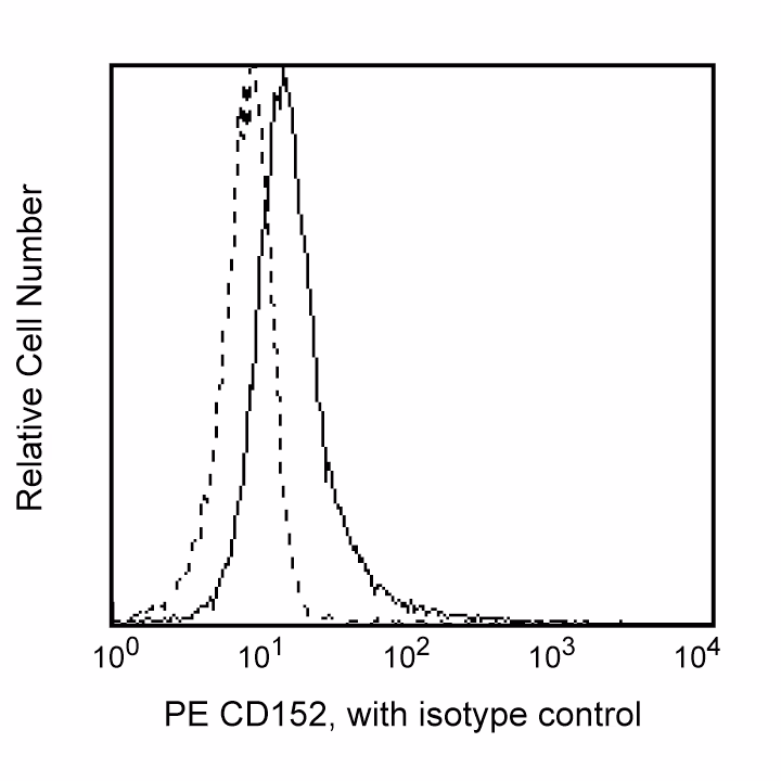
Flow cytometric analysis of CD152 expression on stimulated Human peripheral mononuclear cells. Peripheral blood mononuclear cells were stimulated with Concanavalin A for 3 days, then stained with either PE Mouse IgG2a, κ Isotype Control (Cat. No. 555574; dashed line histogram) or PE Mouse Anti-Human CD152 (Cat. No. 555853/560939/557301; solid line histogram). The fluorescence histograms were derived from gated events with the forward and side light-scattering characteristics of viable activated cells. Flow cytometry was performed on a BD FACScan™ system.
声明 :本官网所有报价均为常温或者蓝冰运输价格,如有产品需要干冰运输,需另外加收干冰运输费。

 危险品化学品经营许可证(不带存储) 许可证编号:沪(杨)应急管危经许[2022]202944(QY)
  营业执照(三证合一)