


 全部商品分类
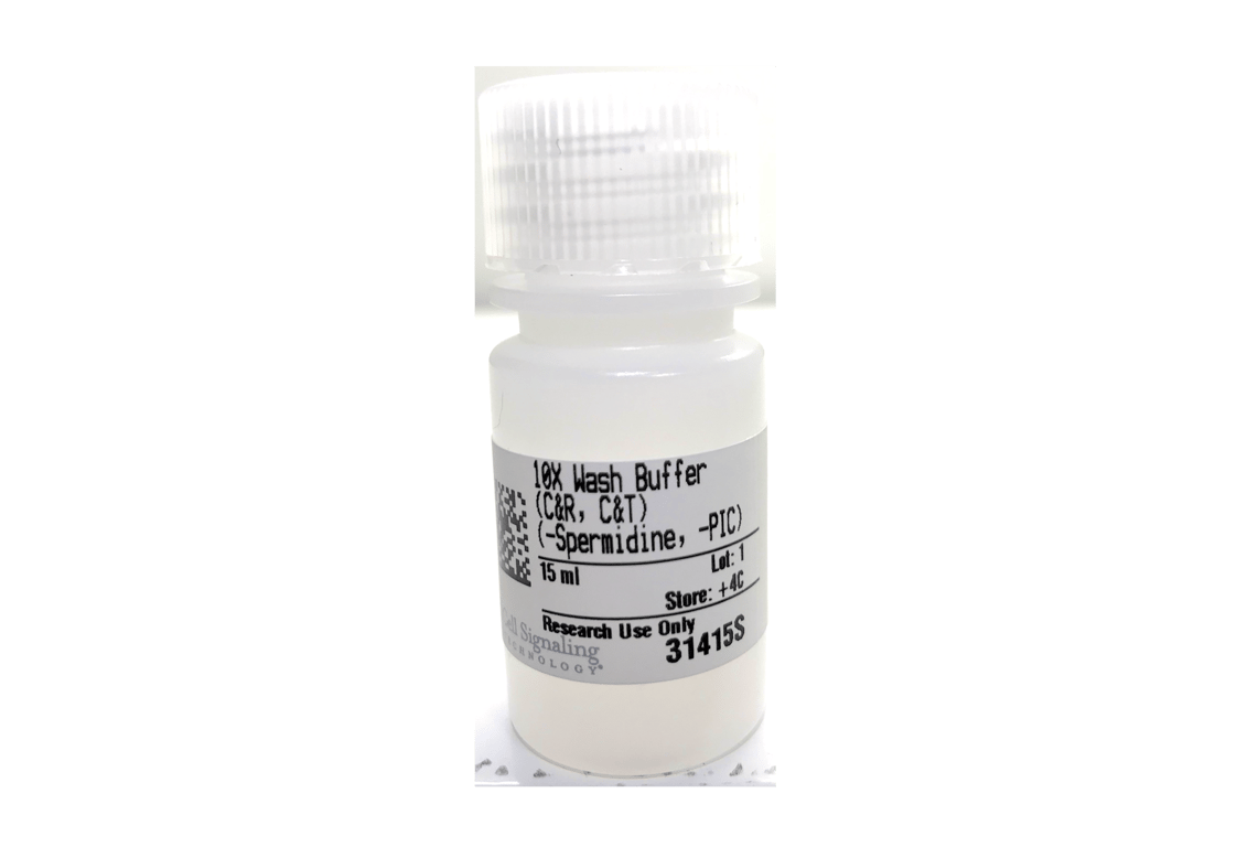


CUT&RUN 10X Wash Buffer

 下载产品说明书 下载COA 下载SDS
 用小程序,查商品更便捷


 收藏
收藏
 对比
对比 咨询
咨询The 10X Wash Buffer (CUT&RUN, CUT&Tag) provides enough reagent to support 24 CUT&RUN or CUT&Tag assays. This product is formulated for optimal performance in the CUT&RUN and CUT&Tag assays and each lot is tested and validated using the CUT&RUN Assay Kit #86652 or the CUT&Tag Assay Kit #77552. This product should be diluted to 1X using nuclease-free water and an appropriate amount of 100X Spermidine #27287 and Protease Inhibitor Cocktail (200X) #7012 should be added right before use. Please keep at room temperature during use to minimize stress on the cells.

Product Usage Information
For the CUT&RUN and CUT&Tag assays, we recommend preparing 2 ml 1X Complete Wash Buffer for each cell line and an additional 100 μl for each reaction or input sample. For example, to prepare 2.5 ml of 1X Complete Wash Buffer, add 250 μl 10X Wash Buffer (CUT&RUN, CUT&Tag), 25 μl 100X Spermidine #27287, and 12.5 μl Protease Inhibitor Cocktail (200X) #7012 to 2,212.5 μl nuclease-free water right before use. Equilibrate it to room temperature to minimize stress on the cells.
Store 10X Wash Buffer (CUT&RUN, CUT&Tag) at 4°C. This product is stable for at least 12 months.
参考图片