


 全部商品分类
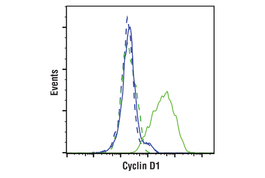


Cyclin D1 (E3P5S) XP ® Rabbit mAb


 下载产品说明书 下载COA 下载SDS
 用小程序,查商品更便捷


 收藏
收藏
 对比
对比 咨询
咨询
Monoclonal antibody is produced by immunizing animals with a synthetic peptide corresponding to residues surrounding Ala284 of human cyclin D1 protein.
Product Usage Information
| Application | Dilution |
|---|---|
| Western Blotting | 1:1000 |
| Immunoprecipitation | 1:50 |
| IHC Leica Bond | 1:250 - 1:1000 |
| Immunohistochemistry (Paraffin) | 1:250 - 1:1000 |
| Immunofluorescence (Immunocytochemistry) | 1:400 - 1:1600 |
| Flow Cytometry (Fixed/Permeabilized) | 1:400 - 1:1600 |
Specificity/Sensitivity
Species Reactivity:
Human, Mouse, Rat, Monkey
Supplied in 10 mM sodium HEPES (pH 7.5), 150 mM NaCl, 100 µg/ml BSA, 50% glycerol and less than 0.02% sodium azide. Store at –20°C. Do not aliquot the antibody.
For a carrier free (BSA and azide free) version of this product see product #66467.
参考图片
Flow cytometric analysis of K-562 cells (blue) and SH-SY5Y cells (green) using Cyclin D1 (E3P5S) XP® Rabbit mAb (solid lines) or a concentration-matched Rabbit (DA1E) mAb IgG XP® Isotype Control #3900 (dashed lines). Anti-rabbit IgG (H+L), F(ab')2 Fragment (Alexa Fluor® 488 Conjugate) #4412 was used as a secondary antibody.
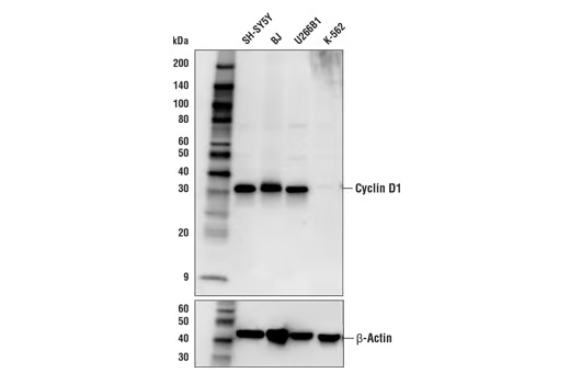
Western blot analysis of extracts from various cell lines using Cyclin D1 (E3P5S) XP® Rabbit mAb (upper) or β-Actin (D6A8) Rabbit mAb #8457 (lower).
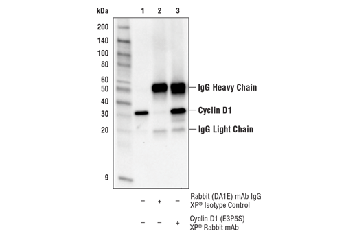
Immunoprecipitation of Cyclin D1 from SH-SY5Y cell extracts. Lane 1 is 10% input, lane 2 is Rabbit (DA1E) mAb IgG XP® Isotype Control #3900, and lane 3 is Cyclin D1 (E3P5S) XP® Rabbit mAb. Western blot analysis was performed usingCyclin D1 (E3P5S) XP® Rabbit mAb as the primary antibody and Anti-rabbit IgG, HRP-linked Antibody #7074 as the secondary antibody.
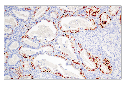
Immunohistochemical analysis of paraffin-embedded human endometrioid adenocarcinoma using Cyclin D1 (E3P5S) XP® Rabbit mAb performed on the Leica® BOND™ Rx.
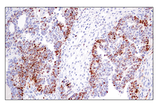
Immunohistochemical analysis of paraffin-embedded human urothelial carcinoma using Cyclin D1 (E3P5S) XP® Rabbit mAb.
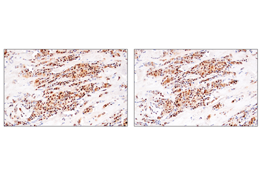
Immunohistochemical analysis of paraffin-embedded human ductal breast carcinoma using Cyclin D1 (E3P5S) XP® Rabbit mAb (left) or Cyclin D1 (92G2) Rabbit mAb #2978 (right). These two antibodies detect independent, unique epitopes on human cyclin D1. The similar staining patterns obtained with both antibodies help to confirm the specificity of the staining.
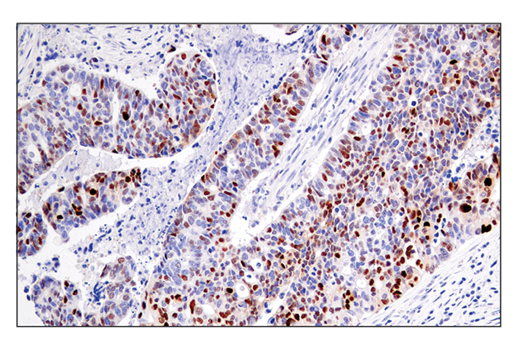
Immunohistochemical analysis of paraffin-embedded human lung adenocarcinoma using Cyclin D1 (E3P5S) XP® Rabbit mAb.
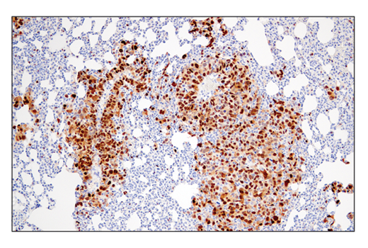
Immunohistochemical analysis of paraffin-embedded 4T1 metastatic tumor in mouse lung using Cyclin D1 (E3P5S) XP® Rabbit mAb.
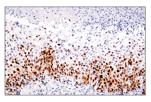
Immunohistochemical analysis of paraffin-embedded human ovarian carcinosarcoma using Cyclin D1 (E3P5S) XP® Rabbit mAb.
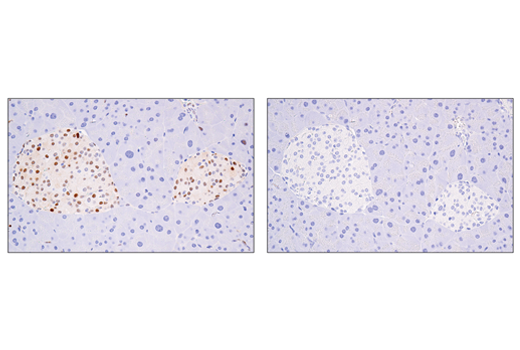
Immunohistochemical analysis of paraffin-embedded mouse pancreas using Cyclin D1 (E3P5S) XP® Rabbit mAb (left) compared to concentration-matched Rabbit (DA1E) mAb IgG XP® Isotype Control #3900 (right).
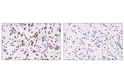
Immunohistochemical analysis of paraffin-embedded SH-SY5Y cell pellet (left, positive) or THP-1 cell pellet (right, negative) using Cyclin D1 (E3P5S) XP® Rabbit mAb.
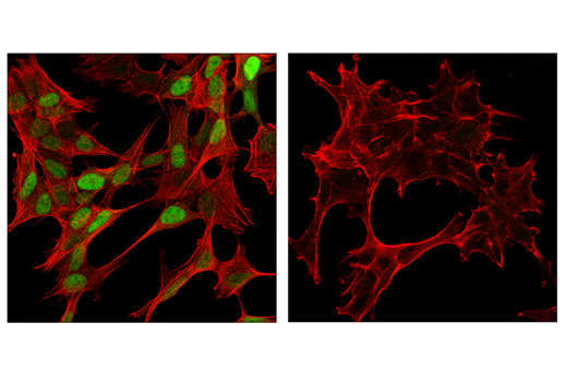
Confocal immunofluorescent analysis of SH-SY5Y cells (left, positive) and 293T cells (right, negative) using Cyclin D1 (E3P5S) XP® Rabbit mAb (green). Actin filaments were labeled with DyLight™ 554 Phalloidin #13054 (red).