Bioimaging, Flow cytometry, Immunofluorescence (Tested During Development)
产品介绍
产品介绍
产品信息
简单描述
DAPI (4',6-Diamidino-2-Phenylindole, Dihydrochloride) is a nucleic acid stain that binds to A-T rich regions of DNA along the minor groove. DAPI is predominantly impermeant to live cells, allowing it to be used as a viability dye in unfixed cells to discriminate intact from membrane-compromised cells. Note, however, that high concentrations of the dye may still enter intact cells. Additionally, DAPI may be used to analyze DNA content in fixed cells, or as a nuclear counterstain in imaging or flow cytometry.
When bound to double-stranded DNA, DAPI has an excitation wavelength maximum of 358 nm and an emission maximum of 461 nm. DAPI is also well excited by the violet laser line (eg, 405 nm). Note that DAPI also binds RNA. Under these conditions, DAPI emits maximally at 500 nm, but with less intensity than when bound to double-stranded DNA.
商品描述
DAPI (4',6-Diamidino-2-Phenylindole, Dihydrochloride) is a nucleic acid stain that binds to A-T rich regions of DNA along the minor groove. DAPI is predominantly impermeant to live cells, allowing it to be used as a viability dye in unfixed cells to discriminate intact from membrane-compromised cells. Note, however, that high concentrations of the dye may still enter intact cells. Additionally, DAPI may be used to analyze DNA content in fixed cells, or as a nuclear counterstain in imaging or flow cytometry.
When bound to double-stranded DNA, DAPI has an excitation wavelength maximum of 358 nm and an emission maximum of 461 nm. DAPI is also well excited by the violet laser line (eg, 405 nm). Note that DAPI also binds RNA. Under these conditions, DAPI emits maximally at 500 nm, but with less intensity than when bound to double-stranded DNA.
克隆号
(RUO)
浓度
1.0 mg/ml
应用
实验应用
Bioimaging, Flow cytometry, Immunofluorescence (Tested During Development)
目标/特异性
DAPI
文献
文献
研发参考(5)
1. Darzynkiewicz Z, Bruno S, Del Bino G, et al. Features of apoptotic cells measured by flow cytometry. Cytometry. 1992; 13(8):795-808. (Methodology: Flow cytometry).
2. Hotz MA, Gong J, Traganos F, and Darzynkiewicz Z. Flow cytometric detection of apoptosis: Comparison of the assays of in situ DNA degradation and chromatin changes. Cytometry. 1994; 15(3):237-244. (Methodology: Flow cytometry).
3. Otto F. DAPI staining of fixed cells for high-resolution flow cytometry of nuclear DNA. Methods Cell Biol. 1990; 33:105-110. (Methodology: Flow cytometry).
4. Shapiro HM. Practical flow cytometry, 3rd ed.. New York: Wiley-Liss; 1995:280-282.
5. Tanious FA, Veal JM, Buczak H, Ratmeyer LS, Wilson WD. DAPI (4',6-diamidino-2-phenylindole) binds differently to DNA and RNA: minor-groove binding at AT sites and intercalation at AU sites. Biochemistry. 1992; 31(12):3103-3112. (Biology).
参考图片
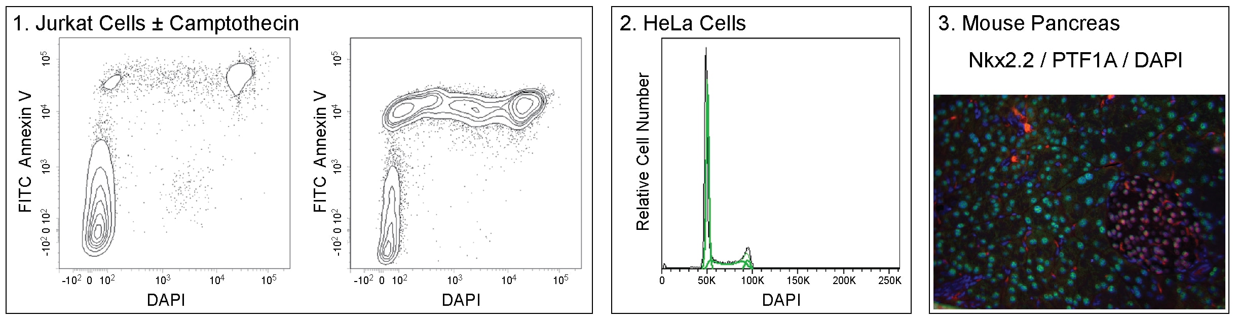
Panel 1. Two-color flow cytometric analysis of Jurkat cell viability. Jurkat cells were treated with DMSO vehicle (Left Plot) or 5 μM Camptothecin (Right Plot) overnight. Cells were resuspended in Annexin V Binding Buffer (Cat. No. 556454) and stained with FITC Annexin V (Cat. No. 556419) and 0.06 μg/mL DAPI. Camptothecin-treated cells show an increased frequency of apoptotic (Annexin V+DAPI-) and dead (Annexin V+DAPI+) cells. Analysis was performed using a BD LSRFortessa™ Cell Analyzer System. Panel 2. Flow cytometric analysis of HeLa cell DNA content. Cultured HeLa cells in log phase growth were harvested using Gibco® Cell Dissociation Buffer (Life Technologies), fixed, and permeabilized using BD Pharmingen™ Transcription Factor Buffer Set (Cat. No. 562574/562725). Cells were resuspended in DPBS with 1 μg/mL DAPI and analyzed using a low BD LSRFortessa™ cytometer flow rate. A 405 nm laser with a 450/50 nm bandpass filter was used to collect data; comparable results were obtained using a 355 nm laser with a 450/50 nm bandpass filter (not shown). Histograms were deconvoluted by FlowJo™ software into G0/G1, S, and G2/M populations. Panel 3. Multicolor immunofluorescence analysis of NKX2.2 and PTF1A expression in mouse pancreas. Following antigen retrieval with BD Retrievagen A Buffer (Cat. No. 550524), a formalin-fixed paraffin-embedded pancreas tissue section was stained with Purified Mouse Anti-Nkx2.2 antibody (Cat. No. 564731, pseudo-colored red), washed, and stained with Alexa Fluor® 488 Goat Anti-Mouse Ig (Life Technologies). After washing, the section was stained with Alexa Fluor® 647 Mouse Anti-PTF1A (pseudo-colored green) and counterstained with DAPI (pseudo-colored blue). The nuclei of islet cells (DAPI+) stain positively for both NKX2.2 and PTF1A. The nuclei of acinar cells and some islet precursor cells stain positive for PTF1A. All images were analyzed using a BD Pathway™ 435 High-Content Bioimager System and merged with BD Attovision™ Software.


 全部商品分类


 下载产品说明书
 用小程序,查商品更便捷


 收藏
收藏
 对比
对比 咨询
咨询