


 全部商品分类


Phospho-DRP1 (Ser616) Antibody

 下载产品说明书 下载COA 下载SDS
 用小程序,查商品更便捷


 收藏
收藏
 对比
对比 咨询
咨询
Polyclonal antibodies are produced by immunizing animals with a synthetic phosphopeptide corresponding to residues surrounding Ser616 of human DRP1. Antibodies are purified by protein A and peptide affinity chromatography.
Product Usage Information
| Application | Dilution |
|---|---|
| Western Blotting | 1:1000 |
| Simple Western™ | 1:10 - 1:50 |
| Immunoprecipitation | 1:50 |
| Immunofluorescence (Immunocytochemistry) | 1:200 - 1:400 |
| Flow Cytometry (Fixed/Permeabilized) | 1:50 - 1:200 |
Specificity/Sensitivity
Species Reactivity:
Human
Supplied in 10 mM sodium HEPES (pH 7.5), 150 mM NaCl, 100 µg/ml BSA and 50% glycerol. Store at –20°C. Do not aliquot the antibody.
参考图片
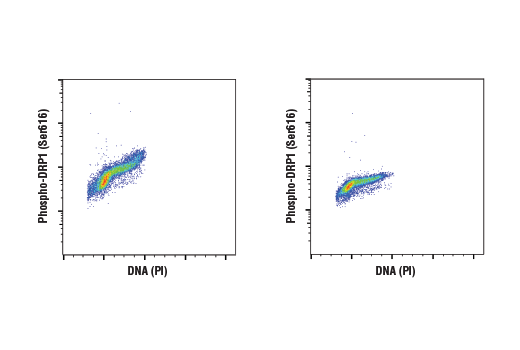
Flow cytometric analysis of Jurkat cells, untreated (left) or λ phosphatase-treated (right), using Phospho-DRP1 (Ser616) Antibody and Propidium Iodide (PI)/RNase Staining Solution #4087 to measure DNA content. Anti-rabbit IgG (H+L), F(ab')2 Fragment (Alexa Fluor® 488 Conjugate) #4412 was used as a secondary antibody.
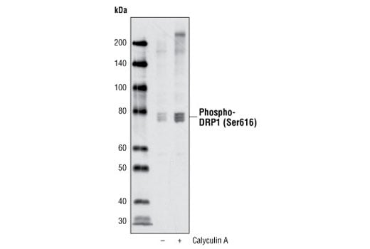
Western blot analysis of extracts from HeLa cells, untreated or treated with Calyculin A #9902, using Phospho-DRP1 (Ser616) Antibody.
Western blot analysis of extracts from HeLa cells, untreated or nocodazole-treated for the indicated times, using Phospho-DRP1 (Ser616) Antibody.
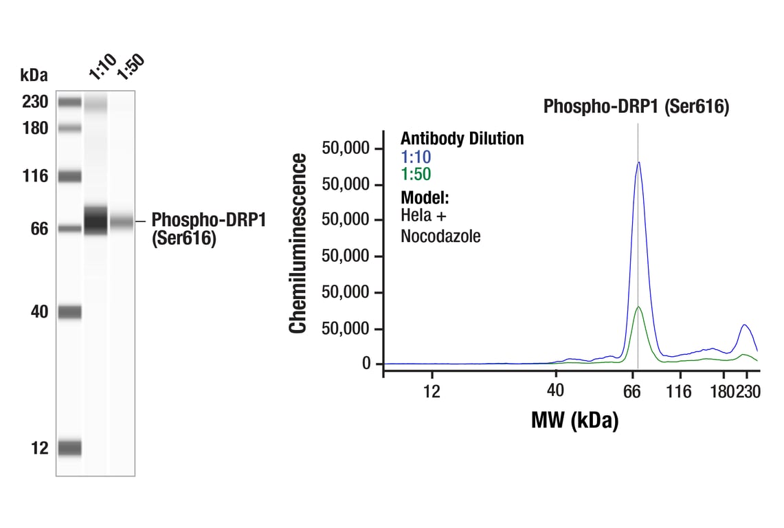
Simple WesternTM analysis of lysates (1 mg/mL) from Hela cells treated with Nocodazole (100ng/mL, 24hr) using Phospho-DRP1 (Ser616) Antibody #3455. The virtual lane view (left) shows a single target band (as indicated) at 1:10 and 1:50 dilutions of primary antibody. The corresponding electropherogram view (right) plots chemiluminescence by molecular weight along the capillary at 1:10 (blue line) and 1:50 (green line) dilutions of primary antibody. This experiment was performed under reducing conditions on the JessTM Simple Western instrument from ProteinSimple, a BioTechne brand, using the 12-230 kDa separation module.
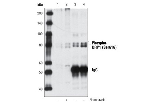
Immunoprecipitation of Phospho-DRP1 (Ser616) from HeLa cell extracts, untreated or nocodazole-treated, using Phospho-DRP1 (Ser616) Antibody followed by western blot using the same antibody. Lanes 1 & 2 are 5% input.
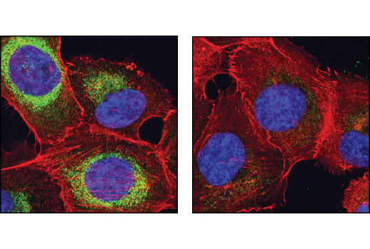
Confocal immunofluorescent analysis of NCI-H1299 cells, untreated (left) or λ-phosphatase-treated (right), using Phospho-DRP1 (Ser616) Antibody (green). Actin filaments have been labeled with DY-554 phallodin (red). Blue pseudocolor = DRAQ5® (fluorescent DNA dye).