


 全部商品分类


 用小程序,查商品更便捷


Monoclonal antibody is produced by immunizing animals with a synthetic peptide corresponding to residues surrounding Pro220 of human SQSTM1 protein.

Product Usage Information
| Application | Dilution |
|---|---|
| Western Blotting | 1:1000 |
| Immunoprecipitation | 1:200 |
| Immunohistochemistry (Paraffin) | 1:400 - 1:1600 |
| Immunofluorescence (Immunocytochemistry) | 1:200 - 1:800 |
Specificity/Sensitivity
Species Reactivity:
Human, Monkey
Supplied in 10 mM sodium HEPES (pH 7.5), 150 mM NaCl, 100 µg/ml BSA, 50% glycerol and less than 0.02% sodium azide. Store at –20°C. Do not aliquot the antibody.
For a carrier free (BSA and azide free) version of this product see product #17274.
参考图片
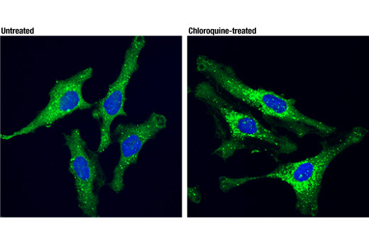
Confocal immunofluorescent analysis of HeLa cells, untreated (left) or treated with chloroquine (50 μM overnight; right), using SQSTM1/p62 (D5L7G) Mouse mAb (green). Blue pseudocolor = DRAQ5® #4084 (fluorescent DNA dye).
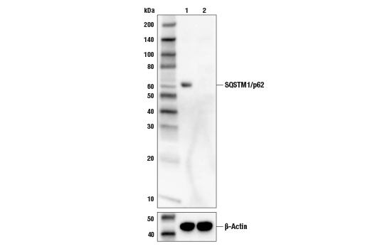
Western blot analysis of extracts from HeLa cells (lane 1) or SQSTM1 knock-out cells (lane 2) using SQSTM1/p62 (D5L7G) Mouse mAb #88588 (upper), and β-actin (D6A8) Rabbit mAb #8457 (lower). The absence of signal in the SQSTM1 knock-out HeLa cells confirms specificity of the antibody for SQSTM1.
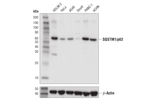
Western blot analysis of extracts from various cell lines using SQSTM1/p62 (D5L7G) Mouse mAb (upper) or β-Actin (D6A8) Rabbit mAb #8457 (lower).
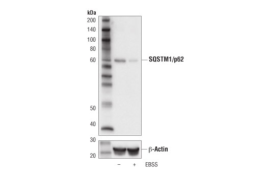
Western blot analysis of extract from HCT 116 cells, untreated (-) or starved with Earles Basic Salt Solution (EBSS; 4 hr; +) using SQSTM1/p62 (D5L7G) Mouse mAb (upper) or β-Actin (D6A8) Rabbit mAb #8457 (lower).
Western blot anlaysis of extracts from HeLa cells, untreated (-) or treated with Chloroquine #14774 (50 μM, overnight; +) using SQSTM1/p62 (D5L7G) Mouse mAb (upper) or β-Actin (D6A8) Rabbit mAb #8457 (lower).
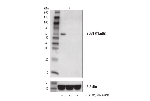
Western blot analysis of extracts from HeLa cells transfected with 100 nM SignalSilence® Control siRNA (Unconjugated) #6568 (-), SignalSilence® SQSTM1/p62 siRNA I #6394 (+), or SignalSilence® SQSTM1/p62 siRNA II #6399 (+), using SQSTM1/p62 (D5L7G) Mouse mAb (upper) or β-Actin (D6A8) Rabbit mAb #8457 (lower).
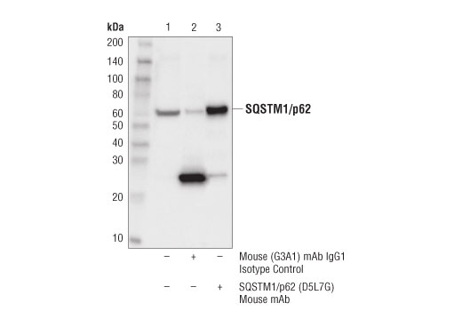
Immunoprecipitation of SQSTM1/p62 protein from PANC-1 cell extracts. Lane 1 is 10% input, lane 2 is Mouse (G3A1) mAb IgG1 Isotype Control #5415, and lane 3 is SQSTM1/p62 (D5L7G) Mouse mAb. Western blot was performed using SQSTM1/p62 (D5L7G) Mouse mAb. A light chain-specific secondary antibody was used to avoid reactivity with heavy chain IgG.
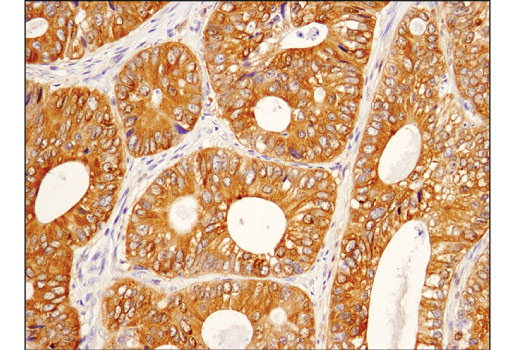
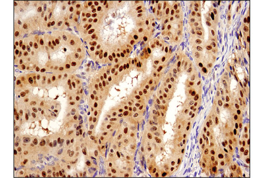
Immunohistochemical analysis of paraffin-embedded human colon carcinoma using SQSTM1/p62 (D5L7G) Mouse mAb.
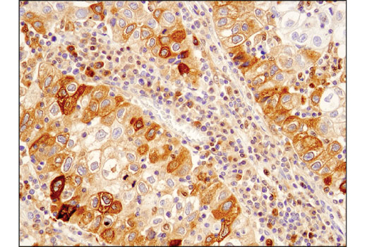
Immunohistochemical analysis of paraffin-embedded human lung carcinoma using SQSTM1/p62 (D5L7G) Mouse mAb.
Immunohistochemical analysis of paraffin-embedded human endometrioid adenocarcinoma using SQSTM1/p62 (D5L7G) Mouse mAb.
Immunohistochemical analysis of paraffin-embedded HDLM-2 (left) and Daudi (right) cell pellets using SQSTM1/p62 (D5L7G) Mouse mAb.
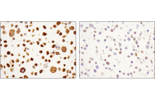
Immunohistochemical analysis of paraffin-embedded human inflamed gall bladder using SQSTM1/p62 (D5L7G) Mouse mAb in the presence of control peptide (left) or antigen-specific peptide (right).