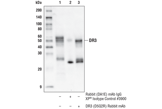
老带新
DR3 (D5Q2R) Rabbit mAb
1/3

DR3 (D5Q2R) Rabbit mAb

分享
品牌: CST
pdf 下载产品说明书
pdf 下载COA
pdf 下载SDS
用小程序,查商品更便捷
收藏
对比对比
咨询咨询
分子量:
55-60
来源宿主:
Rabbit
产品介绍
产品介绍
产品信息
荧光素标记
来源纯化

Monoclonal antibody is produced by immunizing animals with a synthetic peptide corresponding to residues surrounding Ala80 of human DR3 protein. The antigen resides within the extracellular domain of DR3.

宿主
Rabbit
商品描述

Product Usage Information

ApplicationDilution
Western Blotting1:1000
Immunoprecipitation1:100
同种型
Rabbit IgG
分子量
55-60
应用
目标/特异性

Specificity/Sensitivity

DR3 (D5Q2R) Rabbit mAb recognizes endogenous levels of total DR3 protein.

Species Reactivity:

Human

敏感性
Endogenous
背景
背景
The tumor necrosis factor receptor family, which includes TNF-RI, Fas, DR3, DR4, DR5, and DR6, plays an important role in the regulation of apoptosis in various physiological systems (1,2). The receptors are activated by a family of cytokines that include TNF, FasL, and TNF-related apoptosis-inducing ligand (TRAIL). They are characterized by a highly conserved extracellular region containing cysteine-rich repeats and a conserved intracellular region of about 80 amino acids termed the death domain (DD). The DD is important for transducing the death signal by recruiting other DD containing adaptor proteins (FADD, TRADD, RIP) to the death-inducing signaling complex (DISC), resulting in activation of caspases.DR3/WSL-1/Apo-3/TRAMP/LARD is a TNFR family member containing the characteristic extracellular cysteine-repeats, transmembrane region, and an intracellular DD (3-7). DR3 is activated by its ligand Apo-3L/TWEAK to induce apoptosis and activation of NF-κB (8,9). Like TNF-R1, DR3 binds to the DD adaptor protein TRADD, which can then associate with other DD proteins like FADD and RIP as well as members of the TRAF family (3,4). Tissue expression of DR3 is very restricted, primarily seen on the surface of activated thymocytes and lymphocytes and plays an important role in thymocyte negative selection (3,4,10). Studies have also indicated an association with DR3 and rheumatoid arthritis (11,12). 1.Nagata, S. (1997) Cell 88, 355-65. 2.Thorburn, A. (2004) Cell Signal 16, 139-44. 3.Chinnaiyan, A.M. et al. (1996) Science 274, 990-2. 4.Kitson, J. et al. (1996) Nature 384, 372-5. 5.Marsters, S.A. et al. (1996) Curr Biol 6, 1669-76. 6.Bodmer, J.L. et al. (1997) Immunity 6, 79-88. 7.Screaton, G.R. et al. (1997) Proc Natl Acad Sci U S A 94, 4615-9. 8.Marsters, S.A. et al. (1998) Curr Biol 8, 525-8. 9.Kaptein, A. et al. (2000) FEBS Lett 485, 135-41. 10.Wang, E.C. et al. (2001) Mol Cell Biol 21, 3451-61. 11.Osawa, K. et al. (2004) Genes Immun 5, 439-43. 12.Borysenko, C.W. et al. (2005) Biochem Biophys Res Commun 328, 794-9.
研究领域
癌症,细胞生物学,纤维化,神经科学,
翻译后修饰
unmodified
制备和贮存
保存方式

Supplied in 10 mM sodium HEPES (pH 7.5), 150 mM NaCl, 100 µg/ml BSA, 50% glycerol and less than 0.02% sodium azide. Store at –20°C. Do not aliquot the antibody.

数据库链接
Entrez-Gene ID
8718
UniProt ID
Q93038

参考图片

Immunoprecipitation of DR3 from TF-1 extracts. Lane 1 is 10% input, lane 2 is Rabbit (DA1E) mAb IgG XP® Isotype Control #3900, and lane 3 is DR3 (D5Q2R) Rabbit mAb. Western blot was performed using DR3 (D5Q2R) Rabbit mAb. Mouse Anti-rabbit IgG (Conformation Specific) (L27A9) mAb (HRP Conjugate) #5127 was used as a secondary antibody.

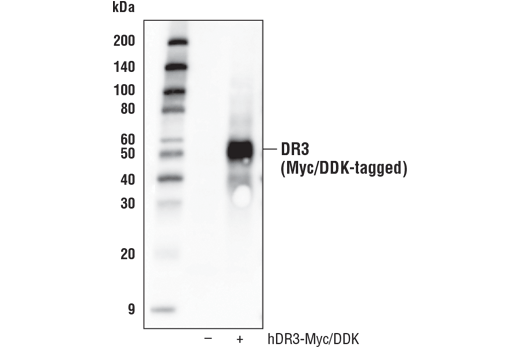
Western blot analysis of extracts from 293T cells, mock transfected (-) or transfected with a construct expressing Myc/DDK-tagged full-length human DR3 (hDR3-Myc/DDK; +), using DR3 (D5Q2R) Rabbit mAb.

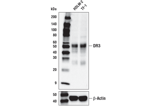
Western blot analysis of extracts from HDLM-2 and TF-1 cell lines using DR3 (D5Q2R) Rabbit mAb (upper) or β-Actin (D6A8) Rabbit mAb #8457 (lower).

声明 :本官网所有报价均为常温或者蓝冰运输价格,如有产品需要干冰运输,需另外加收干冰运输费。
货号:
45901T
一键复制
询价
20μl
100ul
选择数量
当前规格1件起购 
配送至
预计2-3周送达,快递: 免运费,若需干冰额外收费