


 全部商品分类
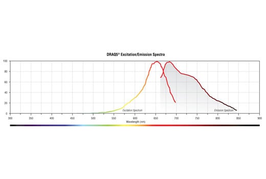


 用小程序,查商品更便捷


Product Usage Information
Please follow CST’s recommended IF and Flow protocols. For both applications, following secondary detection:
Flow Cytometry: Centrifuge cells and resuspend in 0.5 ml DRAQ5® diluted 1:500 (10 µM) in PBS or Antibody Dilution Buffer. Incubate for 5 min at room temperature in the dark before analyzing cells on flow cytometer.
Immunofluorescence: Rinse samples twice in PBS for five minutes each. Dilute DRAQ5® 1:1000 (5 µM) in PBS and incubate for 5 minutes at room temperature in the dark. Rinse samples once in PBS, coat coverslips with anti-fade reagent and examine immediately using appropriate excitation wavelength.

Store at 4°C protected from light.
参考图片
Absorption and fluorescence emission spectra of DRAQ5® in aqueous solution.
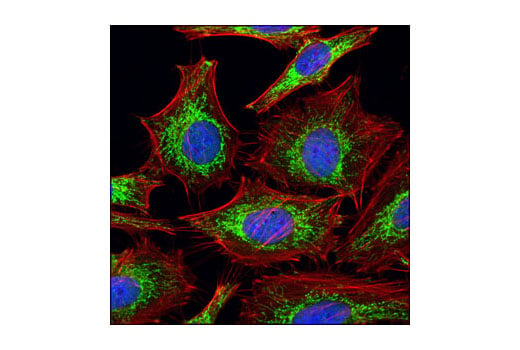
Confocal immunofluorescent analysis of HeLa cells using COX IV (3E11) Rabbit mAb #4850 (green). Actin filaments have been labeled with Alexa Fluor® 555 phalloidin (red). Blue pseudocolor = DRAQ5® (fluorescent DNA dye).
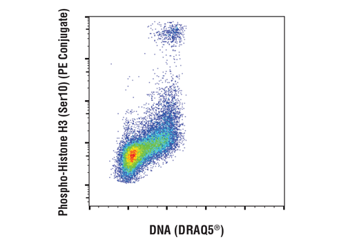
Flow cytometric analysis of Jurkat cells using Phospho-Histone H3 (Ser10) (D7N8E) XP® Rabbit mAb (PE Conjugate) #29237 versus DRAQ5® (DNA content).