老带新
DRP1 (D6C7) Rabbit mAb
1/4

DRP1 (D6C7) Rabbit mAb

分享
品牌: CST
pdf 下载产品说明书
pdf 下载COA
pdf 下载SDS
用小程序,查商品更便捷
收藏
对比对比
咨询咨询
分子量:
78-82
来源宿主:
Rabbit
产品介绍
产品介绍
产品信息
荧光素标记
抗原名称
DRP1
来源纯化

Monoclonal antibody is produced by immunizing animals with a synthetic peptide corresponding to residues near the amino terminus of human DRP1 protein.

宿主
Rabbit
商品描述

Product Usage Information

ApplicationDilution
Western Blotting1:1000
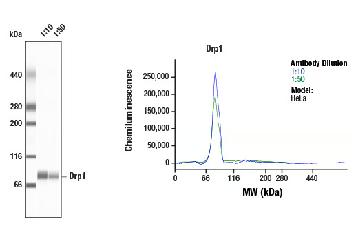
Simple Western™1:10 - 1:50
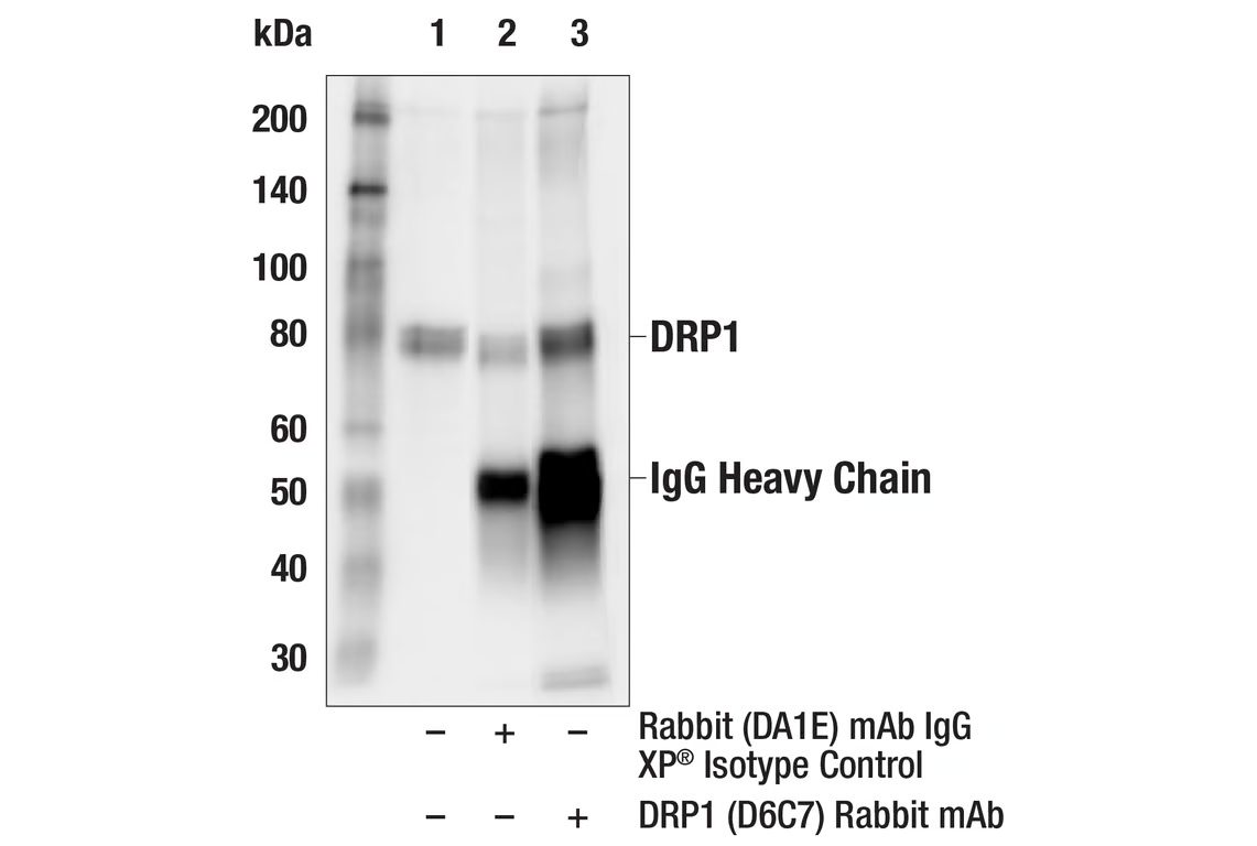
Immunoprecipitation1:100
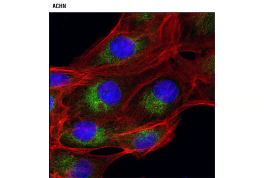
Immunofluorescence (Immunocytochemistry)1:50 - 1:100
同种型
Rabbit IgG
分子量
78-82
内毒素水平
研究领域
细胞生物学,神经科学,
应用
反应种属
Human,Mouse,Rat,Monkey,
预测反应种属
Pig
目标/特异性

Specificity/Sensitivity

DRP1 (D6C7) Rabbit mAb recognizes endogenous levels of total DRP1 protein.

Species Reactivity:

Human, Mouse, Rat, Monkey

敏感性
Endogenous
背景
背景
Dynamin-related protein 1 (DRP1) is a member of the dynamin superfamily of GTPases. Members of this family have diverse cellular functions including vesicle scission, organelle fission, viral resistance, and intracellular trafficking (reviewed in 1). DRP1 affects mitochondrial morphology and is important in mitochondrial and peroxisomal fission in mammalian cells (2-5). The yeast ortholog of DRP1 clusters into a spiral-shaped structure on the mitochondrial membrane at the site of fission (reviewed in 6), and this structure is likely conserved in mammalian cells (3). The division of the mitochondria, which is required for apoptosis, as well as normal cell growth and development is controlled, in part, by the phosphorylation of DRP1 at Ser616 by Cdk1/cyclin B and at Ser637 by protein kinase A (PKA) (reviewed in 6). When phosphorylated at Ser616, DRP1 stimulates mitochondrial fission during mitosis. Conversely, fission is inhibited when DRP1 is phosphorylated at Ser637 (reviewed in 6). Dephosphorylation at Ser637 by calcineurin reverses this inhibition (7). In addition to phosphorylation, sumoylation of DRP1 is also an enhancer of mitochondrial fission (8). Balancing fission and fusion events is essential for proper mitochondrial function. Research studies have demonstrated mitochondrial defects in a variety of neurodegenerative diseases including Alzheimer’s disease, Parkinson’s disease, and Huntington’s disease (reviewed in 6). 1.Praefcke, G.J. and McMahon, H.T. (2004) Nat Rev Mol Cell Biol 5, 133-47. 2.Taguchi, N. et al. (2007) J Biol Chem 282, 11521-9. 3.Smirnova, E. et al. (2001) Mol Biol Cell 12, 2245-56. 4.Smirnova, E. et al. (1998) J Cell Biol 143, 351-8. 5.Koch, A. et al. (2003) J Biol Chem 278, 8597-605. 6.Knott, A.B. et al. (2008) Nat Rev Neurosci 9, 505-18. 7.Cereghetti, G.M. et al. (2008) Proc Natl Acad Sci USA 105, 15803-8. 8.Zunino, R. et al. (2007) J Cell Sci 120, 1178-88.
研究领域
细胞生物学,神经科学,
翻译后修饰
unmodified
制备和贮存
保存方式

Supplied in 10 mM sodium HEPES (pH 7.5), 150 mM NaCl, 100 µg/ml BSA, 50% glycerol and less than 0.02% sodium azide. Store at –20°C. Do not aliquot the antibody.

For a carrier free (BSA and azide free) version of this product see product #73266.

数据库链接
Entrez-Gene ID
10059
UniProt ID
O00429

参考图片

Confocal immunofluorescent analysis of ACHN cells using DRP1 (D6C7) Rabbit mAb (green). Actin filaments were labeled with DY-554 phalloidin (red). Blue pseudocolor = DRAQ5® #4084 (fluorescent DNA dye).

Western blot analysis of extracts from various cell lines using DRP1 (D6C7) Rabbit mAb.

Simple Western™ analysis of lysates (0.1 mg/mL) from HeLa cells using DRP1 (D6C7) Rabbit mAb #8570. The virtual lane view (left) shows the target band (as indicated) at 1:10 and 1:50 dilutions of primary antibody. The corresponding electropherogram view (right) plots chemiluminescence by molecular weight along the capillary at 1:10 (blue line) and 1:50 (green line) dilutions of primary antibody. This experiment was performed under reducing conditions on the Jess™ ​​​​​​​ Simple Western instrument from ProteinSimple, a BioTechne brand, using the 66 - 440 kDa separation module.

Immunoprecipitation of DRP1 from Hela cell extracts. Lane 1 is 10% input, lane 2 is Rabbit (DA1E) mAb IgG XP® Isotype Control #3900, and lane 3 is DRP1 (D6C7) Rabbit mAb. Western blot analysis was performed using DRP1 (D6C7) Rabbit mAb. Anti-rabbit IgG, HRP-linked Antibody #7074 was used as a secondary antibody.

声明 :本官网所有报价均为常温或者蓝冰运输价格,如有产品需要干冰运输,需另外加收干冰运输费。
货号:
8570T
一键复制
询价
优宁维现货
20μl
100ul
选择数量
当前规格1件起购 
配送至
预计3-4个工作日送达,快递: 免运费,若需干冰额外收费