


 全部商品分类
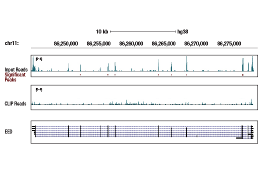


EED (E4L6E) XP ® Rabbit mAb

 下载产品说明书 下载COA 下载SDS
 用小程序,查商品更便捷


 收藏
收藏
 对比
对比 咨询
咨询
Monoclonal antibody is produced by immunizing animals with a synthetic peptide corresponding to residues near the amino terminus of human EED protein.
Product Usage Information
For optimal ChIP and ChIP-seq results, use 5 μl of antibody and 10 μg of chromatin (approximately 4 x 10⁶ cells) per IP. This antibody has been validated using SimpleChIP® Enzymatic Chromatin IP Kits. The CUT&RUN dilution was determined using CUT&RUN Assay Kit #86652. The CUT&Tag dilution was determined using CUT&Tag Assay Kit #77552.
| Application | Dilution |
|---|---|
| Western Blotting | 1:1000 |
| Simple Western™ | 1:10 - 1:50 |
| Immunoprecipitation | 1:50 |
| Immunohistochemistry (Paraffin) | 1:50 - 1:200 |
| Immunofluorescence (Immunocytochemistry) | 1:50 - 1:200 |
| Flow Cytometry (Fixed/Permeabilized) | 1:50 - 1:200 |
| Chromatin IP | 1:100 |
| Chromatin IP-seq | 1:100 |
| CUT&RUN | 1:100 |
| CUT&Tag | 1:100 |
| eCLIP | 1:200 |
Specificity/Sensitivity
Species Reactivity:
Human, Mouse, Rat, Monkey
Supplied in 10 mM sodium HEPES (pH 7.5), 150 mM NaCl, 100 µg/ml BSA, 50% glycerol and less than 0.02% sodium azide. Store at –20°C. Do not aliquot the antibody.
For a carrier free (BSA and azide free) version of this product see product #53069.
参考图片
Enhanced cross-linking and immunoprecipitation (eCLIP) was performed with RNA from K-562 cells and EED (E4L6E) XP® Rabbit mAb using a protocol based on the RBP-eCLIP method from Eclipsebio. The figure shows binding across the EED transcript. Data is kindly provided by the laboratory of Dr. Gene Yeo and used with permission.
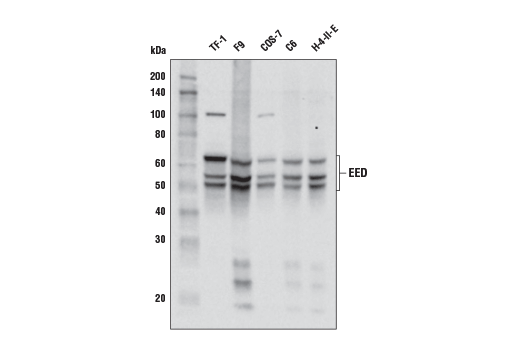
Western blot analysis of extracts from various cell lines using EED (E4L6E) XP® Rabbit mAb.
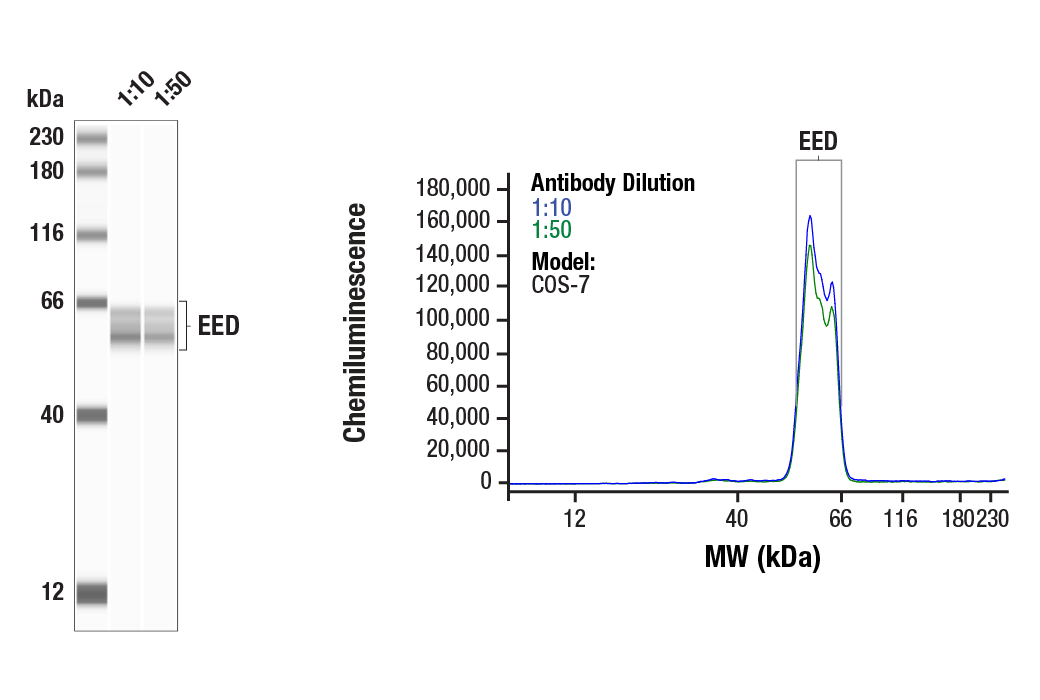
Simple Western™ analysis of lysates (1 mg/mL) from COS-7 cells using EED (E4L6E) XP® Rabbit mAb #85322. The virtual lane view (left) shows the target bands (as indicated) at 1:10 and 1:50 dilutions of primary antibody. The corresponding electropherogram view (right) plots chemiluminescence by molecular weight along the capillary at 1:10 (blue line) and 1:50 (green line) dilutions of primary antibody. This experiment was performed under reducing conditions on the Jess™ Simple Western instrument from ProteinSimple, a BioTechne brand, using the 12-230 kDa separation module.
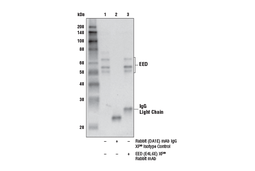
Immunoprecipitation of EED from TF-1 cell extracts. Lane 1 is 10% input, lane 2 is Rabbit (DA1E) mAb IgG XP® Isotype Control #3900, and lane 3 is EED (E4L6E) XP® Rabbit mAb. Western blot analysis was performed using EED (E4L6E) XP® Rabbit mAb. Mouse Anti-Rabbit IgG (Light-Chain Specific) (D4W3E) mAb (HRP Conjugate) #93702 was used as the secondary antibody.
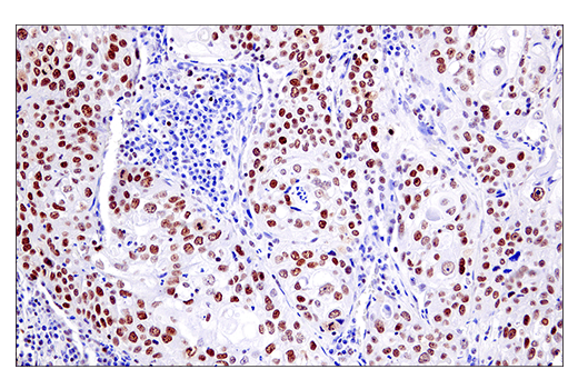
Immunohistochemical analysis of paraffin-embedded human squamous cell lung carcinoma using EED (E4L6E) XP® Rabbit mAb.
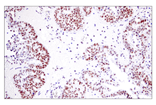
Immunohistochemical analysis of paraffin-embedded human endometrioid adenocarcinoma using EED (E4L6E) XP® Rabbit mAb.
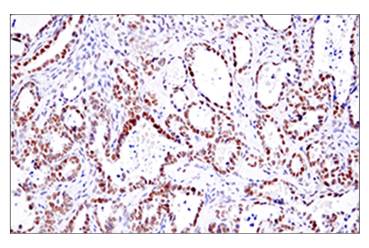
Immunohistochemical analysis of paraffin-embedded human ovarian clear cell carcinoma using EED (E4L6E) XP® Rabbit mAb.
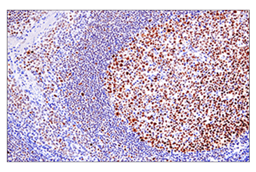
Immunohistochemical analysis of paraffin-embedded human tonsil using EED (E4L6E) XP® Rabbit mAb.
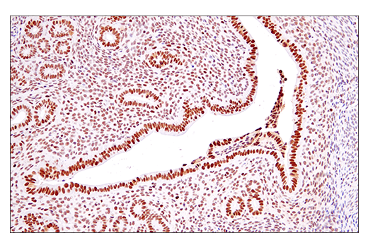
Immunohistochemical analysis of paraffin-embedded mouse endometrium using EED (E4L6E) XP® Rabbit mAb.
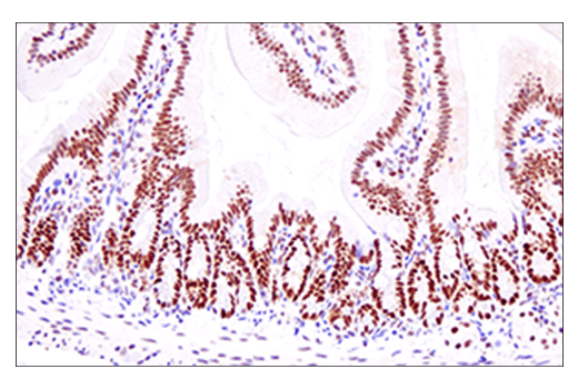
Immunohistochemical analysis of paraffin-embedded mouse small intestine using EED (E4L6E) XP® Rabbit mAb.
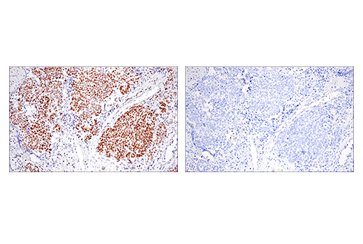
Immunohistochemical analysis of paraffin-embedded human ovarian serous carcinoma using EED (E4L6E) XP® Rabbit mAb (left) compared to concentration-matched Rabbit (DA1E) mAb IgG XP® Isotype Control #3900 (right).
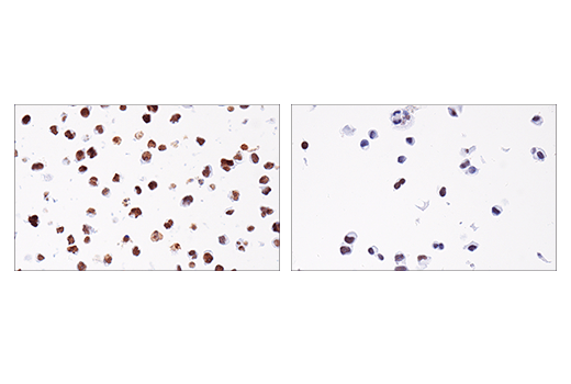
Immunohistochemical analysis of paraffin-embedded Daudi cell pellet (left, high-expressing) or HCC1419 cell pellet (right, low-expressing) using EED (E4L6E) XP® Rabbit mAb.
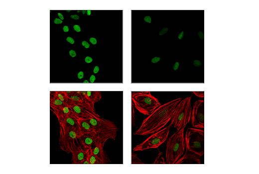
Confocal immunofluorescent analysis of OVCAR8 cells (left, high-expressing) or A-498 cells (right, low-expressing), using EED (E4L6E) XP® Rabbit mAb (green). Actin filaments were labeled with DyLight™ 554 Phalloidin #13054 (red).
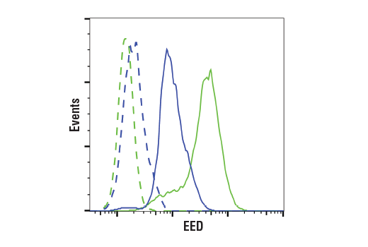
Flow cytometric analysis of U-87 MG cells (blue, moderate) and TF-1 cells (green, positive) using EED (E4L6E) XP® Rabbit mAb (solid lines) or a concentration-matched Rabbit (DA1E) mAb IgG XP® Isotype Control #3900 (dashed lines). Anti-rabbit IgG (H+L), F(ab')2 Fragment (Alexa Fluor® 488 Conjugate) #4412 was used as a secondary antibody.
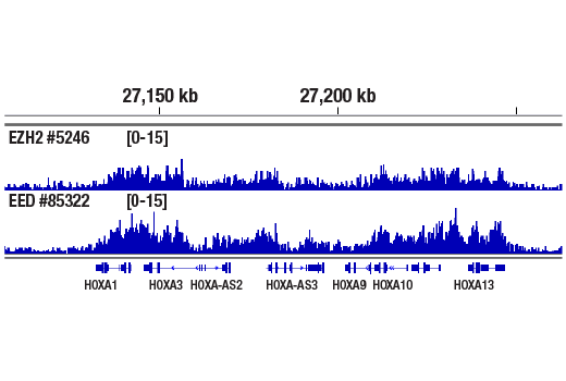
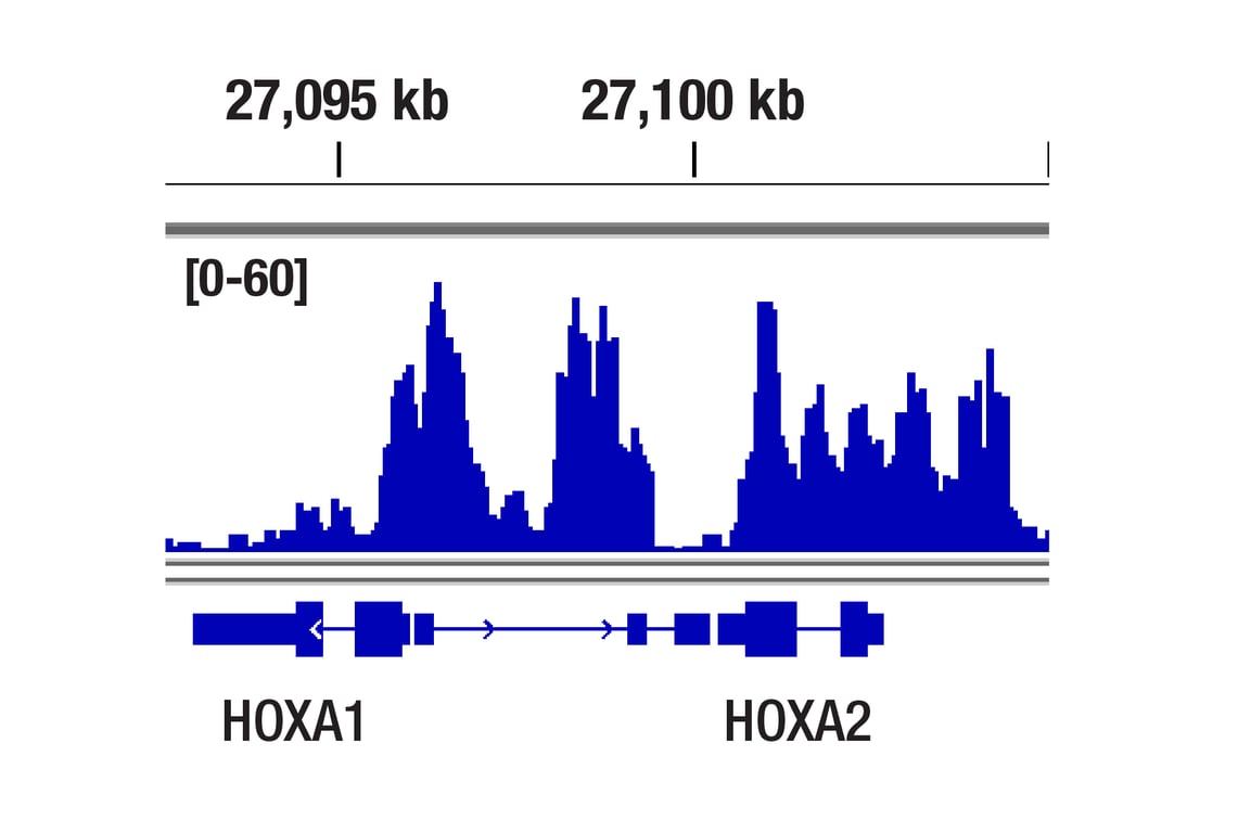
Chromatin immunoprecipitations were performed with cross-linked chromatin from NCCIT and EED (E4L6E) XP® Rabbit mAb or Ezh2 (D2C9) XP® Rabbit mAb #5246, using SimpleChIP® Plus Enzymatic Chromatin IP Kit (Magnetic Beads) #9005. DNA Libraries were prepared using DNA Library Prep Kit for Illumina #56795. Ezh2 and EED are both components of the Polycomb PRC2 complex. The figure shows binding across HOXA1, a known target gene of Ezh2 and EED (see additional figure containing ChIP-qPCR data).
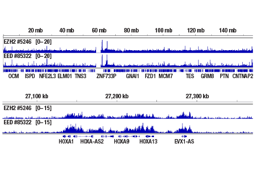
Chromatin immunoprecipitations were performed with cross-linked chromatin from NCCIT and EED (E4L6E) XP® Rabbit mAb or Ezh2 (D2C9) XP® Rabbit mAb #5246, using SimpleChIP Plus Enzymatic Chromatin IP Kit (Magnetic Beads) #9005. DNA Libraries were prepared using DNA Library Prep Kit for Illumina #56795. Ezh2 and EED are both components of the Polycomb PRC2 complex. The figure shows binding across chromosome 7 (upper), including HOXA1 (lower), a known target gene of Ezh2 and EED (see additional figure containing ChIP-qPCR data).
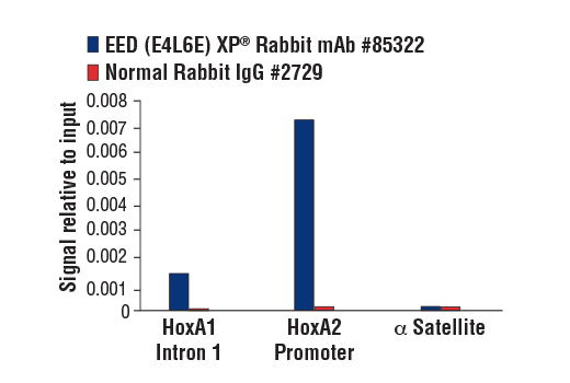
Chromatin immunoprecipitations were performed with cross-linked chromatin from NCCIT and either EED (E4L6E) XP® Rabbit mAb or Normal Rabbit IgG #2729 using SimpleChIP® Plus Enzymatic Chromatin IP Kit (Magnetic Beads) #9005. The enriched DNA was quantified by real-time PCR using SimpleChIP® Human HoxA1 Intron 1 Primers #7707, SimpleChIP® Human HoxA2 Promoter Primers #5517, and SimpleChIP® Human α Satellite Repeat Primers #4486. The amount of immunoprecipitated DNA in each sample is represented as signal relative to the total amount of input chromatin, which is equivalent to one.
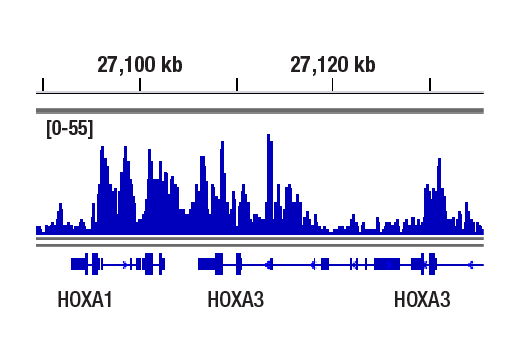
CUT&RUN was performed with NCCIT cells and EED (E4L6E) XP® Rabbit mAb, using CUT&RUN Assay Kit #86652. DNA library was prepared using DNA Library Prep Kit for Illumina Systems (ChIP-seq, CUT&RUN) #56795. The figure shows binding across HOXA3 gene.
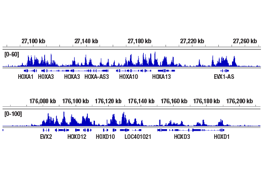
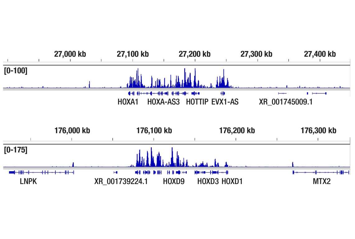
CUT&RUN was performed with NCCIT cells and EED (E4L6E) XP® Rabbit mAb, using CUT&RUN Assay Kit #86652. DNA library was prepared using DNA Library Prep Kit for Illumina Systems (ChIP-seq, CUT&RUN) #56795. The figures show binding across HOXA (upper) and HOXD (lower).
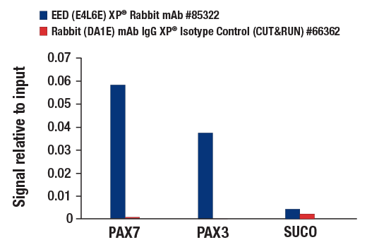
CUT&RUN was performed with NCCIT cells and either EED (E4L6E) XP® Rabbit mAb or Rabbit (DA1E) mAb IgG XP® Isotype Control (CUT&RUN) #66362, using CUT&RUN Assay Kit #86652. The enriched DNA was quantified by real-time PCR using human PAX7 promoter primers, human PAX3 promoter primers, and human SUCO promoter primers. The amount of immunoprecipitated DNA in each sample is represented as signal relative to the total amount of input chromatin, which is equivalent to one.
CUT&Tag was performed with NCCIT cells and EED (E4L6E) XP® Rabbit mAb, using CUT&Tag Assay Kit #77552. DNA library was prepared using CUT&Tag Dual Index Primers and PCR Master Mix for Illumina Systems #47415. The figure shows binding across HOXA1, a known target gene of EED (see our ChIP-qPCR figure).
CUT&Tag was performed with NCCIT cells and EED (E4L6E) XP® Rabbit mAb, using CUT&Tag Assay Kit #77552. DNA library was prepared using CUT&Tag Dual Index Primers and PCR Master Mix for Illumina Systems #47415. The figures show binding across the HOXA gene cluster (upper), and the HOXD gene cluster (lower), which are known target genes of EED (see our ChIP-qPCR figure).