


 全部商品分类
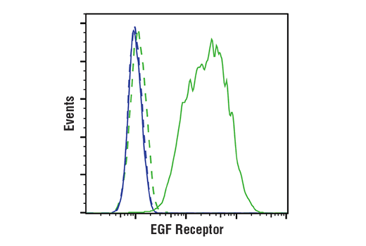


EGF Receptor (D38B1) XP ® Rabbit mAb

 下载产品说明书 下载COA 下载SDS
 用小程序,查商品更便捷


 收藏
收藏
 对比
对比 咨询
咨询
Monoclonal antibody is produced by immunizing animals with a fusion protein containing the cytoplasmic domain of human EGF receptor.
Product Usage Information
| Application | Dilution |
|---|---|
| Western Blotting | 1:1000 |
| Simple Western™ | 1:10 - 1:50 |
| Immunoprecipitation | 1:100 |
| IHC Leica Bond | 1:50 |
| Immunohistochemistry (Paraffin) | 1:50 |
| Immunofluorescence (Immunocytochemistry) | 1:50 - 1:200 |
| Flow Cytometry (Fixed/Permeabilized) | 1:50 - 1:200 |
Specificity/Sensitivity
Species Reactivity:
Human, Mouse, Monkey
Supplied in 10 mM sodium HEPES (pH 7.5), 150 mM NaCl, 100 µg/ml BSA, 50% glycerol and less than 0.02% sodium azide. Store at –20°C. Do not aliquot the antibody.
For a carrier-free (BSA and azide free) version of this product see product #26038.
参考图片
Flow cytometric analysis of Jurkat cells (blue) and A431 cells (green) using EGF Receptor (D38B1) XP® Rabbit mAb #4267 (solid lines) or a concentration-matched Rabbit (DA1E) mAb IgG XP® Isotype Control #3900 (dashed lines). Anti-rabbit IgG (H+L), F(ab')2 Fragment (Alexa Fluor® 488 Conjugate) #4412 was used as a secondary antibody.
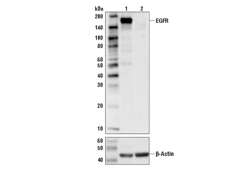
Western blot analysis of extracts from control Hela cells (lane 1), or EGFR knockout Hela cells (lane 2) using EGF Receptor (D38B1) XP® Rabbit mAb #4267, (upper) or #8457 β-Actin (D6A8) Rabbit mAb (lower). The absence of signal in EGFR-knockout Hela cells confirms specificity of the antibody for EGFR.
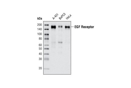
Western blot analysis of extracts from A-431, BxPC3 and HeLa cells using EGF Receptor (D38B1) XP® Rabbit mAb.
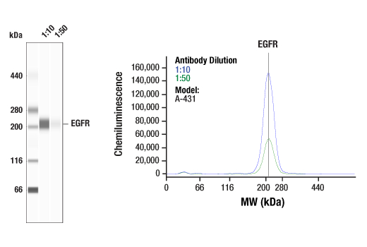
Simple Western™ analysis of lysates (0.1 mg/mL) from A-431 cells using EGF Receptor (D38B1) XP® Rabbit mAb #4267. The virtual lane view (left) shows a single target band (as indicated) at 1:10 and 1:50 dilutions of primary antibody. The corresponding electropherogram view (right) plots chemiluminescence by molecular weight along the capillary at 1:10 (blue line) and 1:50 (green line) dilutions of primary antibody. This experiment was performed under reducing conditions on the Jess™ Simple Western instrument from ProteinSimple, a BioTechne brand, using the 66-440 kDa separation module.
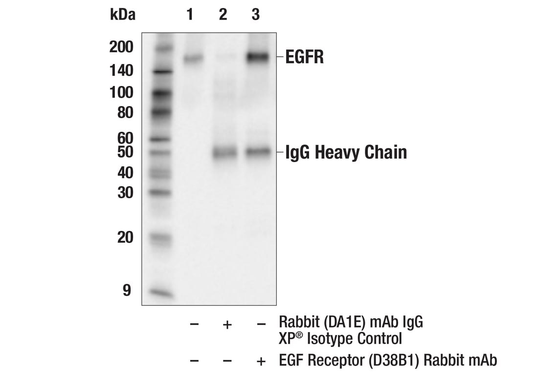
Immunoprecipitation of EGFR protein from HeLa cell extracts. Lane 1 is 10% input, lane 2 is Rabbit (DA1E) mAb IgG XP® Isotype Control #3900, and lane 3 is EGF Receptor (D38B1) XP® Rabbit mAb. Western blot analysis was performed using EGF Receptor (D38B1) XP® Rabbit mAb. Anti-rabbit IgG, HRP-linked Antibody #7074 was used as a secondary antibody.
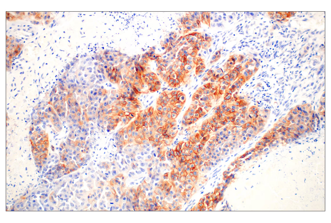
Immunohistochemical analysis of paraffin-embedded human lung carcinoma using EGF Receptor (D38B1) Rabbit mAb performed on the Leica® BOND™ Rx.
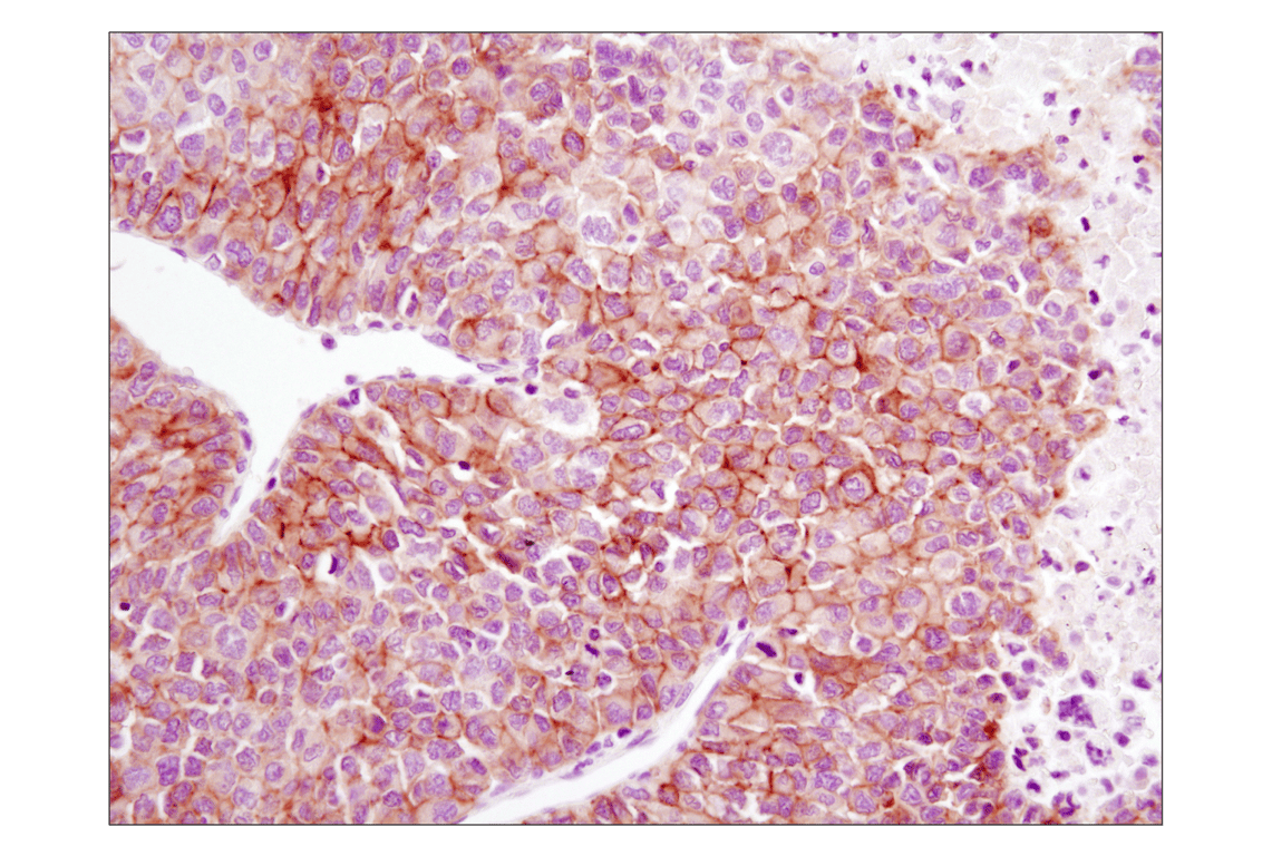
Immunohistochemical analysis of paraffin-embedded human hepatocellular carcinoma using EGF Receptor (D38B1) XP® Rabbit mAb.
Immunohistochemical analysis of paraffin-embedded human lung carcinoma using EGF Receptor (D38B1) XP® Rabbit mAb.
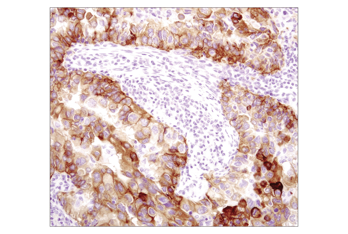
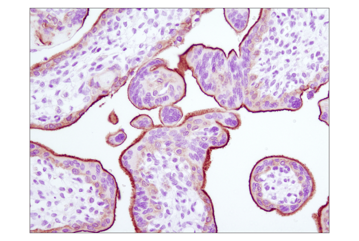
Immunohistochemical analysis of paraffin-embedded human placenta using EGF Receptor (D38B1) XP® Rabbit mAb.
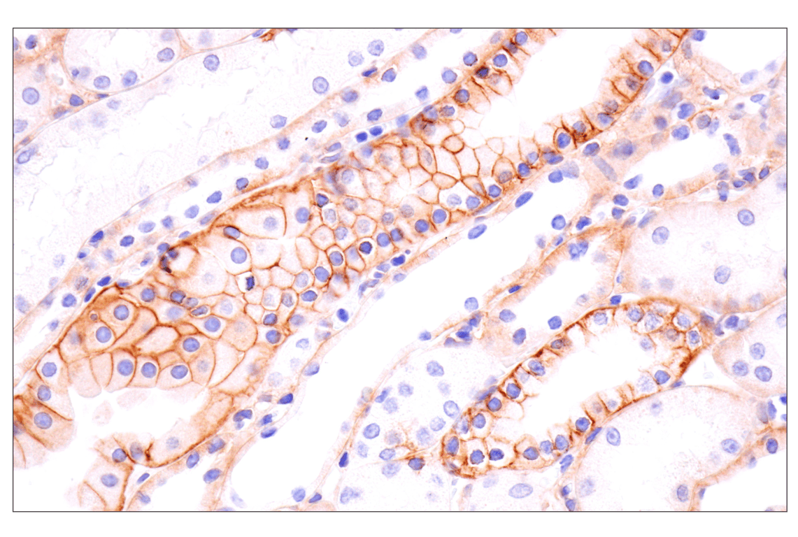
Immunohistochemical analysis of paraffin-embedded rhesus monkey kidney using EGF Receptor (D38B1) Rabbit mAb.
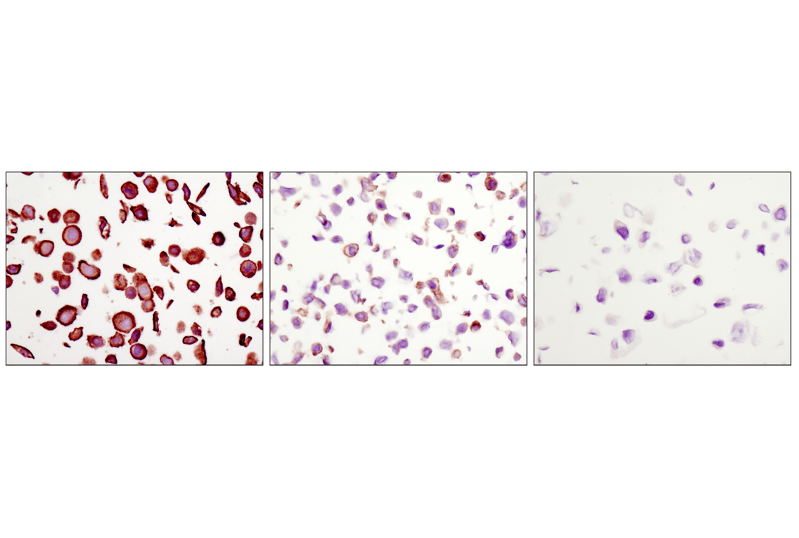
Immunohistochemical analysis of paraffin-embedded MDA-MB-468 (amplified EGFR, left), HT-29 (low EGFR, middle) and CAMA-1 (EGFR negative, right) cells using EGF Receptor (D38B1) XP® Rabbit mAb.
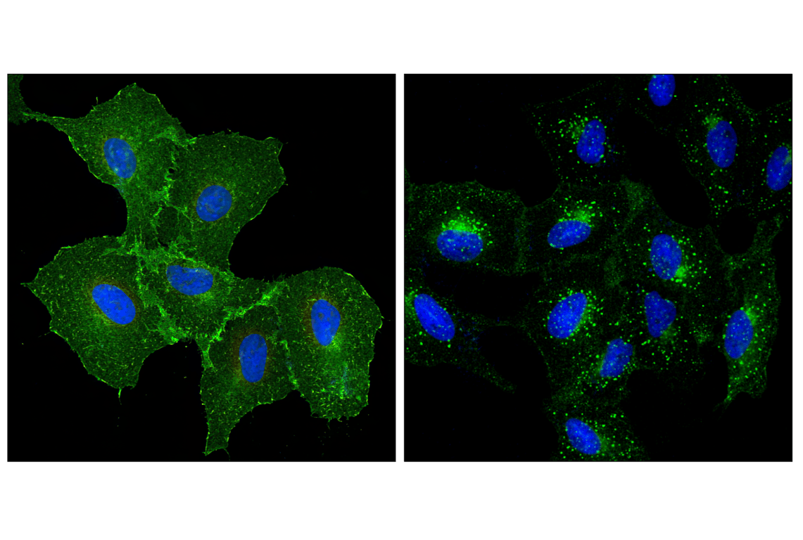
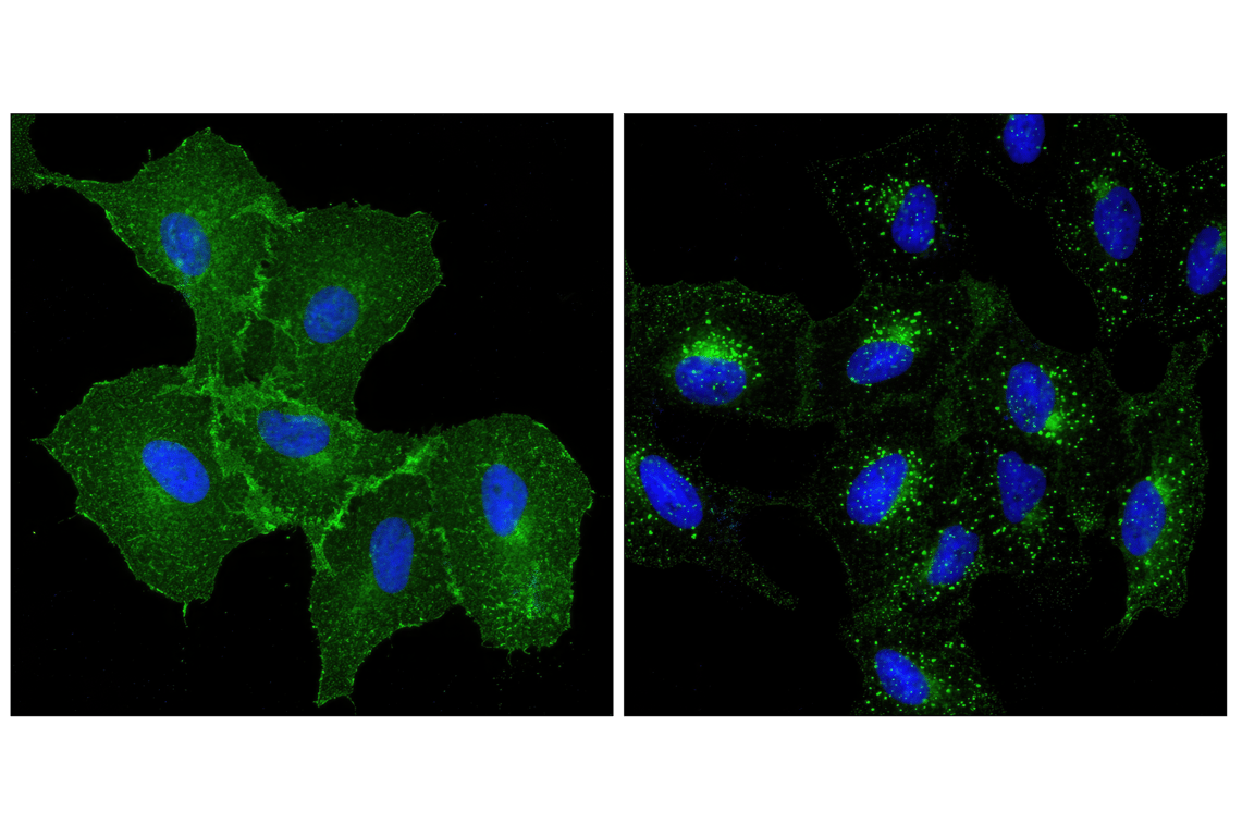
Confocal immunofluorescent analysis of A549 cells, untreated (left) or treated with human epidermal growth factor (right), using EGF Receptor (D38B1) XP® Rabbit mAb (green). Blue pseudocolor = DRAQ5® #4084 (fluorescent DNA dye).
Confocal immunofluorescent analysis of A549 cells, untreated (left) or treated with human epidermal growth factor (right), using EGF Receptor (D38B1) XP® Rabbit mAb (green). Blue pseudocolor = DRAQ5® #4084 (fluorescent DNA dye).