


 全部商品分类
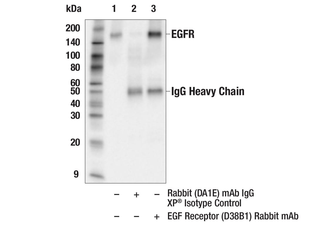


PhosphoPlus ® EGFR (Tyr1068) Antibody Duet

 下载产品说明书 下载SDS
 用小程序,查商品更便捷


 收藏
收藏
 对比
对比 咨询
咨询
PhosphoPlus® Duets from Cell Signaling Technology (CST) provide a means to assess protein activation status. Each Duet contains an activation-state and total protein antibody to your target of interest. These antibodies have been selected from CST's product offering based upon superior performance in specified applications.
参考图片
Immunoprecipitation of EGFR protein from HeLa cell extracts. Lane 1 is 10% input, lane 2 is Rabbit (DA1E) mAb IgG XP® Isotype Control #3900, and lane 3 is EGF Receptor (D38B1) XP® Rabbit mAb. Western blot analysis was performed using EGF Receptor (D38B1) XP® Rabbit mAb. Anti-rabbit IgG, HRP-linked Antibody #7074 was used as a secondary antibody.
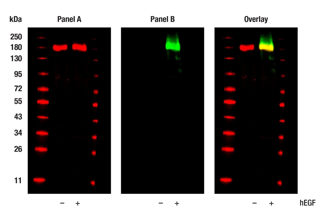
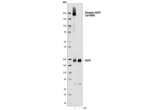
Western blot analysis of extracts from serum-starved A431 cells, untreated (-) or treated with hEGF (100 ng/ml, 5 min; +), using EGF Receptor (1F4) Mouse mAb #2239 (Panel A) and Phospho-EGF Receptor (Tyr1068) (D7A5) XP® Rabbit mAb #3777 (Panel B). Anti-rabbit IgG (H+L) (DyLight 800 4X PEG Conjugate) #5151 (green) and Anti-mouse IgG (H+L) (DyLight 680 Conjugate) #5470 (red) were used as secondary antibodies.
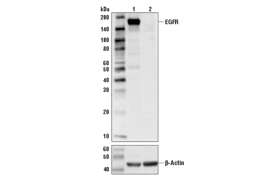
Western blot analysis of extracts from control Hela cells (lane 1), or EGFR knockout Hela cells (lane 2) using EGF Receptor (D38B1) XP® Rabbit mAb #4267, (upper) or #8457 β-Actin (D6A8) Rabbit mAb (lower). The absence of signal in EGFR-knockout Hela cells confirms specificity of the antibody for EGFR.
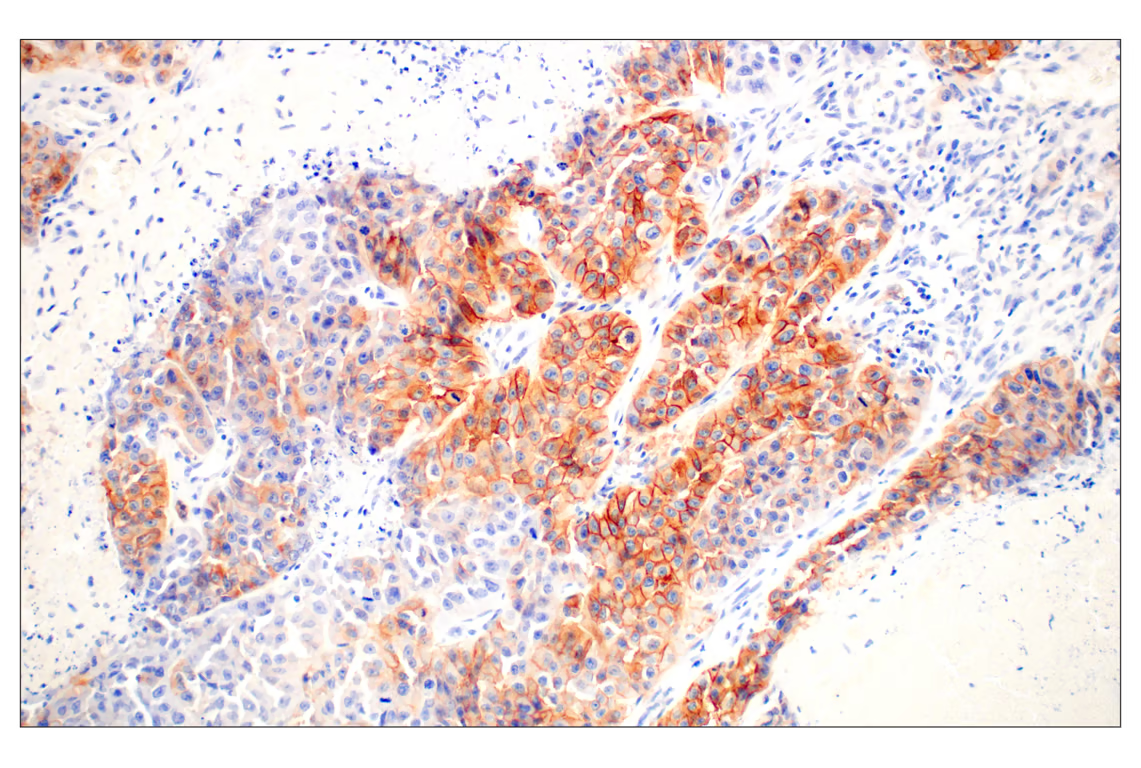
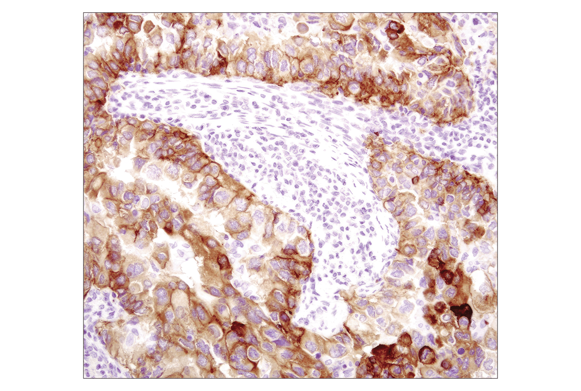
Immunohistochemical analysis of paraffin-embedded human lung carcinoma using EGF Receptor (D38B1) Rabbit mAb performed on the Leica® BOND™ Rx.
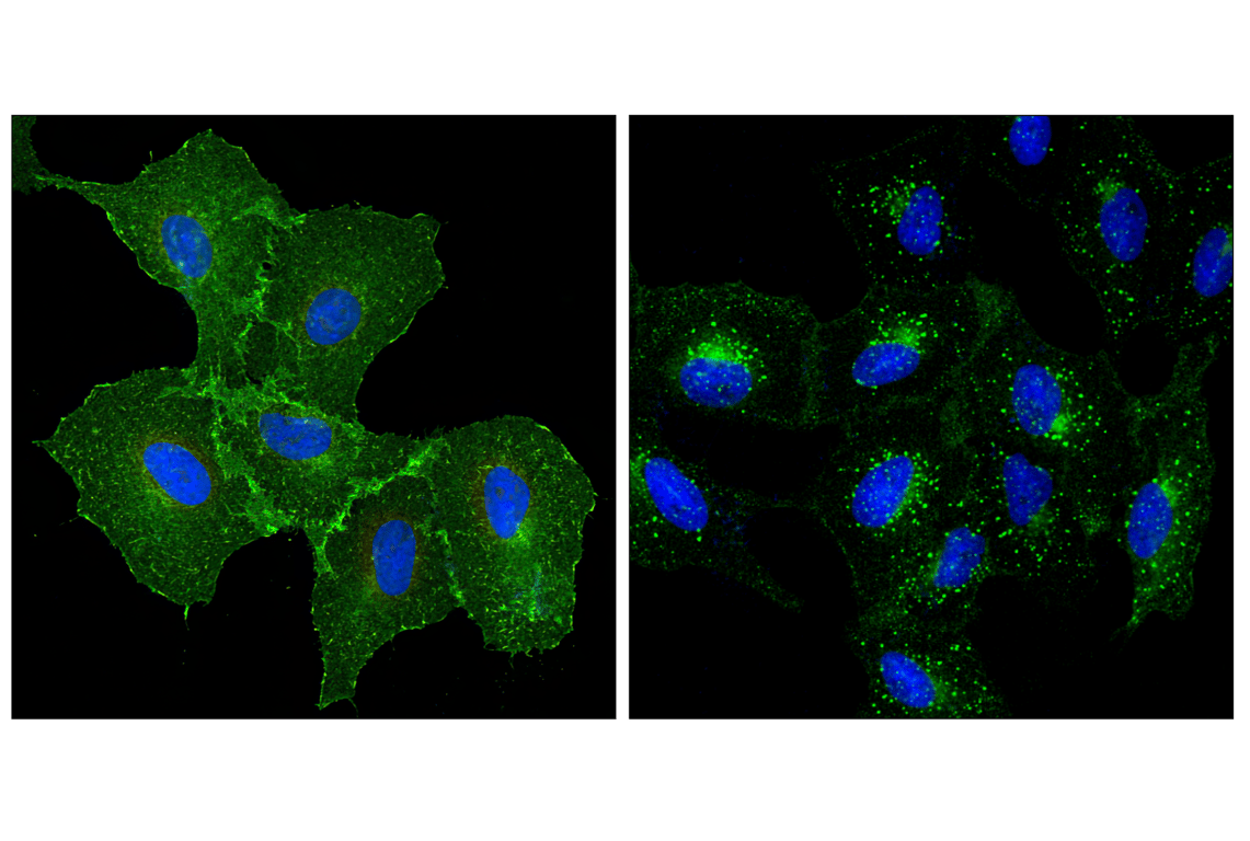
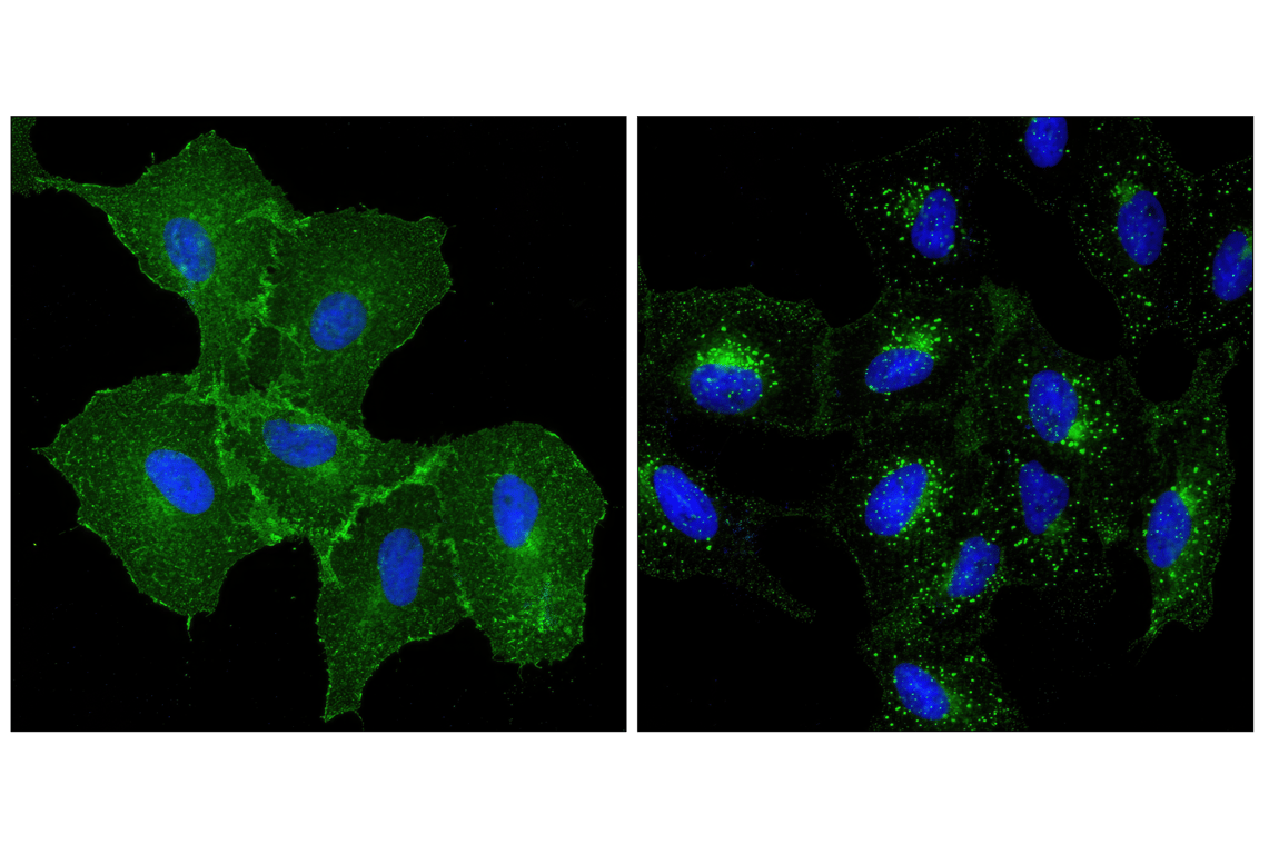
Confocal immunofluorescent analysis of A549 cells, untreated (left) or treated with human epidermal growth factor (right), using EGF Receptor (D38B1) XP® Rabbit mAb (green). Blue pseudocolor = DRAQ5® #4084 (fluorescent DNA dye).
Western blot analysis of extracts of BxPC-3 cells, untreated or EGF-stimulated, using Phospho-EGF Receptor (Tyr1068) (D7A5) XP® Rabbit mAb (upper) and EGF Receptor Antibody #2232 (lower).
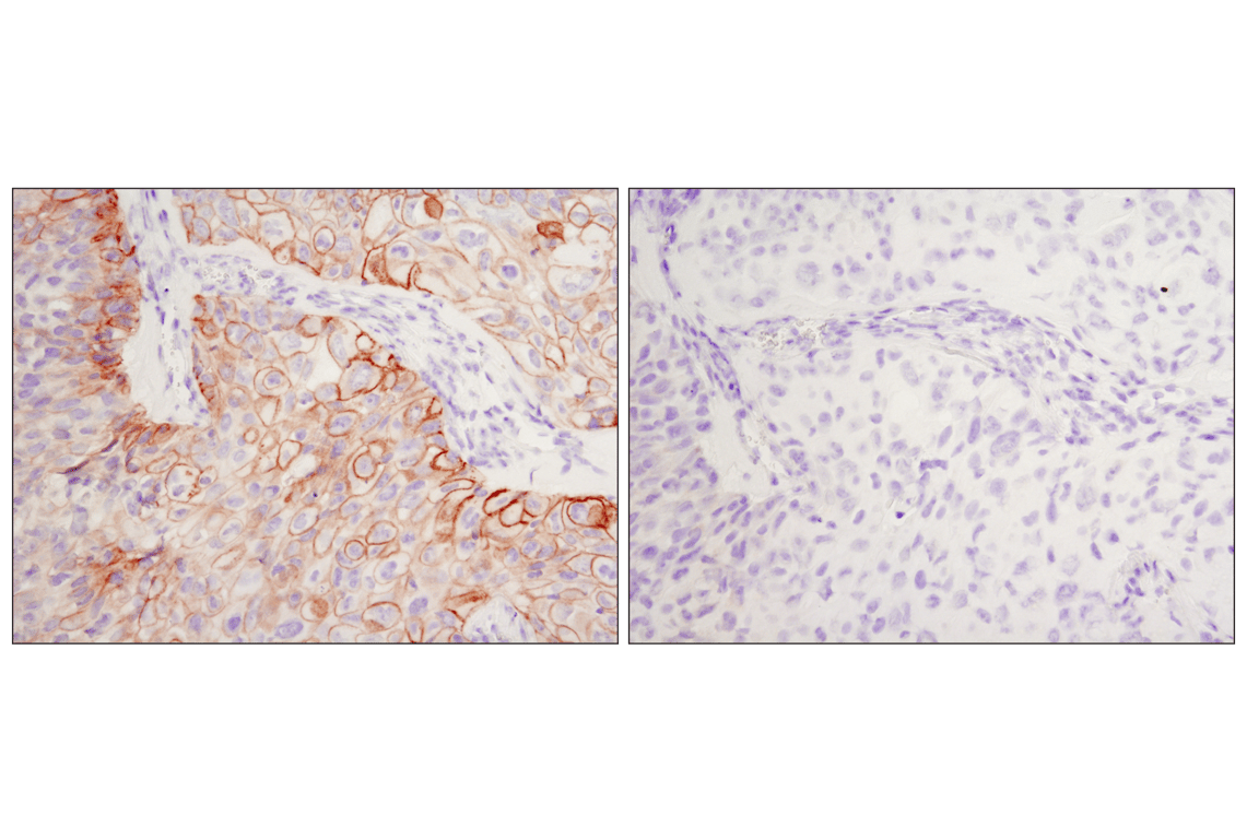
Immunohistochemical analysis of paraffin-embedded HCC827 xenograft, control (left) or λ phosphatase-treated (right), using Phospho-EGF Receptor (Tyr1068) (D7A5) XP® Rabbit mAb.
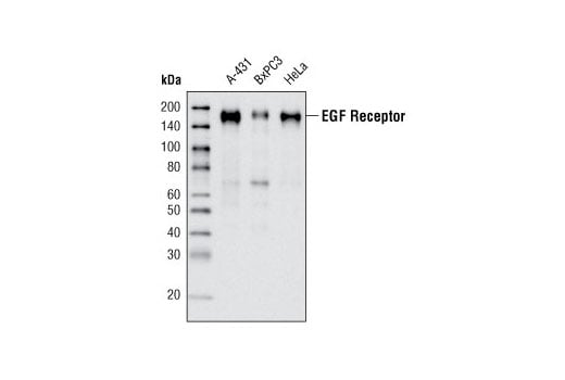
Western blot analysis of extracts from A-431, BxPC3 and HeLa cells using EGF Receptor (D38B1) XP® Rabbit mAb.
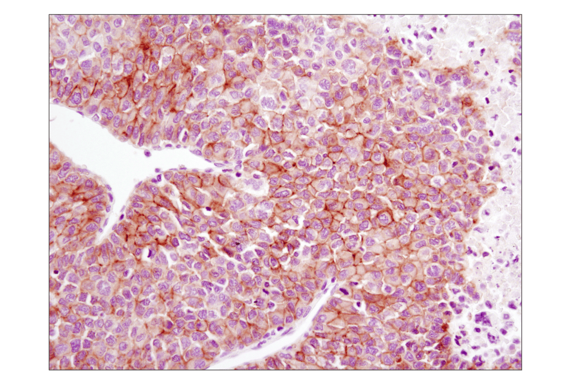
Immunohistochemical analysis of paraffin-embedded human hepatocellular carcinoma using EGF Receptor (D38B1) XP® Rabbit mAb.
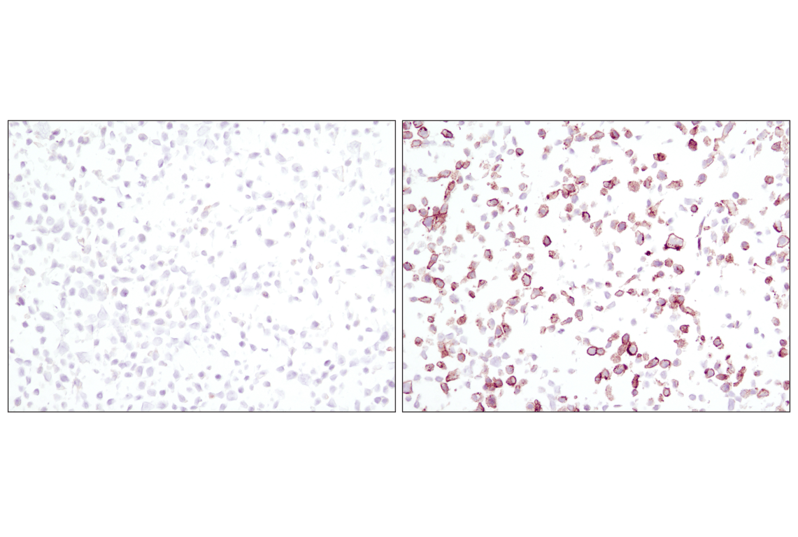
Immunohistochemical analysis using Phospho-EGF Receptor (Tyr1068) (D7A5) XP® Rabbit mAb on SignalSlide™ Phospho-EGF Receptor IHC Controls #8102 (paraffin-embedded KYSE450 cell pellets, untreated (left) or EGF-treated (right)).
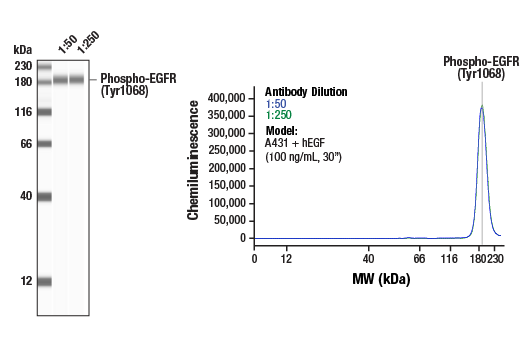
Simple Western™ analysis of lysates (0.1 mg/mL) from A431 cells treated with human EGF (100 ng/mL, 30”) using Phospho-EGF Receptor (Tyr1068) (D7A5) XP® Rabbit mAb #3777. The virtual lane view (left) shows a single target band (as indicated) at 1:50 and 1:250 dilutions of primary antibody. The corresponding electropherogram view (right) plots chemiluminescence by molecular weight along the capillary at 1:50 (blue line) and 1:250 (green line) dilutions of primary antibody. This experiment was performed under reducing conditions on the Jess™ Simple Western instrument from ProteinSimple, a BioTechne brand, using the 12-230 kDa separation module.
Immunohistochemical analysis of paraffin-embedded human lung carcinoma using EGF Receptor (D38B1) XP® Rabbit mAb.
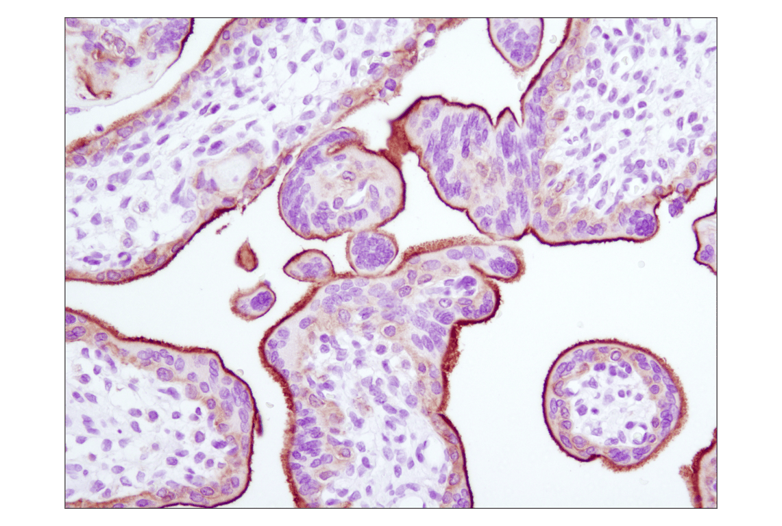
Immunohistochemical analysis of paraffin-embedded human placenta using EGF Receptor (D38B1) XP® Rabbit mAb.
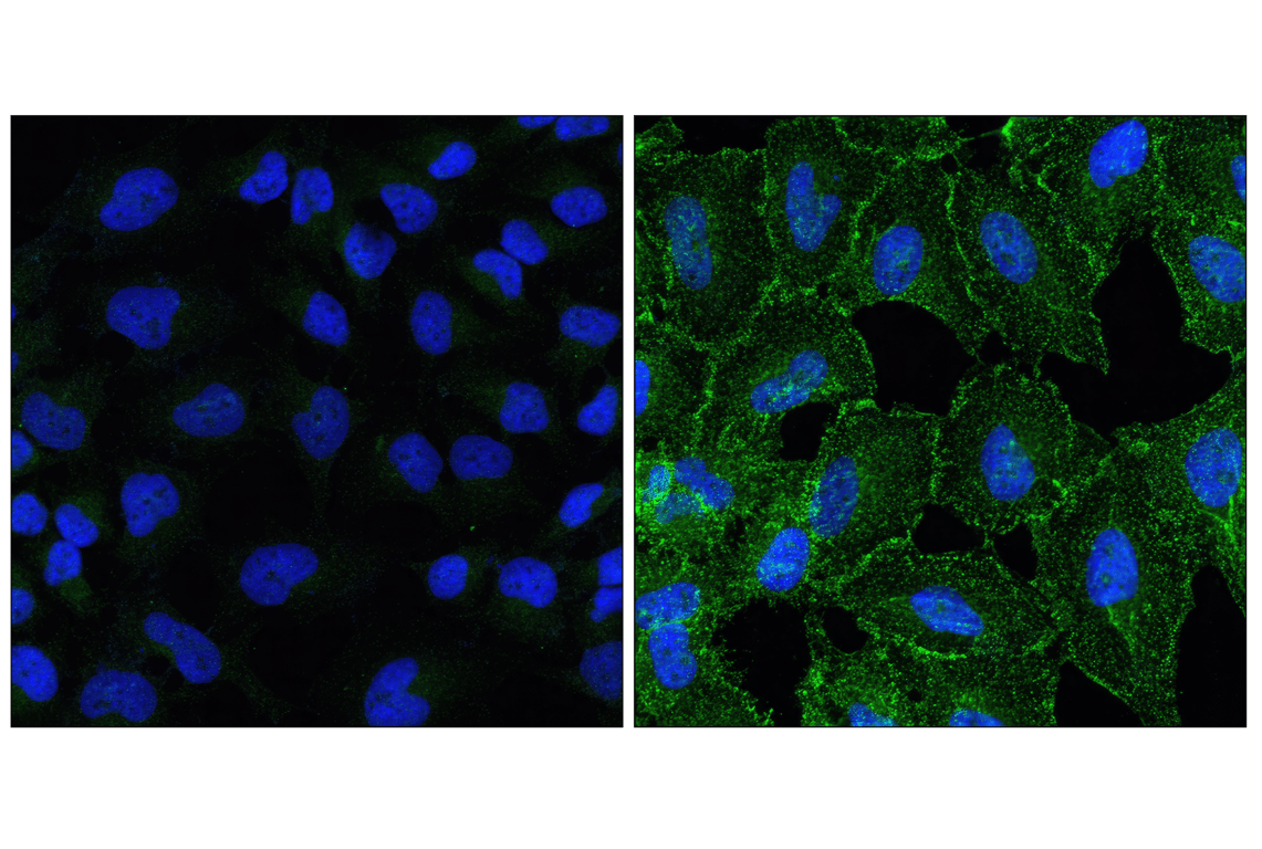
Confocal immunofluorescent analysis of HeLa cells, untreated (left) or EGF-treated (right), using Phospho-EGF Receptor (Tyr1068) (D7A5) XP® Rabbit mAb (green) and DRAQ5® #4084 (blue pseudocolor).
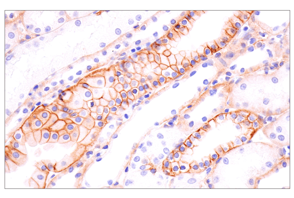
Immunohistochemical analysis of paraffin-embedded rhesus monkey kidney using EGF Receptor (D38B1) Rabbit mAb.
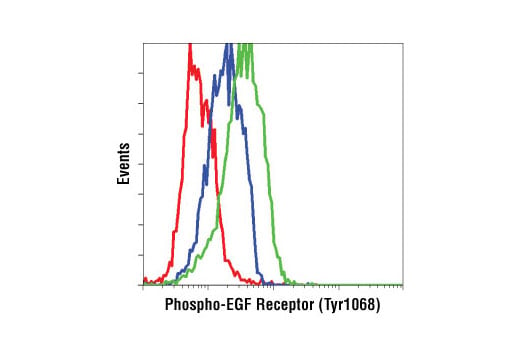
Flow cytometric analysis of A549 cells, untreated (blue) or EGF-treated (green), using Phospho-EGF Receptor (Tyr1068) (D7A5) XP® Rabbit mAb compared to concentration matched XP® Rabbit (DA1E) mAb IgG Isotype Control #3900 (red).
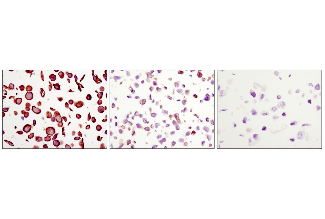
Immunohistochemical analysis of paraffin-embedded MDA-MB-468 (amplified EGFR, left), HT-29 (low EGFR, middle) and CAMA-1 (EGFR negative, right) cells using EGF Receptor (D38B1) XP® Rabbit mAb.
Confocal immunofluorescent analysis of A549 cells, untreated (left) or treated with human epidermal growth factor (right), using EGF Receptor (D38B1) XP® Rabbit mAb (green). Blue pseudocolor = DRAQ5® #4084 (fluorescent DNA dye).
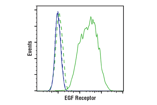
Flow cytometric analysis of Jurkat cells (blue) and A431 cells (green) using EGF Receptor (D38B1) XP® Rabbit mAb #4267 (solid lines) or a concentration-matched Rabbit (DA1E) mAb IgG XP® Isotype Control #3900 (dashed lines). Anti-rabbit IgG (H+L), F(ab')2 Fragment (Alexa Fluor® 488 Conjugate) #4412 was used as a secondary antibody.
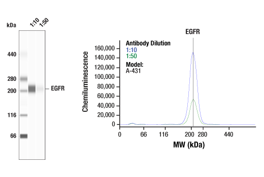
Simple Western™ analysis of lysates (0.1 mg/mL) from A-431 cells using EGF Receptor (D38B1) XP® Rabbit mAb #4267. The virtual lane view (left) shows a single target band (as indicated) at 1:10 and 1:50 dilutions of primary antibody. The corresponding electropherogram view (right) plots chemiluminescence by molecular weight along the capillary at 1:10 (blue line) and 1:50 (green line) dilutions of primary antibody. This experiment was performed under reducing conditions on the Jess™ Simple Western instrument from ProteinSimple, a BioTechne brand, using the 66-440 kDa separation module.