The TU66 monoclonal antibody specifically recognizes human CD39 which is also known as Ectonucleoside triphosphate diphosphohydrolase 1 (NTPDase 1), Ecto-ATP diphosphohydrolase 1 (Ecto-ATPDase 1), or Ecto-apyrase. CD39 is an integral membrane glycoprotein with two transmembrane domains, N- and C-terminal cytoplasmic tails, and an extracellular region that contains the NTPDase 1 active site. CD39 is encoded by
ENTPD1
which belongs to the ectoenzyme family. CD39 is variably expressed on activated T cells and B cells, regulatory T cells (Treg), dendritic cells, Langerhans cells, NK cells, monocytes, macrophages, endothelial cells, and granulocytes. CD39 acts on extracellular nucleoside triphosphates and diphosphates including ATP and ADP that are hydrolyzed into AMP. Through cell surface CD73 (Ecto-5'-nucleotidase), regulatory T cells can act on extracellular AMP to generate immunosuppressive adenosine. CD39 is involved in the control of the extracellular pool of phosphorylated nucleosides, the suppression of inflammation and immunity, and the regulation of platelet activation.
商品描述
TU66
The TU66 monoclonal antibody specifically recognizes human CD39 which is also known as Ectonucleoside triphosphate diphosphohydrolase 1 (NTPDase 1), Ecto-ATP diphosphohydrolase 1 (Ecto-ATPDase 1), or Ecto-apyrase. CD39 is an integral membrane glycoprotein with two transmembrane domains, N- and C-terminal cytoplasmic tails, and an extracellular region that contains the NTPDase 1 active site. CD39 is encoded by
ENTPD1
which belongs to the ectoenzyme family. CD39 is variably expressed on activated T cells and B cells, regulatory T cells (Treg), dendritic cells, Langerhans cells, NK cells, monocytes, macrophages, endothelial cells, and granulocytes. CD39 acts on extracellular nucleoside triphosphates and diphosphates including ATP and ADP that are hydrolyzed into AMP. Through cell surface CD73 (Ecto-5'-nucleotidase), regulatory T cells can act on extracellular AMP to generate immunosuppressive adenosine. CD39 is involved in the control of the extracellular pool of phosphorylated nucleosides, the suppression of inflammation and immunity, and the regulation of platelet activation.
同种型
Mouse IgG2b, κ
克隆号
克隆 TU66 (also known as Tü 66, Tü66) (RUO)
产品详情
APC
Allophycocyanin (APC), is part of the BD family of phycobiliprotein dyes. This fluorochrome is a multimeric fluorescent phycobiliprotein with excitation maximum (Ex Max) of 651 nm and an emission maximum (Em Max) at 660 nm. APC is designed to be excited by the Red (627-640 nm) laser and detected using an optical filter centered near 660 nm (e.g., a 660/20 nm bandpass filter). Please ensure that your instrument’s configurations (lasers and optical filters) are appropriate for this dye.
APC
Red 627-640 nm
651 nm
660 nm
应用
实验应用
Flow cytometry (Routinely Tested)
推荐用量
20 µl
反应种属
Human (QC Testing)
目标/特异性
CD39 (ENTPD1)
背景
别名
ENTPD1; NTPDase-1; Ecto-ATPase 1; Ecto-ATPDase 1
制备和贮存
存储溶液
Aqueous buffered solution containing BSA and ≤0.09% sodium azide.
保存方式
Aqueous buffered solution containing BSA and ≤0.09% sodium azide.
文献
文献
研发参考(4)
1. Borsellino G, Kleinewietfeld M, Di Mitri D, et al. Expression of ectonucleotidase CD39 by Foxp3+ Treg cells: hydrolysis of extracellular ATP and immune suppression.. Blood. 2007. (Biology).
2. Duensing S, Kirshner H, Atzpodien J. CD39 as a novel marker of in vivo immune activation. Blood. 1994; 83(12):3826-3827. (Biology).
3. Knapp W. W. Knapp .. et al., ed. Leucocyte typing IV : white cell differentiation antigens. Oxford New York: Oxford University Press; 1989:1-1182.
4. Schlossman SF. Stuart F. Schlossman .. et al., ed. Leucocyte typing V : white cell differentiation antigens : proceedings of the fifth international workshop and conference held in Boston, USA, 3-7 November, 1993. Oxford: Oxford University Press; 1995.
数据库链接
Entrez-Gene ID
953
参考图片
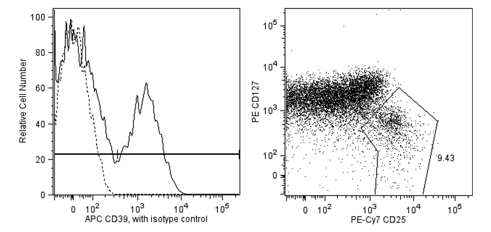
Flow cytometric analysis of APC anti-human CD39 on peripheral blood. Human peripheral blood was stained simultaneously with FITC anti-human CD4 (clone RPA-T4, Cat. No. 5555346), PE-Cy7 anti-human CD25 (Clone M-A251, Cat. No. 557741), PE anti-human CD127 (clone hIL-7R-M21, Cat No. 557938) and APC anti-human CD39 (clone TU66) or an APC conjugated mouse IgG2b, κ isotype control (clone 27-35, Cat. No. 555745). Cells were then lysed and CD39 expression examined. CD39 expression is shown on regulatory T cells (solid line) versus isotype control (dotted line,left panel). RegulatoryT cells were identified from the gated events based on light scattering characteristics of lymphocytes and fluorescence characteristics of CD4+ cells shown as CD25bright,CD127dim population. (right panel). Flow cytometry was performed on a BD FACSCanto™ System.
Flow cytometric analysis of APC anti-human CD39 on peripheral blood. Human peripheral blood was stained simultaneously with FITC anti-human CD4 (clone RPA-T4, Cat. No. 5555346), PE-Cy7 anti-human CD25 (Clone M-A251, Cat. No. 557741), PE anti-human CD127 (clone hIL-7R-M21, Cat No. 557938) and APC anti-human CD39 (clone TU66) or an APC conjugated mouse IgG2b, κ isotype control (clone 27-35, Cat. No. 555745). Cells were then lysed and CD39 expression examined. CD39 expression is shown on regulatory T cells (solid line) versus isotype control (dotted line,left panel). RegulatoryT cells were identified from the gated events based on light scattering characteristics of lymphocytes and fluorescence characteristics of CD4+ cells shown as CD25bright,CD127dim population. (right panel). Flow cytometry was performed on a BD FACSCanto™ System.


 全部商品分类


 下载产品说明书
 用小程序,查商品更便捷


 收藏
收藏
 对比
对比 咨询
咨询
