


 全部商品分类
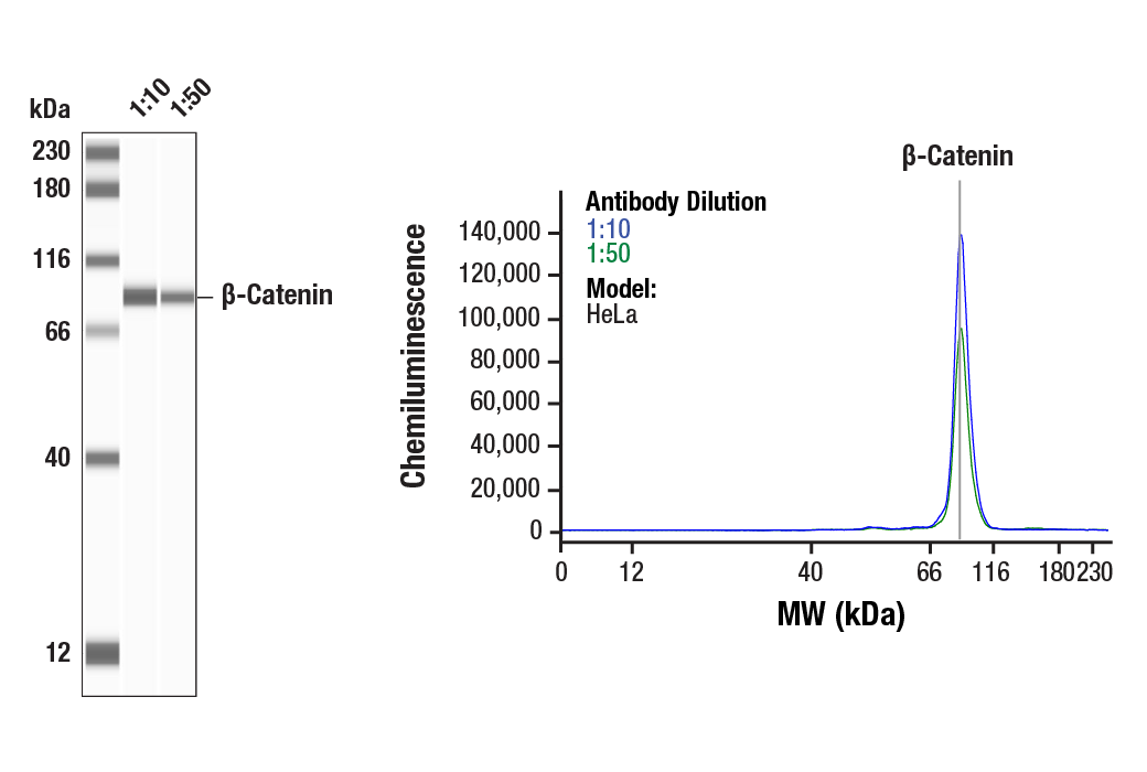


 下载产品说明书 下载SDS
 用小程序,查商品更便捷


 收藏
收藏
 对比
对比 咨询
咨询
The Epithelial-Mesenchymal Transition (EMT) Antibody Sampler Kit provides an economical means of evaluating EMT. The kit contains enough primary antibody to perform two western blots per primary.
参考图片
Simple Western™ analysis of lysates (0.1 mg/mL) from HeLa cells using β-Catenin (D10A8) XP® Rabbit mAb #8480. The virtual lane view (left) shows the target band (as indicated) at 1:10 and 1:50 dilutions of primary antibody. The corresponding electropherogram view (right) plots chemiluminescence by molecular weight along the capillary at 1:10 (blue line) and 1:50 (green line) dilutions of primary antibody. This experiment was performed under reducing conditions on the Jess™ Simple Western instrument from ProteinSimple, a BioTechne brand, using the 12-230 kDa separation module.
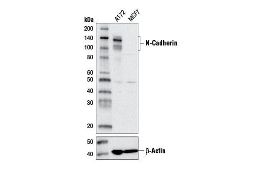
Western blot analysis of extracts from A172 and MCF7 cells using N-Cadherin (D4R1H) XP® Rabbit mAb (upper) or β-Actin (D6A8) Rabbit mAb #8457 (lower).
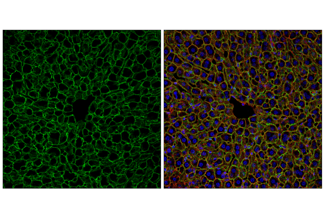
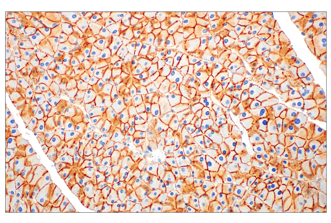
Confocal immunofluorescent analysis of fixed frozen mouse liver labeled with N-Cadherin (D4R1H) XP® Rabbit mAb (green), DyLight 554 Phalloidin #13054 (red) and ProLong® Gold Antifade Reagent with DAPI #8961 (blue).
Confocal immunofluorescent analysis of fixed frozen mouse choroid plexus labeled with N-Cadherin (D4R1H) XP® Rabbit mAb (green) and ProLong® Gold Antifade Reagent with DAPI #8961 (blue).
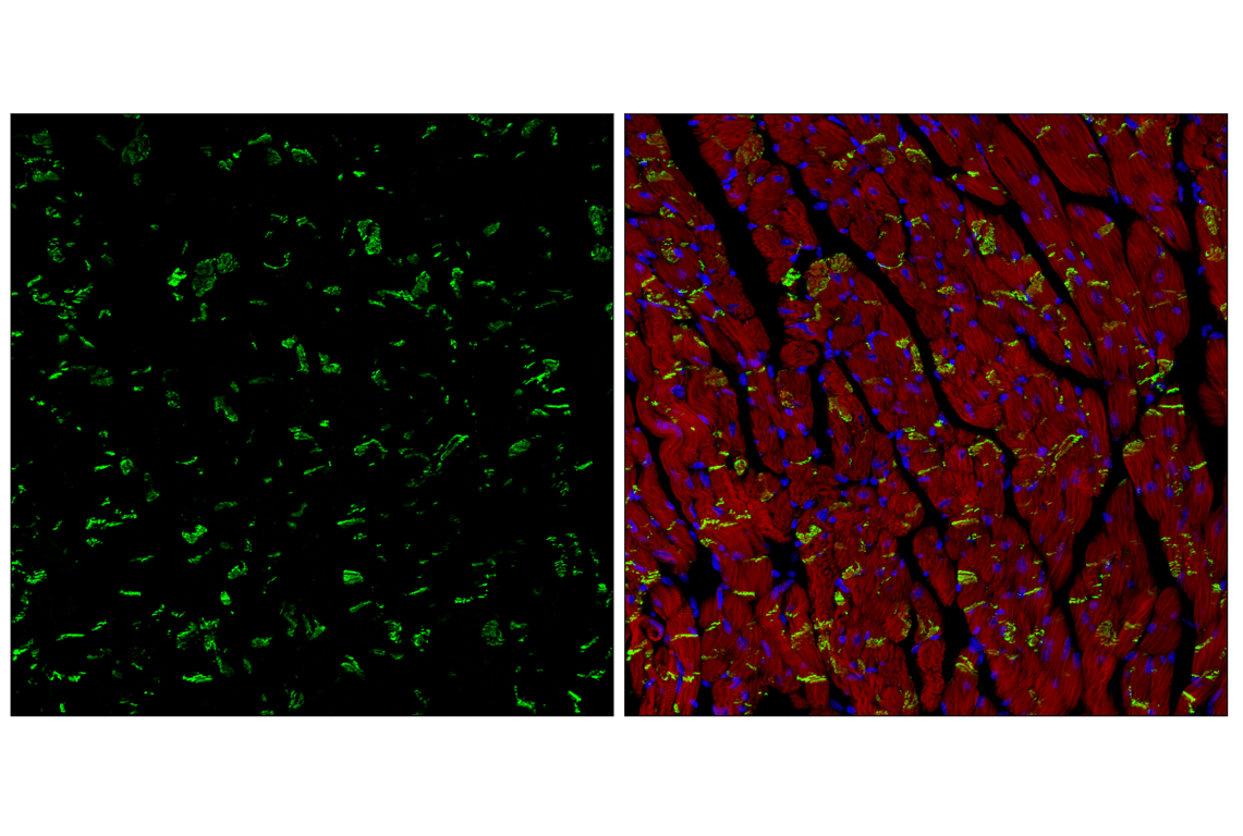
Confocal immunofluorescent analysis of fixed frozen mouse heart labeled with N-Cadherin (D4R1H) XP® Rabbit mAb (green), DyLight 554 Phalloidin #13054 (red) and ProLong® Gold Antifade Reagent with DAPI #8961 (blue).
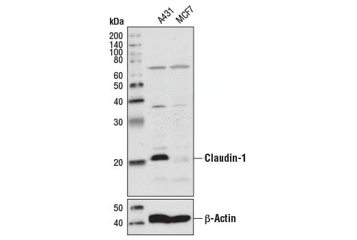
Western blot analysis of extracts from A431 and MCF7 cells using Claudin-1 (D5H1D) XP® Rabbit mAb (upper) or β-Actin (D6A8) Rabbit mAb #8457 (lower).
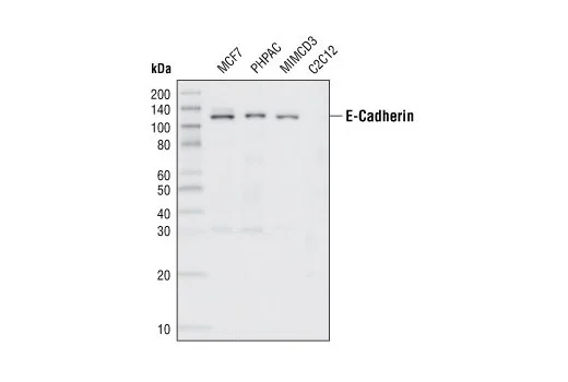
Western blot analysis of extracts from various cell lines, using E-Cadherin (24E10) Rabbit mAb.
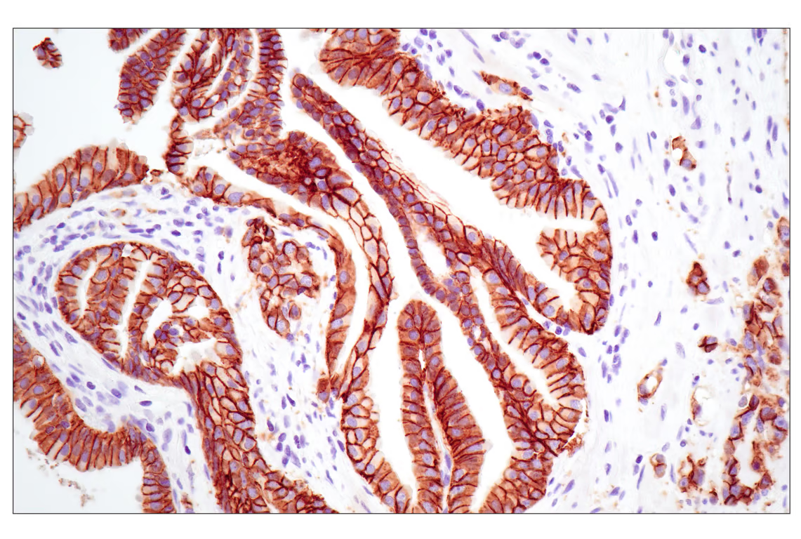
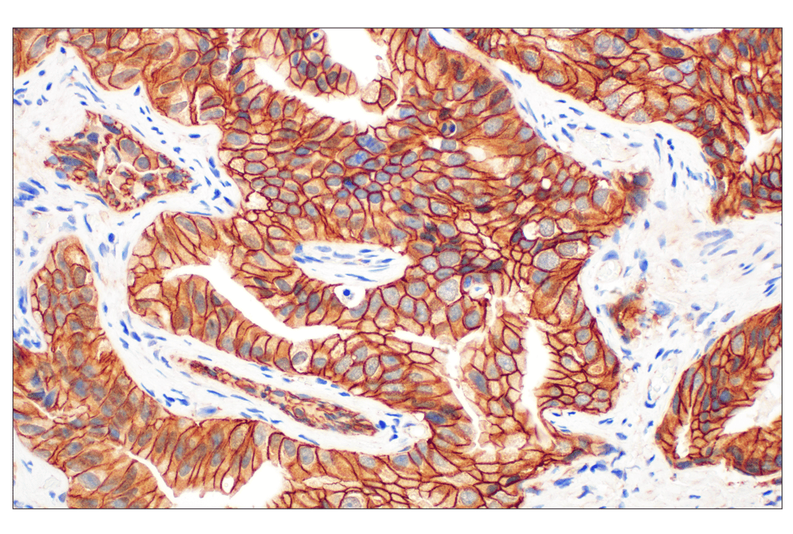
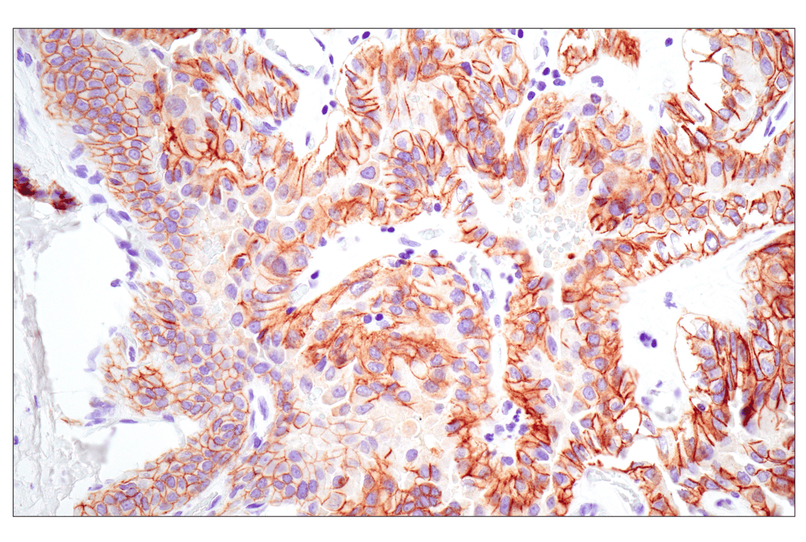
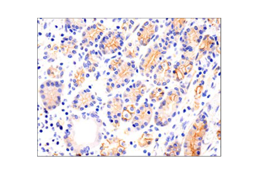
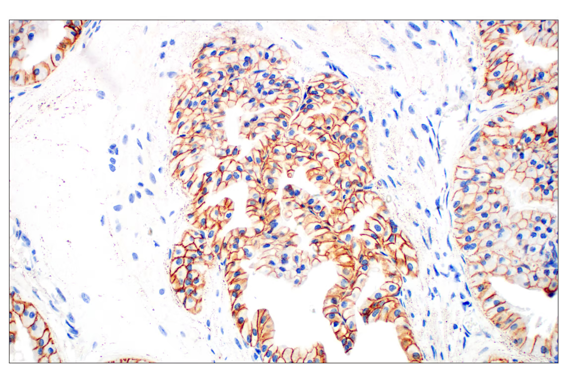
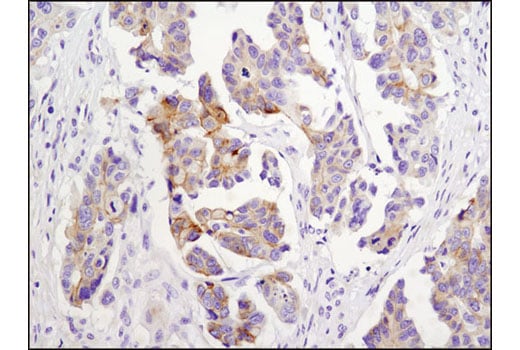
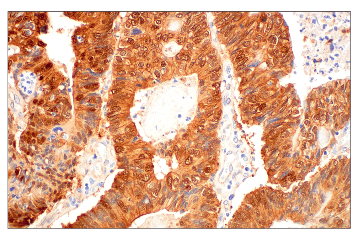
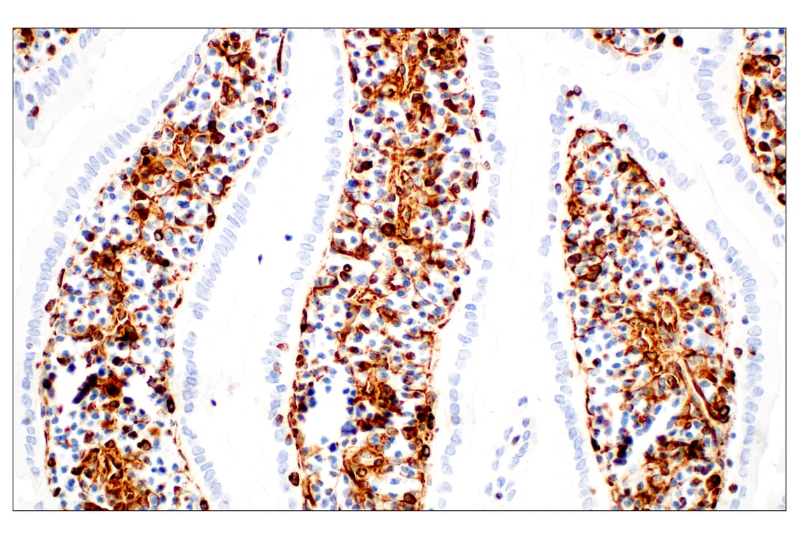
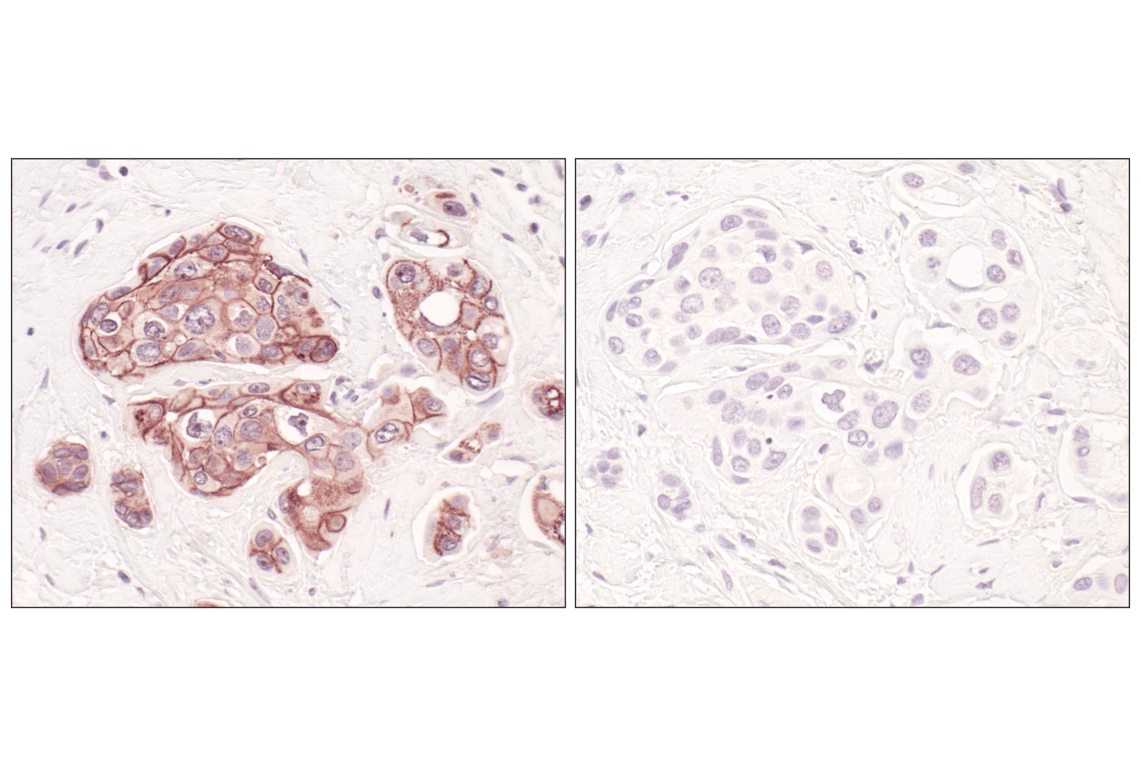
Immunohistochemical analysis of paraffin-embedded human prostate adenocarcinoma using E-Cadherin (24E10) Rabbit mAb performed on the Leica BOND Rx.
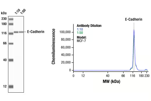
Simple Western™ analysis of lysates (0.1 mg/mL) from MCF-7 cells using E-Cadherin (24E10) Rabbit mAb #3195. The virtual lane view (left) shows a single target band (as indicated) at 1:10 and 1:50 dilutions of primary antibody. The corresponding electropherogram view (right) plots chemiluminescence by molecular weight along the capillary at 1:10 (blue line) and 1:50 (green line) dilutions of primary antibody. This experiment was performed under reducing conditions on the Jess™ Simple Western instrument from ProteinSimple, a BioTechne brand, using the 12-230 kDa separation module.
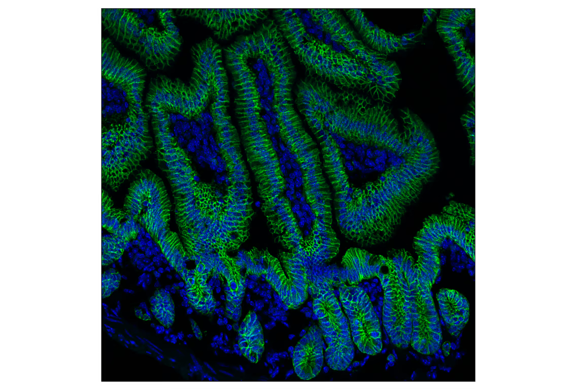
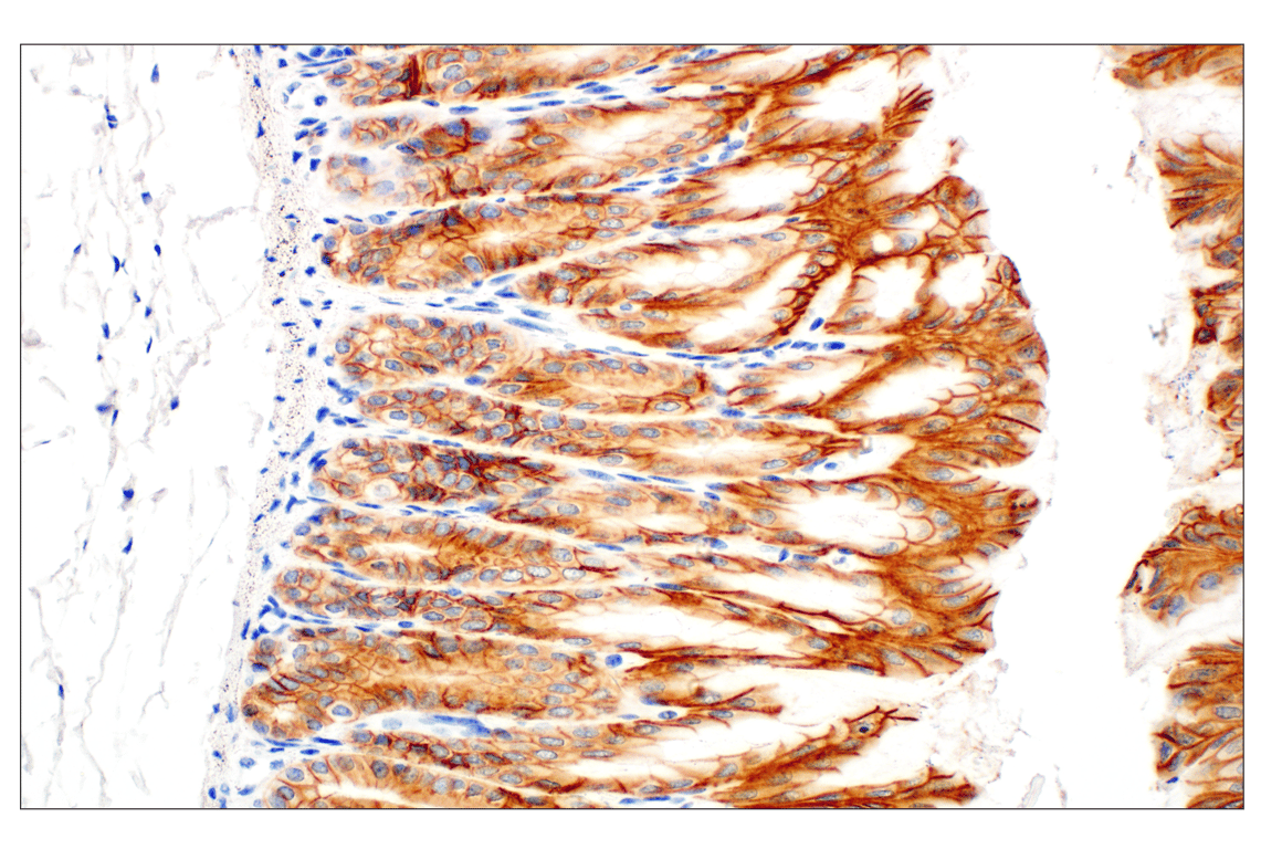
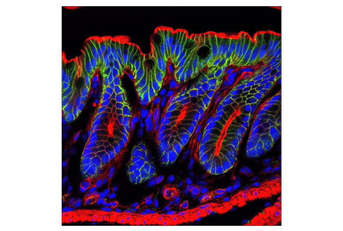
Confocal immunofluorescent analysis of fixed frozen mouse small intestine using E-Cadherin (24E10) Rabbit mAb (green) and ProLong Gold Antifade Reagent with DAPI #8961 (blue).
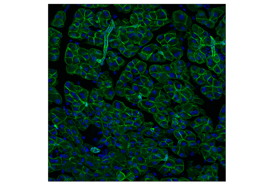
Confocal immunofluorescent analysis of fixed frozen mouse pancreas using E-Cadherin (24E10) Rabbit mAb (green) and ProLong Gold Antifade Reagent with DAPI #8961 (blue).
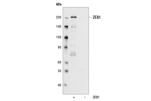
Western blot analysis of extracts from COS cells, mock transfected or transfected with a construct expressing human ZEB1, using ZEB1 (D80D3) Rabbit mAb.
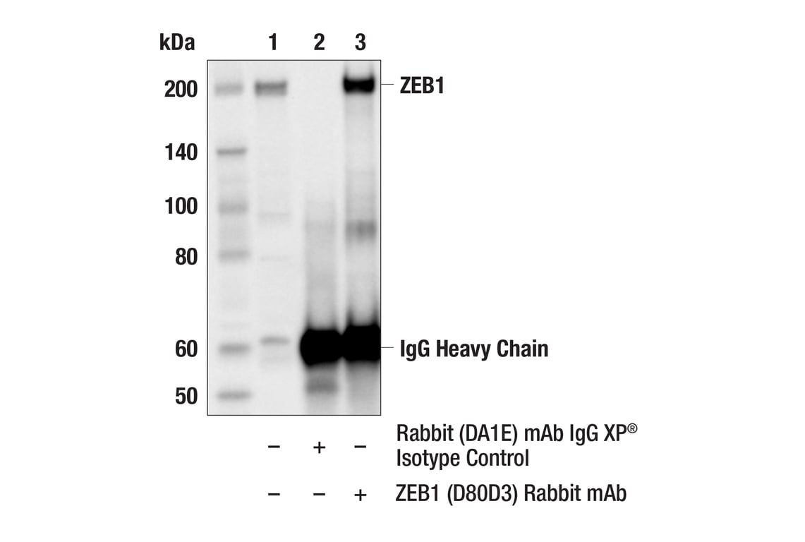
Immunoprecipitation of ZEB1 protein from Jurkat cell extracts. Lane 1 is 10% input, lane 2 is Rabbit (DA1E) mAb IgG XP® Isotype Control #3900, and lane 3 is ZEB1 (D80D3) Rabbit mAb. Western blot analysis was performed using ZEB1 (D80D3) Rabbit mAb.
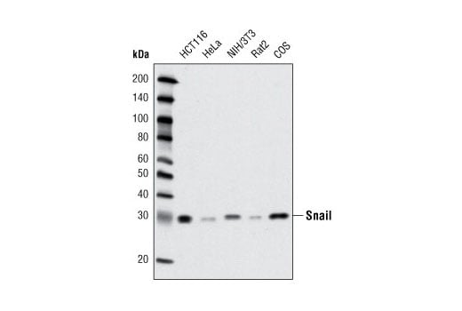
Western blot analysis of extracts from various cell lines using Snail (C15D3) Rabbit mAb.
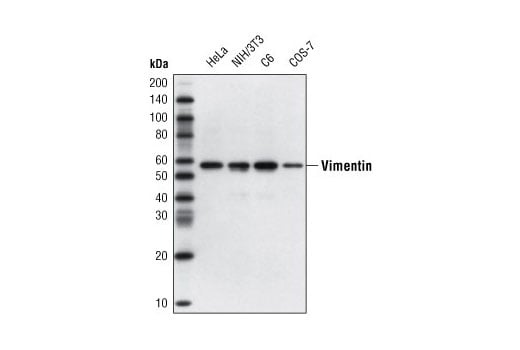
Western blot analysis of extracts from various cell lines using Vimentin (D21H3) XP® Rabbit mAb.
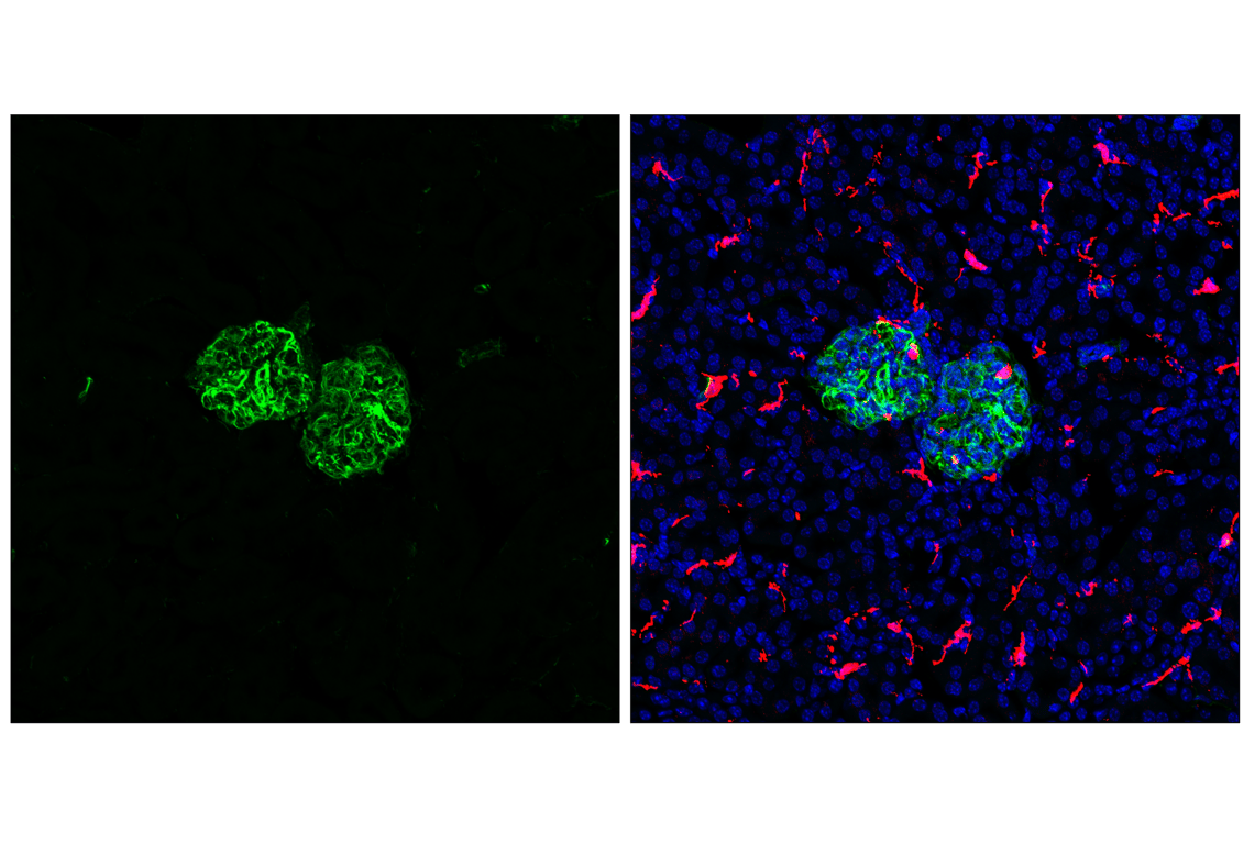
Confocal immunofluorescent analysis of fixed frozen mouse kidney labeled with Vimentin (D21H3) XP® Rabbit mAb (left, green) and co-labeled with F4/80 (BM8.1) Rat mAb (right, red), and ProLong® Gold Antifade Reagent with DAPI #8961 (right, blue).
Confocal immunofluorescent analysis of fixed frozen mouse colon labeled with Vimentin (D21H3) XP® Rabbit mAb (left, green) and co-labeled with F4/80 (BM8.1) Rat mAb (right, red), and ProLong® Gold Antifade Reagent with DAPI #8961 (right, blue).
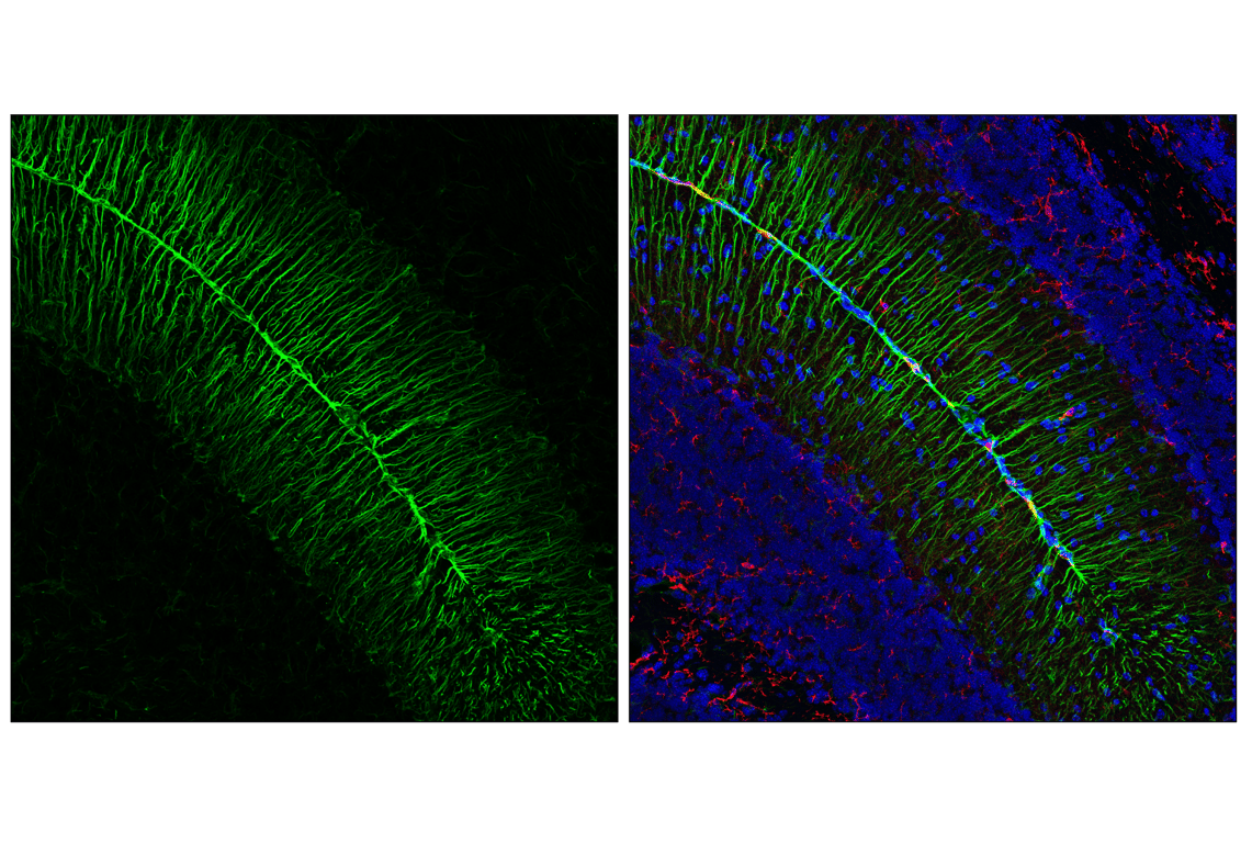
Confocal immunofluorescent analysis of fixed frozen mouse cerebellum labeled with Vimentin (D21H3) XP® Rabbit mAb (left, green) and co-labeled with F4/80 (BM8.1) Rat mAb (right, red), and ProLong® Gold Antifade Reagent with DAPI #8961 (right, blue).
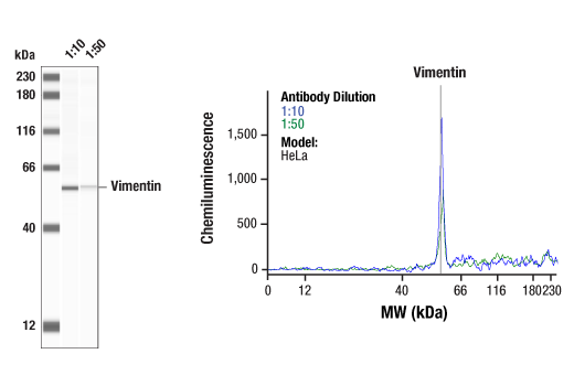
Simple Western™ analysis of lysates (1 mg/mL) from HeLa cells using Vimentin (D21H3) XP ® Rabbit mAb #5741. The virtual lane view (left) shows a single target band (as indicated) at 1:10 and 1:50 dilutions of primary antibody. The corresponding electropherogram view (right) plots chemiluminescence by molecular weight along the capillary at 1:10 (blue line) and 1:50 (green line) dilutions of primary antibody. This experiment was performed under reducing conditions on the Jess™ Simple Western instrument from ProteinSimple, a BioTechne brand, using the 12-230 kDa separation module.
Confocal immunofluorescent analysis of fixed frozen mouse cerebellum labeled with Vimentin (D21H3) XP® Rabbit mAb (left, green) and co-labeled with F4/80 (BM8.1) Rat mAb (right, red), and ProLong® Gold Antifade Reagent with DAPI #8961 (right, blue).
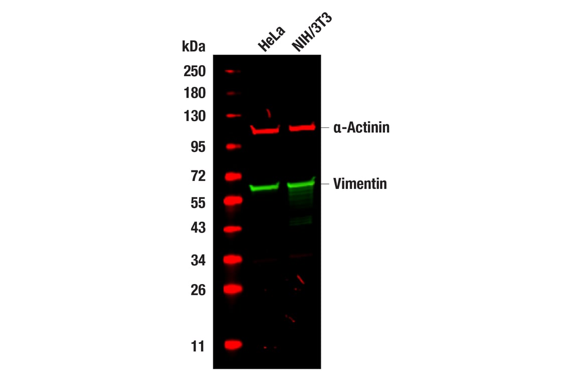
Western blot analysis of extracts from HeLa and NIH/3T3 cells using Vimentin (D21H3) XP® Rabbit mAb #5741 (green), and α-Actinin (E7U1O) Mouse mAb #69758 (red). Anti-rabbit IgG (H+L) (DyLight 800 4X PEG Conjugate) #5151 (green) and Anti-mouse IgG (H+L) (DyLight 680 Conjugate) #5470 (red) were used as secondary antibodies.
After the primary antibody is bound to the target protein, a complex with HRP-linked secondary antibody is formed. The LumiGLO® is added and emits light during enzyme catalyzed decomposition.
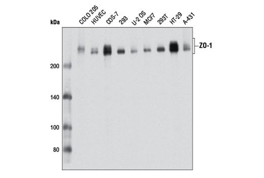
Western blot analysis of extracts from various cell lines using ZO-1 (D7D12) Rabbit mAb.
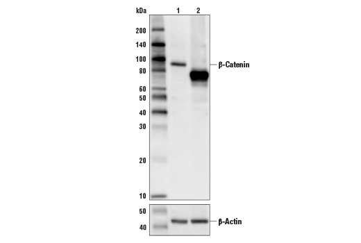
Western blot analysis of extracts from control HeLa cells (lane 1) or HeLa cells with an apparent in-frame truncation mutation in the gene encoding β-Catenin (lane 2) using β-Catenin (D10A8) XP® Rabbit mAb, #8480 (upper) or β-actin (D6A8) Rabbit mAb #8457 (lower). The change in β-Catenin molecular weight in the mutated HeLa cells is consistent with an in-frame deletion.
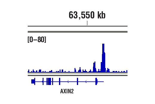
CUT&RUN was performed with HCT 116 cells and β-Catenin (D10A8) XP® Rabbit mAb, using CUT&RUN Assay Kit #86652. DNA library was prepared using DNA Library Prep Kit for Illumina® (ChIP-seq, CUT&RUN) #56795. The figure shows binding across Axin2, a known target gene of β-Catenin (see additional figure containing CUT&RUN-qPCR data).
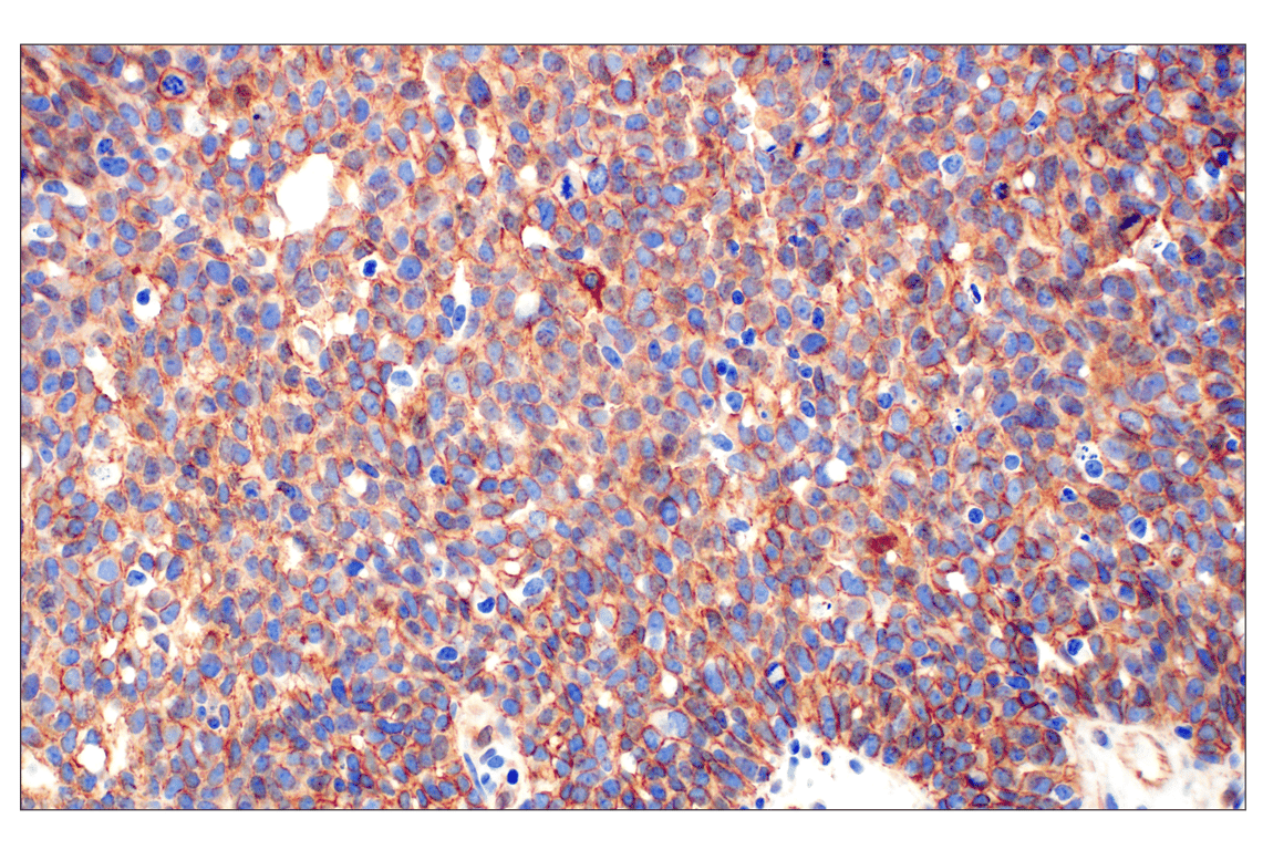
Immunohistochemical analysis of paraffin-embedded human prostate adenocarcinoma using ß-Catenin (D10A8) XP® Rabbit mAb performed on the Leica® BOND™ Rx.
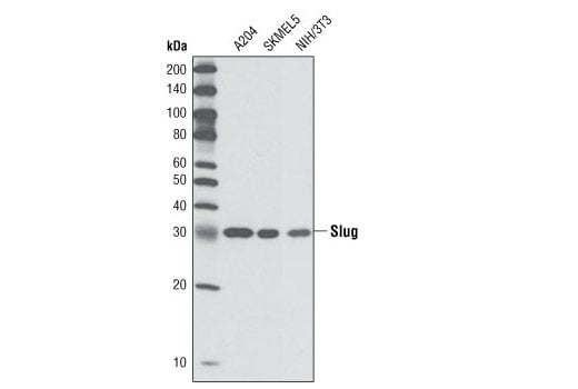
Western blot analysis of extracts from A204, SKMEL5, and NIH/3T3 cells using Slug (C19G7) Rabbit mAb.
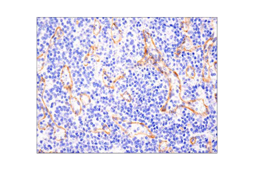
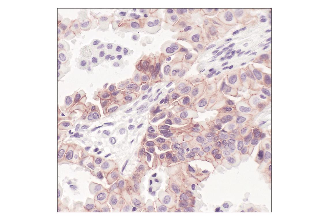
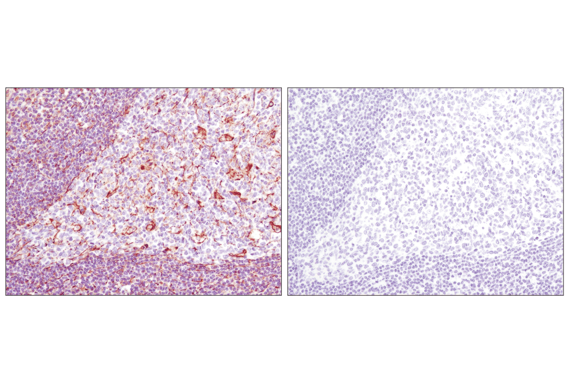
Immunohistochemical analysis of paraffin-embedded human non-Hodgkin Lymphoma using N-Cadherin (D4R1H) XP® Rabbit mAb performed on the Leica® BOND™ Rx.
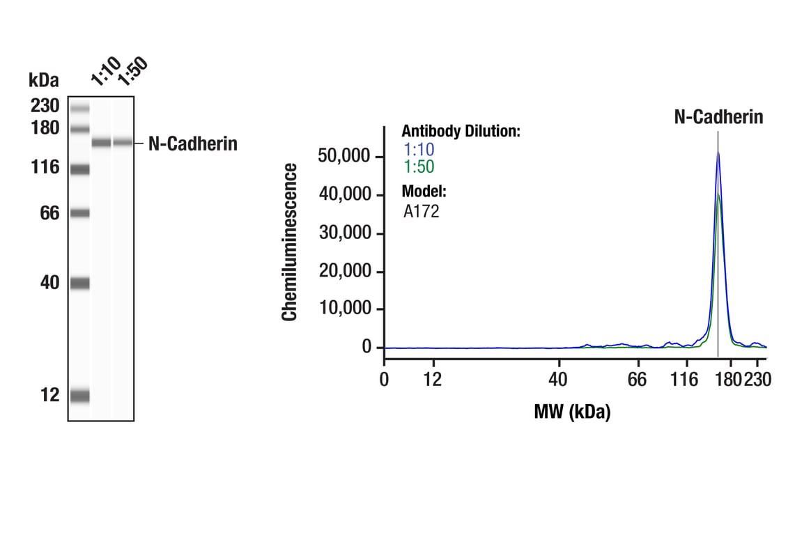
Simple Western™ analysis of lysates (0.1 mg/mL) from A172 lysate using N-Cadherin (D4R1H) XP® Rabbit mAb #13116. The virtual lane view (left) shows the target band (as indicated) at 1:10 and 1:50 dilutions of primary antibody. The corresponding electropherogram view (right) plots chemiluminescence by molecular weight along the capillary at 1:10 (blue line) and 1:50 (green line) dilutions of primary antibody. This experiment was performed under reducing conditions on the Jess™ Simple Western instrument from ProteinSimple, a BioTechne brand, using the 12-230 kDa separation module.
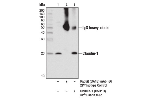
Immunoprecipitation of claudin-1 from A-431 cell extracts using Rabbit (DA1E) mAb IgG XP® Isotype Control #3900 (lane 2) or Claudin-1 (D5H1D) XP® Rabbit mAb (lane 3). Lane 1 is 10% input. Western blot was performed using Claudin-1 (D5H1D) Rabbit mAb.
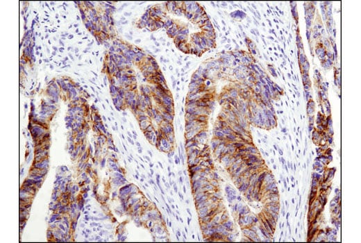
Immunohistochemical analysis of paraffin-embedded human papillary thyroid carcinoma using E-Cadherin (24E10) Rabbit mAb performed on the Leica BOND Rx.
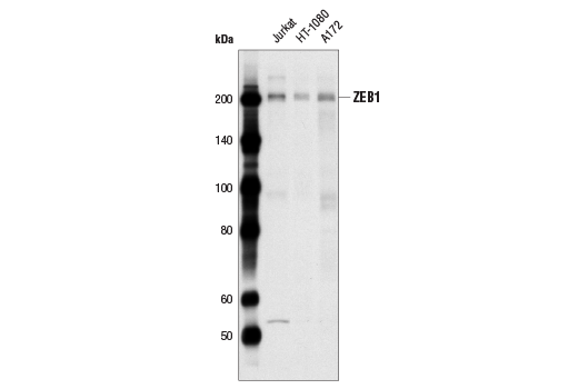
Western blot analysis of extracts from Jurkat, HT1080 and A172 cells using ZEB1 (D80D3) Rabbit mAb.
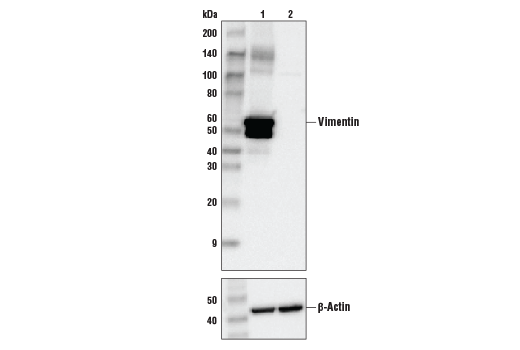
Western blot analysis of extracts from control HeLa cells (lane 1) or Vimentin knockout HeLa cells (lane 2) using Vimentin (D21H3) XP® Rabbit mAb #5741 (upper) or β-Actin (13E5) Rabbit mAb #4970 (lower). The absence of signal in the Vimentin knockout HeLa cells confirms specificity of the antibody for Vimentin.
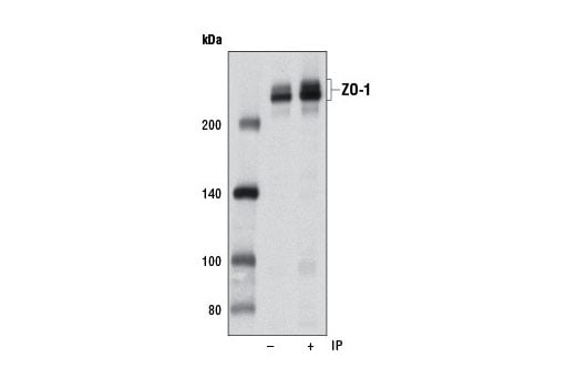
Immunoprecipitation and western blot analysis of extracts from Hep G2 cells using ZO-1 (D7D12) Rabbit mAb. Lane 1 is 10% input.
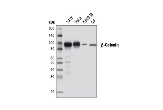
Western blot analysis of extracts from various cell lines using β-Catenin (D10A8) XP® Rabbit mAb.
CUT&RUN was performed with HCT 116 cells and β-Catenin (D10A8) XP® Rabbit mAb, using CUT&RUN Assay Kit #86652. DNA Libraries were prepared using DNA Library Prep Kit for Illumina® (ChIP-seq, CUT&RUN) #56795. The figures show binding across chromosome 17 (upper), including Axin2 (lower), a known target gene of β-Catenin (see additional figure containing CUT&RUN-qPCR data).
Immunohistochemical analysis of paraffin-embedded human colon adenocarcinoma using ß-Catenin (D10A8) XP® Rabbit mAb performed on the Leica® BOND™ Rx.
Immunohistochemical analysis of paraffin-embedded human granulosa cell tumor of the ovary using N-Cadherin (D4R1H) XP® Rabbit mAb performed on the Leica® BOND™ Rx.
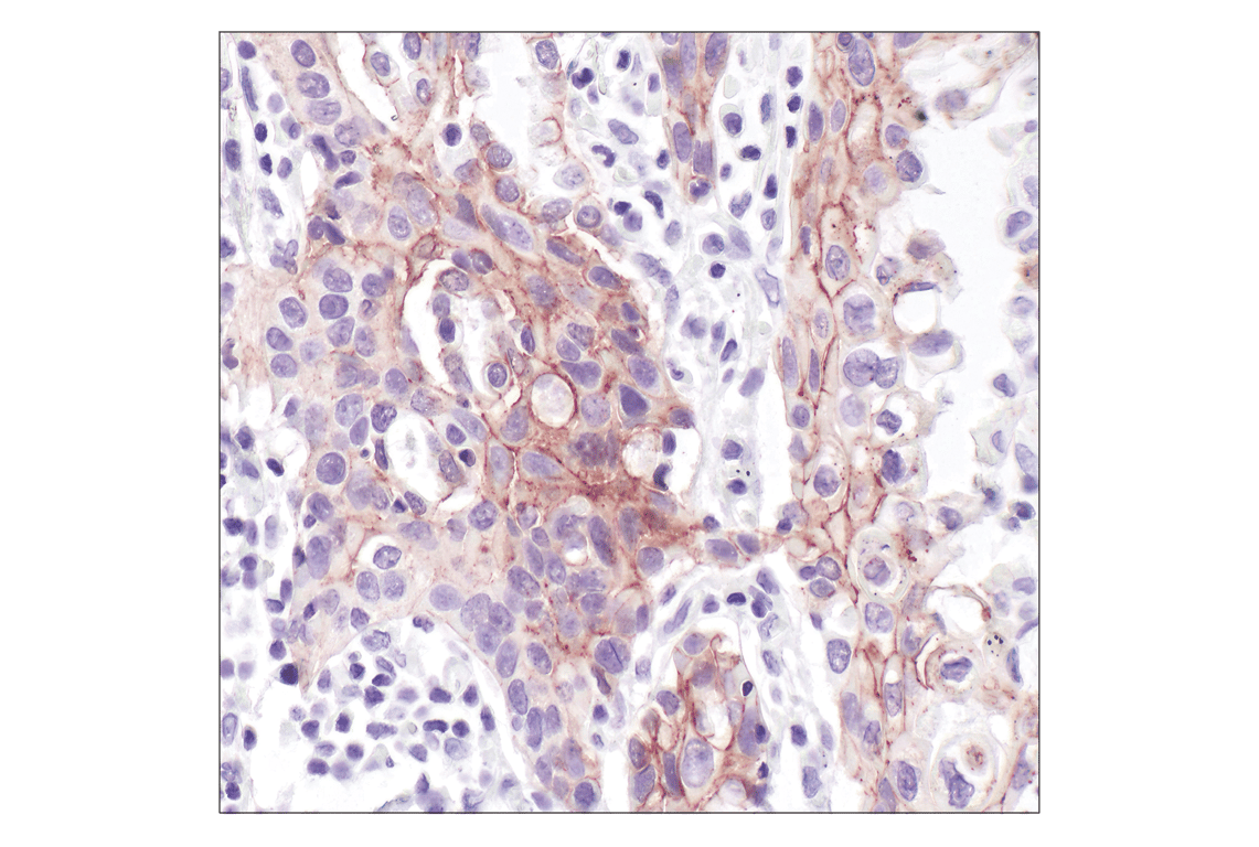
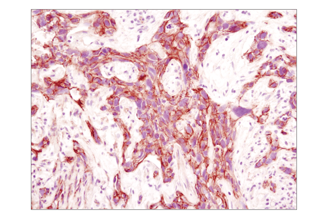
Immunohistochemical analysis of paraffin-embedded human colon carcinoma using Claudin-1 (D5H1D) XP® Rabbit mAb.
Immunohistochemical analysis of paraffin-embedded human lung carcinoma, using E-Cadherin (24E10) Rabbit mAb.
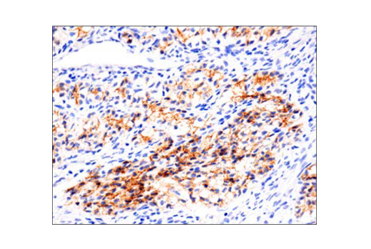
Immunohistochemical analysis of paraffin-embedded human squamous cell lung carcinoma using Vimentin (D21H3) XP® Rabbit mAb performed on the Leica® BOND™ Rx.
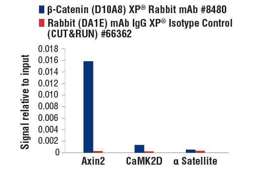
CUT&RUN was performed with HCT 116 cells and either β-Catenin (D10A8) XP® Rabbit mAb or Rabbit (DA1E) mAb IgG XP® Isotype Control (CUT&RUN) #66362, using CUT&RUN Assay Kit #86652. The enriched DNA was quantified by real-time PCR using SimpleChIP® Human Axin2 Intron 1 Primers #8973, SimpleChIP® Human CaMK2D Intron 3 Primers #5111 and SimpleChIP® Human α Satellite Repeat Primers #4486. The amount of immunoprecipitated DNA in each sample is represented as signal relative to the total amount of input chromatin, which is equivalent to one.
Immunohistochemical analysis of paraffin-embedded human serous adenocarcinoma of the ovary using ß-Catenin (D10A8) XP® Rabbit mAb performed on the Leica® BOND™ Rx.
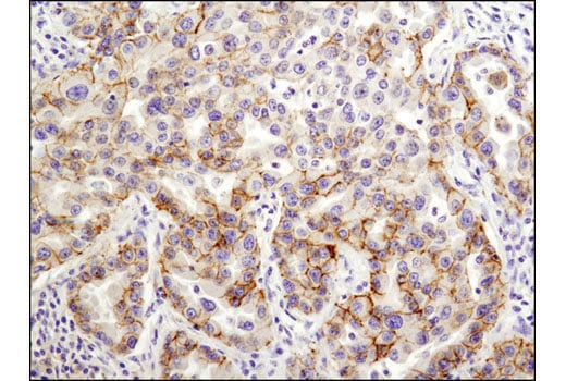
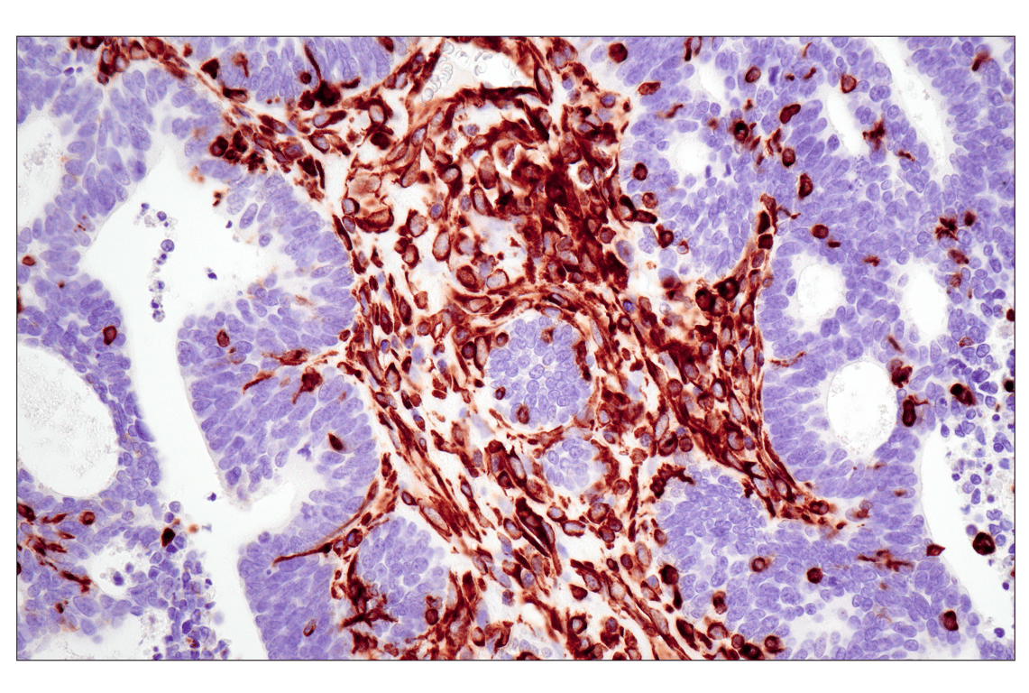
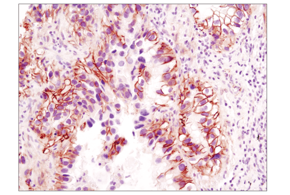
Immunohistochemical analysis of paraffin-embedded human gastric carcinoma using N-Cadherin (D4R1H) XP® Rabbit mAb performed on the Leica® BOND™ Rx.
Immunohistochemical analysis of paraffin-embedded human lung carcinoma using Claudin-1 (D5H1D) XP® Rabbit mAb.
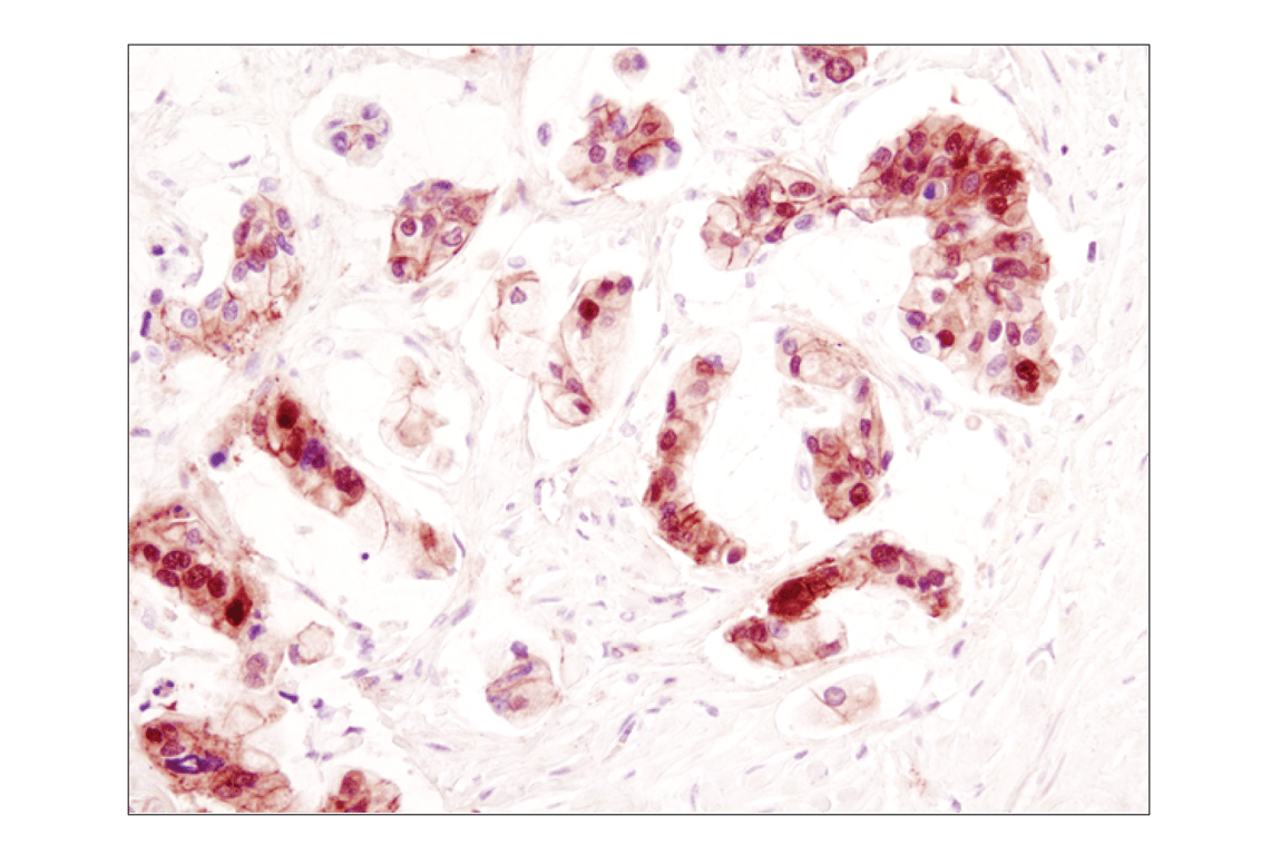
Immunohistochemical analysis of paraffin-embedded human metastatic adenocarcinoma in lymph node, using E-Cadherin (24E10) Rabbit mAb.
Immunohistochemical analysis of paraffin-embedded human endometrioid adenocarcinoma using Vimentin (D21H3) XP® Rabbit mAb performed on the Leica® BOND™ Rx.
Immunohistochemical analysis of paraffin-embedded human colon adenocarcinoma using ß-Catenin (D10A8) XP® Rabbit mAb.
Confocal immunofluorescent analysis of A204 cells (left) and PANC-1 cells (right) using Slug (C19G7) Rabbit mAb (green). Actin filaments have been labeled with DY554 phalloidin (red).
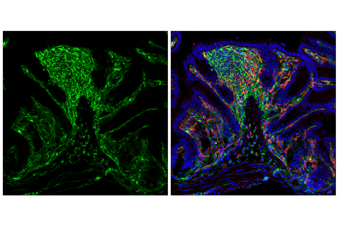
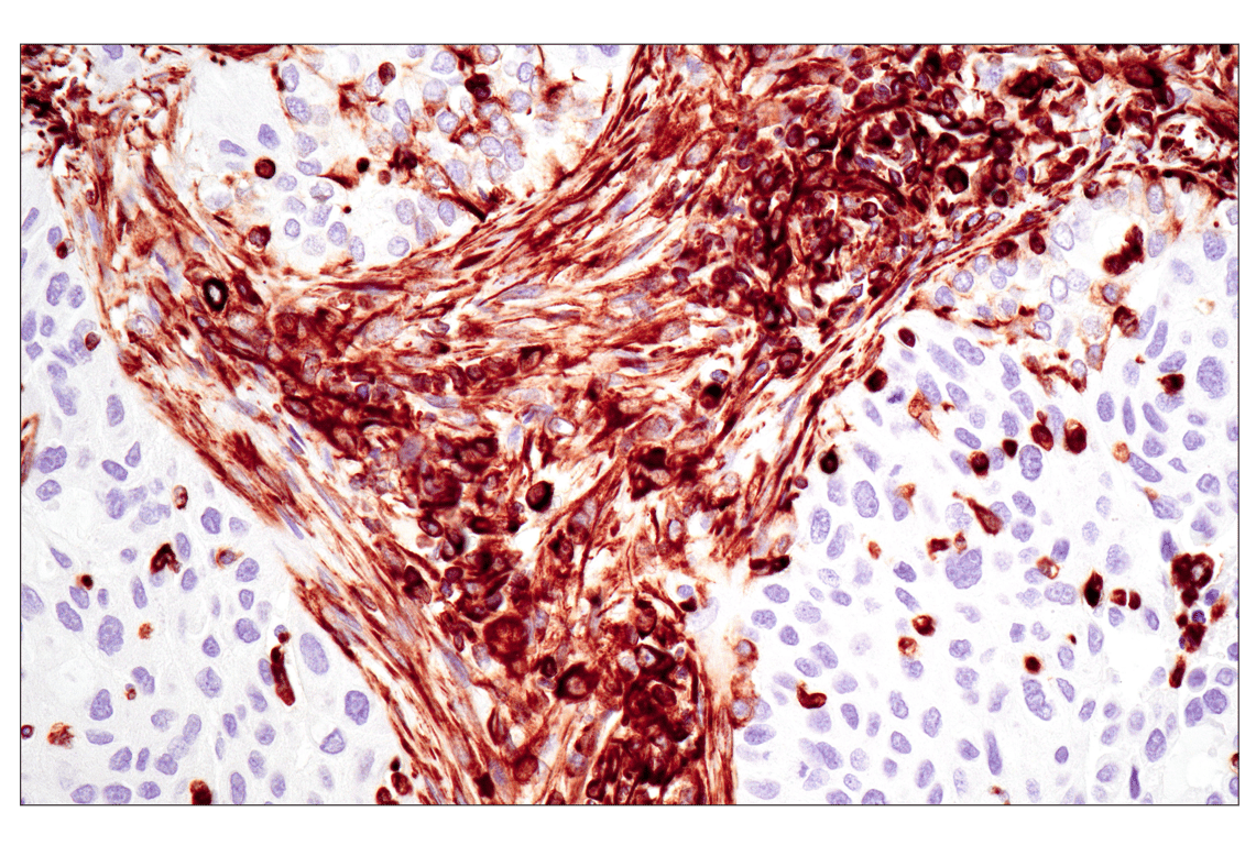
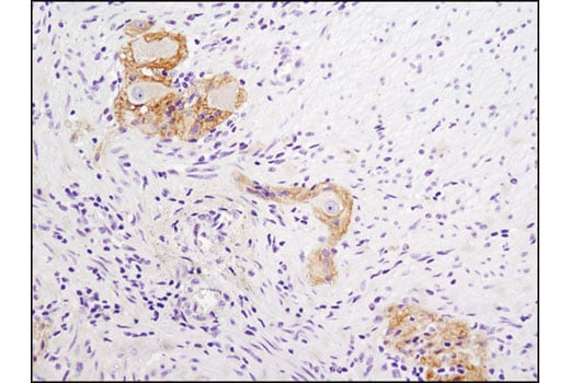
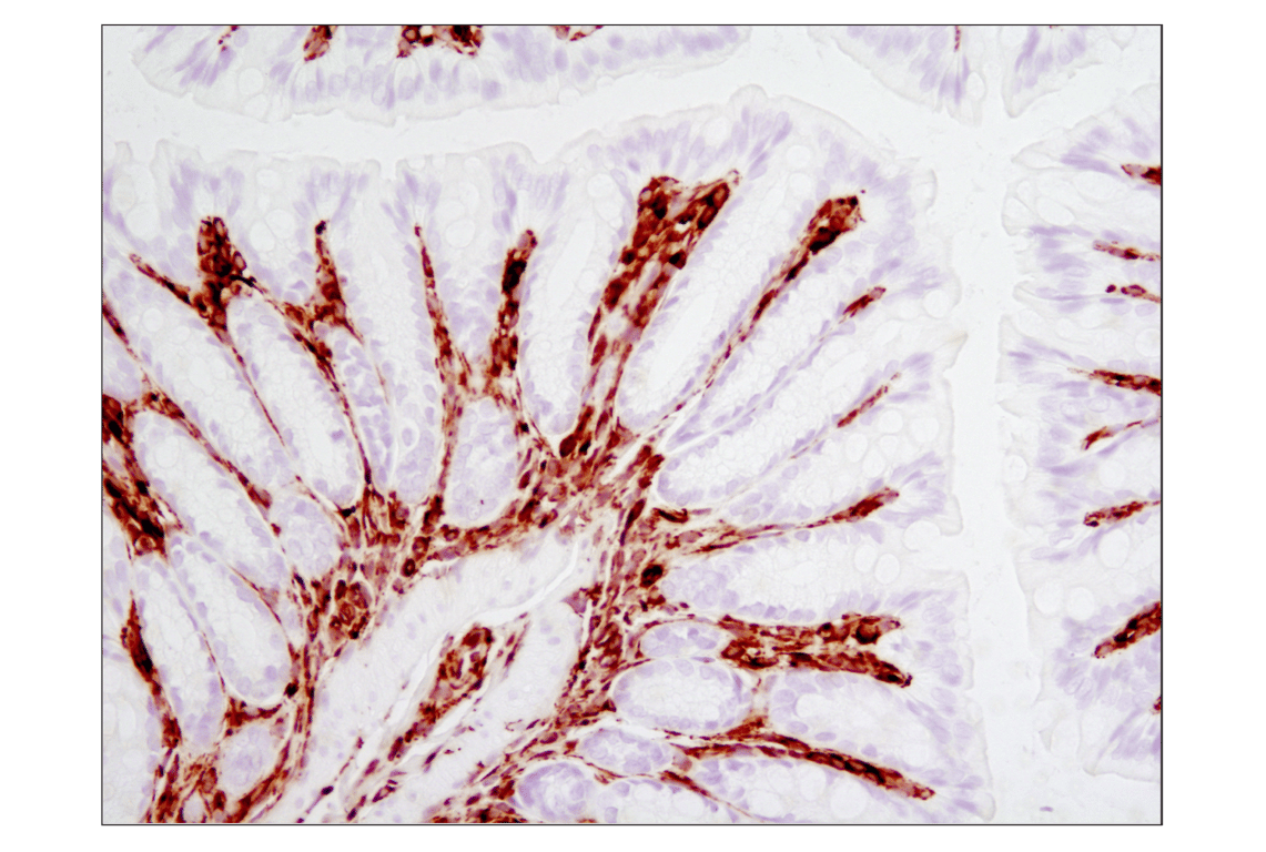
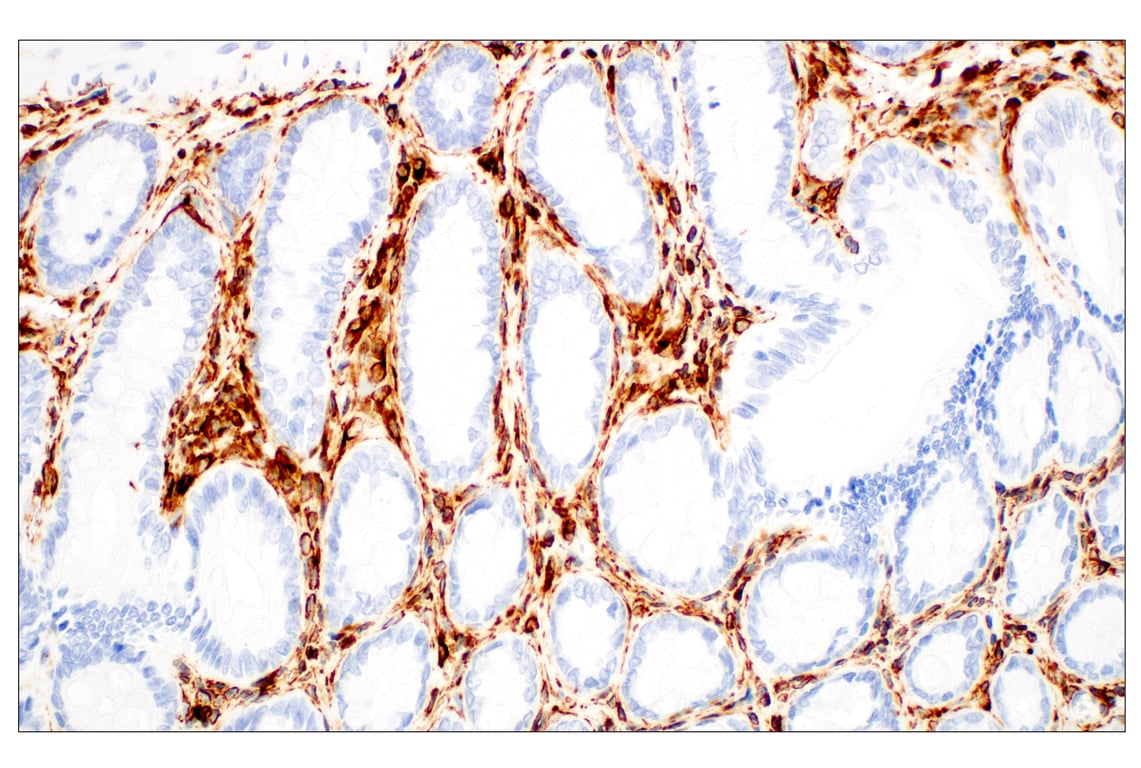
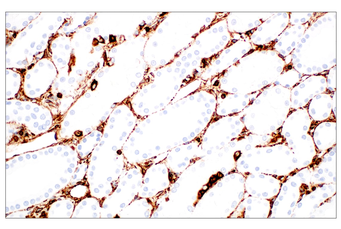
Immunohistochemical analysis of paraffin-embedded human colon using N-Cadherin (D4R1H) XP® Rabbit mAb. Note staining of myenteric plexus.
Immunohistochemical analysis of paraffin-embedded cell pellets, A-431 (left) and MCF7 (right), using Claudin-1 (D5H1D) XP® Rabbit mAb.
Immunohistochemical analysis of paraffin-embedded mouse prostate using E-Cadherin (24E10) Rabbit mAb.
Immunohistochemical analysis of paraffin-embedded human breast carcinoma using Vimentin (D21H3) XP® Rabbit mAb.
Immunohistochemical analysis of paraffin-embedded human lung carcinoma using β-Catenin (D10A8) XP® Rabbit mAb.
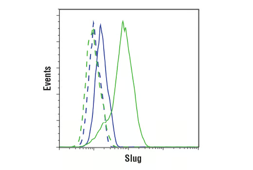
Flow cytometric analysis of PANC-1 cells (blue) and A204 cells (green) using Slug (C19G7) Rabbit mAb (solid lines) or a concentration-matched Rabbit (DA1E) mAb IgG XP® Isotype Control #3900 (dashed lines). Anti-rabbit IgG (H+L), F(ab')2 Fragment (Alexa Fluor® 488 Conjugate) #4412 was used as a secondary antibody.
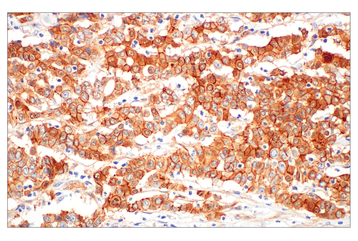
Immunohistochemical analysis of paraffin-embedded human ovarian carcinoma using N-Cadherin (D4R1H) XP® Rabbit mAb.
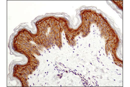
Immunohistochemical analysis of paraffin-embedded human skin using Claudin-1 (D5H1D) XP® Rabbit mAb.
Immunohistochemical analysis of paraffin-embedded mouse pancreas using E-Cadherin (24E10) Rabbit mAb.
Immunohistochemical analysis of paraffin-embedded mouse colon using Vimentin (D21H3) XP® Rabbit mAb.
Immunohistochemical analysis of paraffin-embedded human colon adenocarcinoma using ß-Catenin (D10A8) XP® Rabbit mAb.
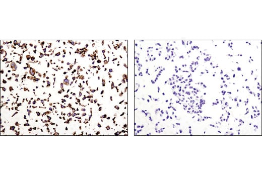
Immunohistochemical analysis of paraffin-embedded A172 (positive, left) and MCF7 (negative, right) cell pellets using N-Cadherin (D4R1H) XP® Rabbit mAb.
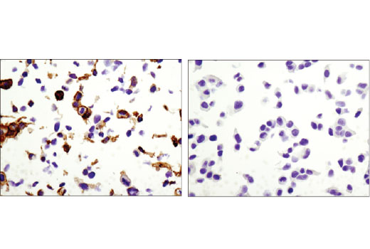
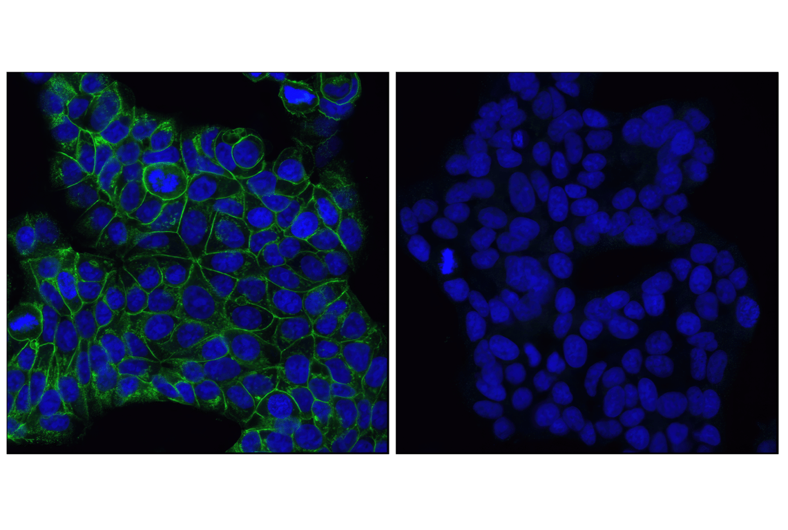
Confocal immunofluorescent images of MCF7 cells using E-Cadherin (24E10) Rabbit mAb (green, left) compared to an isotype control (right). Blue pseudocolor = DRAQ5® (fluorescent DNA dye).
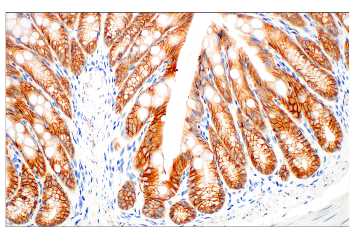
Immunohistochemical analysis of paraffin-embedded mouse small intestine using E-Cadherin (24E10) Rabbit mAb.
Immunohistochemical analysis of paraffin-embedded rat colon using Vimentin (D21H3) XP® Rabbit mAb.
Immunohistochemical analysis of paraffin-embedded human colon carcinoma using β-Catenin (D10A8) XP® Rabbit mAb.
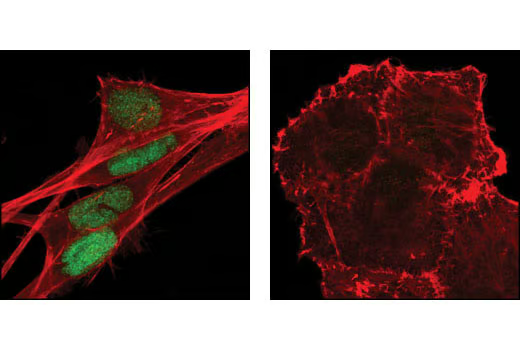
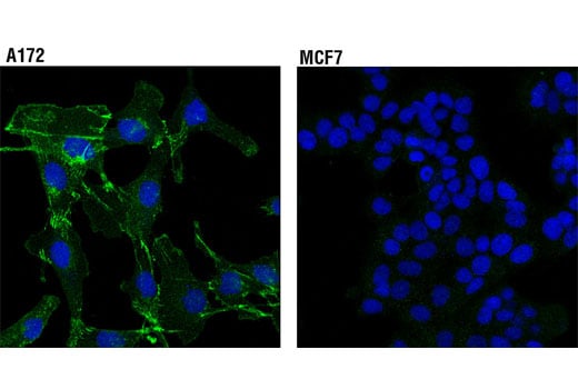
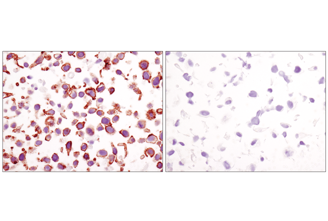
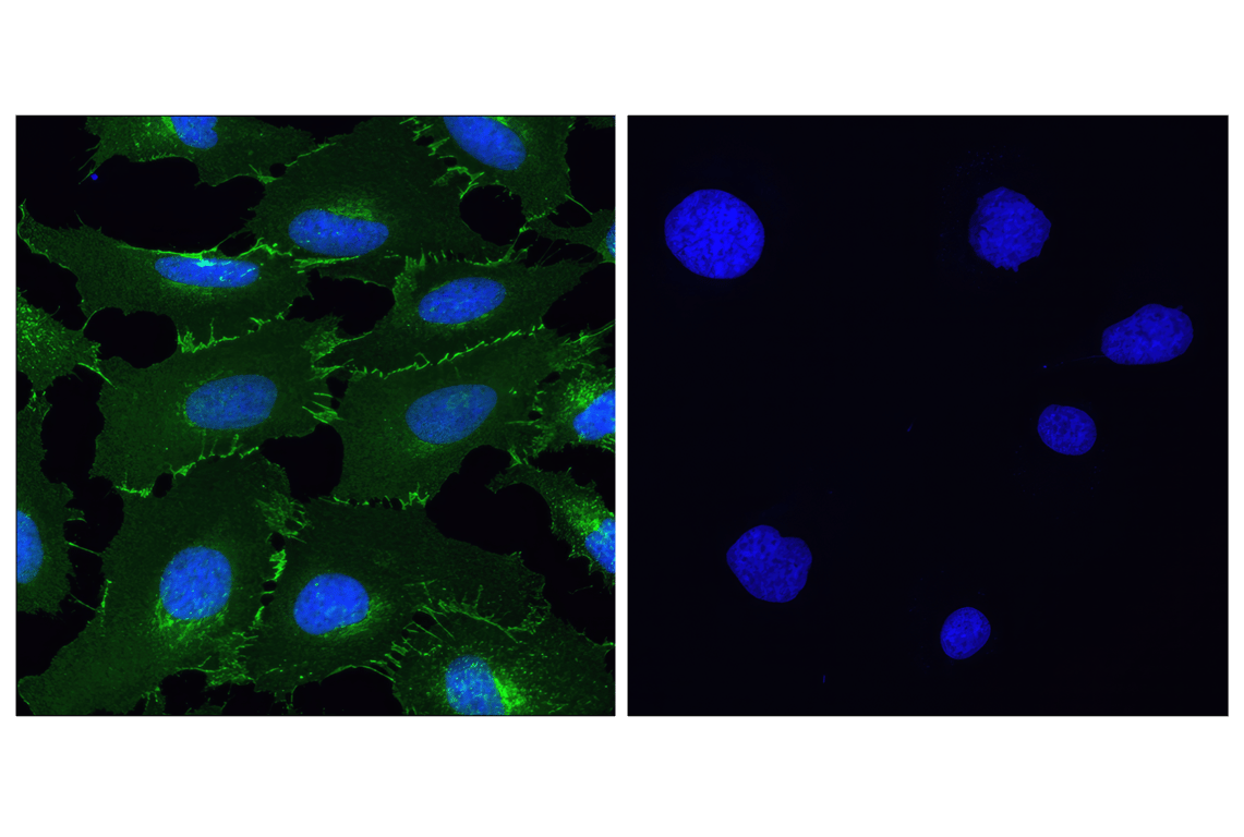
Confocal immunofluorescent analysis of A172 (positive, left) and MCF7 (negative, right) cells using N-Cadherin (D4R1H) XP® Rabbit mAb (green). Blue pseudocolor= DRAQ5® #4084 (fluorescent DNA dye).
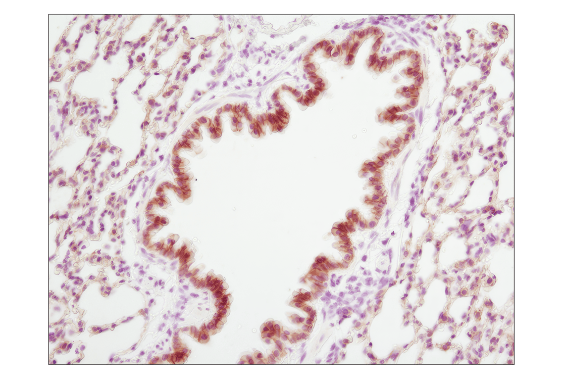
Immunohistochemical analysis of paraffin-embedded mouse lung using E-Cadherin (24E10) Rabbit mAb.
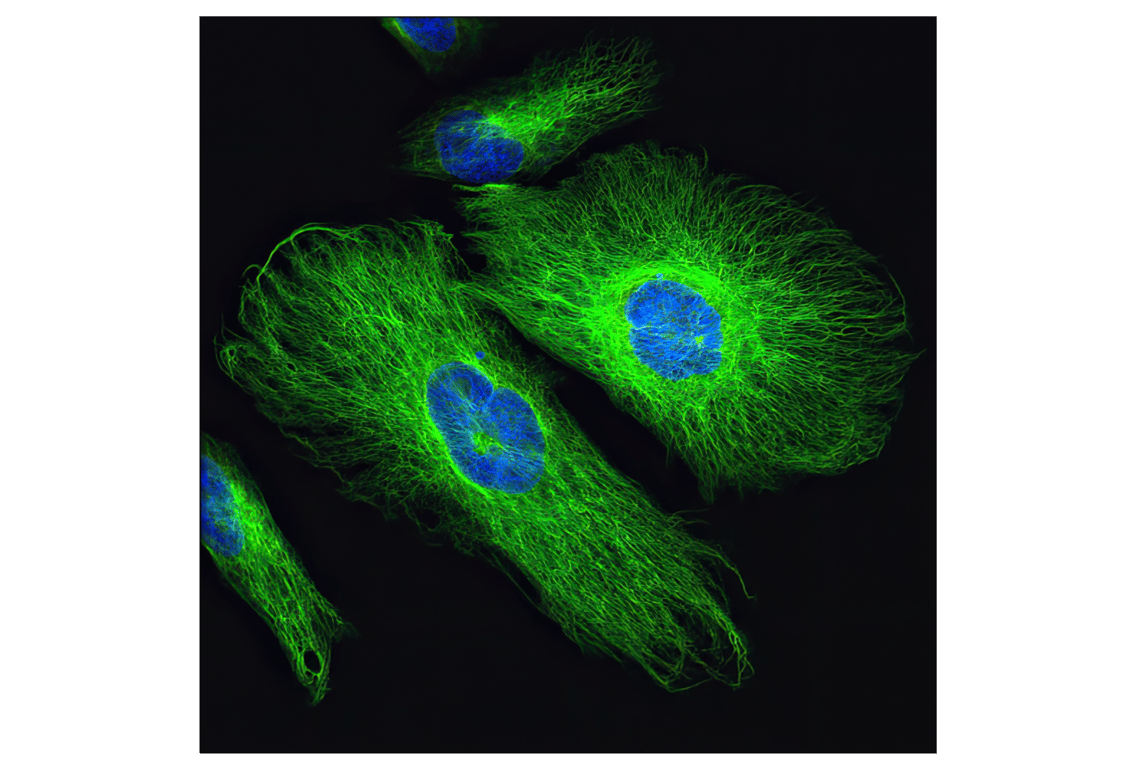
Confocal immunofluorescent analysis of SNB19 cells using Vimentin (D21H3) Rabbit mAb (green). Blue pseudocolor = DRAQ5® #4084 (fluorescent DNA dye).
Immunohistochemical analysis of paraffin-embedded rhesus kidney using Vimentin (D21H3) XP® Rabbit mAb.
Immunohistochemical analysis of paraffin-embedded human breast carcinoma using β-Catenin (D10A8) XP® Rabbit mAb.
Immunohistochemical analysis of paraffin-embedded mouse stomach using E-Cadherin (24E10) Rabbit mAb.
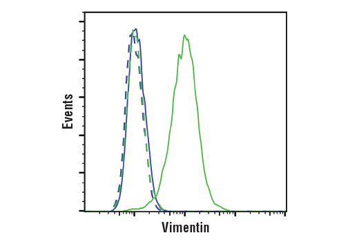
Flow cytometric analysis of MCF7 cells (blue, negative) and HeLa cells (green, positive) using Vimentin (D21H3) XP® Rabbit mAb(solid lines) or a concentration-matched Rabbit (DA1E) mAb IgG XP® Isotype Control #3900 (dashed lines). Anti-rabbit IgG (H+L), F(ab')2 Fragment (Alexa Fluor® 488 Conjugate) #4412 was used as a secondary antibody.
Immunohistochemical analysis of paraffin-embedded Syrian hamster small intestine using Vimentin (D21H3) XP® Rabbit mAb.
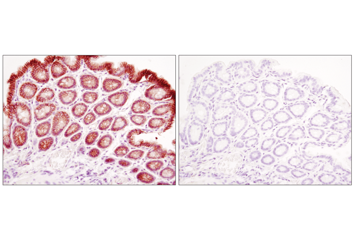
Immunohistochemical analysis of paraffin-embedded mouse colon using β-Catenin (D10A8) XP® Rabbit mAb in the presence of control peptide (left) or antigen-specific peptide (right).
Immunohistochemical analysis of paraffin-embedded human breast carcinoma, using E-Cadherin (24E10) Rabbit mAb in the presence of control peptide (left) or E-Cadherin Blocking Peptide #1056 (right).
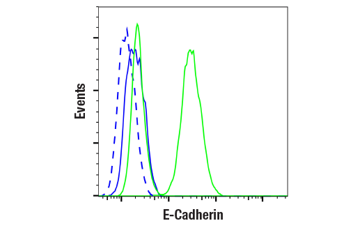
Flow cytometric analysis of Jurkat cells (blue, negative) and MCF7 cells (green, positive) using E-Cadherin (24E10) Rabbit mAb (solid lines) or a concentration-matched Rabbit (DA1E) mAb IgG XP® Isotype Control #3900 (dashed lines). Anti-rabbit IgG (H+L), F(ab')₂ Fragment (Alexa Fluor® 488 Conjugate) #4412 was used as a secondary antibody.
Immunohistochemical analysis of paraffin-embedded human tonsil using Vimentin (D21H3) XP® Rabbit mAb in the presence of control peptide (left) or antigen-specific peptide (right).
Immunohistochemical analysis of paraffin-embedded cell pellets, HeLa (left) or NCI-H28 (right), using β-Catenin (D10A8) XP® Rabbit mAb.
Confocal immunofluorescent analysis of mouse colon using β-Catenin (D10A8) XP® Rabbit mAb (green). Actin filaments were labeled with DY-554 phalloidin (red). Blue pseudocolor = DRAQ5® #4084 (fluorescent DNA dye).
Confocal immunofluorescent analysis of HeLa (left) and NCI-H28 (right) cells using β-Catenin (D10A8) XP® Rabbit mAb (green). Blue pseudocolor = DRAQ5® #4084 (fluorescent DNA dye).
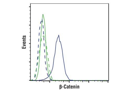
Flow cytometric analysis of NCI-H28 cells (green) and HeLa cells (blue) using β-Catenin (D10A8) XP® Rabbit mAb (solid lines) or concentration-matched Rabbit Isotype Control #3900 (dashed lines). Anti-rabbit IgG (H+L), F(ab')2 Fragment (Alexa Fluor® 488 Conjugate) #4412 was used as secondary antibody.
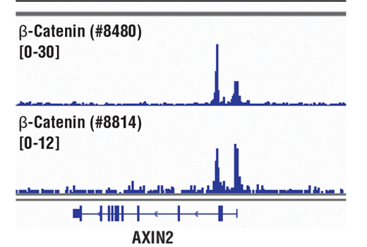
Chromatin immunoprecipitations were performed with cross-linked chromatin from HCT116 cells and either β-Catenin (D10A8) XP® Rabbit mAb or Non-phospho (Active) β-Catenin (Ser33/37/Thr41) (D13A1) Rabbit mAb #8814, using SimpleChIP® Enzymatic Chromatin IP Kit (Magnetic Beads) #9005. DNA Libraries were prepared using DNA Library Prep Kit for Illumina® (ChIP-seq, CUT&RUN) #56795. The figure shows binding across AXIN2, a known target gene of β-Catenin (see additional figure containing ChIP-qPCR data).
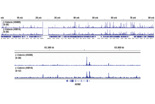
Chromatin immunoprecipitations were performed with cross-linked chromatin from HCT116 cells and either β-Catenin (D10A8) XP® Rabbit mAb or Non-phospho (Active) β-Catenin (Ser33/37/Thr41) (D13A1) Rabbit mAb #8814, using SimpleChIP® Enzymatic Chromatin IP Kit (Magnetic Beads) #9005. DNA Libraries were prepared using DNA Library Prep Kit for Illumina® (ChIP-seq, CUT&RUN) #56795. The figure shows binding across chromosome 17 (upper), including AXIN2 (lower), a known target gene of β-Catenin (see additional figure containing ChIP-qPCR data).
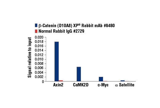
Chromatin immunoprecipitations were performed with cross-linked chromatin from HCT 116 cells and either β-Catenin (D10A8) XP® Rabbit mAb or Normal Rabbit IgG #2729 using SimpleChIP® Enzymatic Chromatin IP Kit (Magnetic Beads) #9003. The enriched DNA was quantified by real-time PCR using SimpleChIP® Human Axin2 Intron 1 Primers #8973, SimpleChIP® Human CaMK2D Intron 3 Primers #5111, human c-Myc promoter primers, and SimpleChIP® Human α Satellite Repeat Primers #4486. The amount of immunoprecipitated DNA in each sample is represented as signal relative to the total amount of input chromatin, which is equivalent to one.
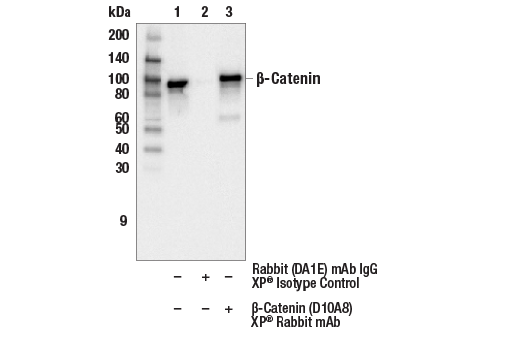
Immunoprecipitation of β-Catenin from HeLa cell extracts. Lane 1 is 10% input, lane 2 is precipitated with Rabbit (DA1E) mAb IgG XP® Isotype Control #3900, and lane 3 is β-Catenin (D10A8) XP® Rabbit mAb, #8480. Western blot was performed using β-Catenin (15B8) Mouse mAb, #37447.