


 全部商品分类


ERK1/ERK2 ANTIBODY

 下载产品说明书 下载COA 下载SDS
 用小程序,查商品更便捷


 收藏
收藏
 对比
对比 咨询
咨询种属反应
已发表种属
宿主/亚型
分类
类型
抗原
偶联物
形式
浓度
纯化类型
保存液
内含物
保存条件
运输条件
RRID
产品详细信息
It is not recommended to aliquot this antibody.
靶标信息
ERK1 and ERK2 are widely expressed and are involved in the regulation of meiosis, mitosis, and postmitotic functions in differentiated cells. Many different stimuli, including growth factors, cytokines, virus infection, ligands for heterotrimeric guanine nucleotide-binding protein (G protein)-coupled receptors and transforming agents, activate the ERK1 and ERK2 pathways. When growth factors bind to the receptor tyrosine kinase, Ras interacts with Raf, the serine/threonine protein kinase and activates it as well. Once actived, Raf phosphorylates serine residue in 2 further kinases, MEK1/2, which in turn phosphorylates tyrosine/threonine in extracellular-signal regulated kinase (ERK) 1/2. Upon activation, the ERKs either phosphorylate a number of cytoplasmic targets or migrate to the nucleus, where they phosphorylate and activate a number of transcription factors such as c-Fos and Elk-1.
仅用于科研。不用于诊断过程。未经明确授权不得转售。
生物信息学
蛋白别名: ERK-1; ERK-2; erk1 erk2; ERK1b; ERK2; ERT1; ERT2; Extracellular signal-regulated kinase 1; Extracellular signal-regulated kinase 2; extracellular signal-regulated kinase-1; extracellular signal-regulated kinase-2; extracellular signal-related kinase 1; extracellular-signal-regulated kinase 2; Insulin-stimulated MAP2 kinase; MAP kinase 1; MAP kinase 2; MAP kinase 3; MAP kinase isoform p42; MAP kinase isoform p44; MAPK 1; MAPK 2; MAPK 3; Microtubule-associated protein 2 kinase; mitogen activated protein kinase 1; mitogen-activated 3; Mitogen-activated protein kinase 1; Mitogen-activated protein kinase 2; Mitogen-activated protein kinase 3; MNK1; p42; p42 MAP Kinase; p42-MAPK; p44 MAP kinase; p44-ERK1; p44-MAPK; pp42/MAP kinase; protein kinase; protein tyrosine kinase ERK2
基因别名: 9030612K14Rik; AA407128; AU018647; C78273; ERK; ERK-1; ERK-2; ERK1; ERK2; ERT1; ERT2; Esrk1; fi06b09; HS44KDAP; HUMKER1A; I79_009500; I79_018350; Mapk; MAPK1; MAPK2; MAPK3; MNK1; Mtap2k; p38; p40; p41; p41mapk; p42-MAPK; P42MAPK; p44; p44-ERK1; p44-MAPK; P44ERK1; P44MAPK; PRKM1; PRKM2; PRKM3; wu:fi06b09; zERK1; zERK2
参考图片
Immunofluorescence analysis of ERK1/ERK2 was performed using 70% confluent log phase RSC96 cells. The cells were fixed with 4% paraformaldehyde for 10 minutes, permeabilized with 0.1% Triton™ X-100 for 15 minutes, and blocked with 1% BSA for 1 hour at room temperature. The cells were labeled with ERK1/ERK2 Rabbit Polyclonal Antibody (Product # 82380) at 1:100 dilution in 0.1% BSA, incubated at 4 degree Celsius overnight and then labeled with Goat anti-Rabbit IgG (Heavy Chain) Superclonal™ Secondary Antibody, Alexa Fluor® 488 conjugate (Product # A27034) at a dilution of 1:2000 for 45 minutes at room temperature (Panel a: green). Nuclei (Panel b: blue) were stained with SlowFade® Gold Antifade Mountant with DAPI (Product # S36938). F-actin (Panel c: red) was stained with Rhodamine Phalloidin (Product # R415, 1:300). Panel d represents the merged image showing cytoplasmic localization. Panel e represents control cells with no primary antibody to assess background. The images were captured at 60X magnification.
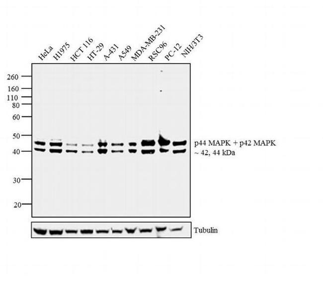
Western blot analysis was performed on whole cell extracts (30 µg lysate) of HeLa (Lane 1), H1975 (Lane 2), HCT 116 (Lane 3), HT-29 (Lane 4), A-431 (Lane 5), A549 (Lane 6), MDA-MB-231 (Lane 7), RSC96 (Lane 8), PC-12 (Lane 9) and NIH/3T3 (Lane 10). The blot was probed with Rabbit Anti-ERK1/ERK2 Polyclonal Antibody (Product # 82380, 1:1000 dilution) and detected by chemiluminescence using Goat anti-Rabbit IgG (Heavy Chain) Superclonal™ Secondary Antibody, HRP conjugate (Product # A27036, 0.25 µg/mL, 1:4000 dilution). Two bands at 42, 44 kDa corresponding to ERK1/ERK2 was observed across the cell lines tested. Known quantity of protein samples were electrophoresed using Novex® NuPAGE® 4-12 % Bis-Tris gel (Product # NP0321BOX), XCell SureLock™ Electrophoresis System (Product # EI0002) and Novex® Sharp Pre-Stained Protein Standard (Product # LC5800). Resolved proteins were then transferred onto a nitrocellulose membrane with iBlot® 2 Dry Blotting System (Product # IB21001). The membrane was probed with the relevant primary and secondary Antibody following blocking with 5 % skimmed milk. Chemiluminescent detection was performed using Pierce™ ECL Western Blotting Substrate (Product # 32106).
Knockdown of ERK1/ERK2 was achieved by transfecting MCF7 cells with ERK1/ERK2 specific validated siRNAs (Silencer® select Product # s11140, s11137 and s11138). Western blot analysis (Fig a) was performed using whole cell lysates from the ERK1 knockdown cells (lane 4), ERK1/ERK2 knock down cells (lane 3), non-specific scrambled siRNA transfected cells (lane 2) and untransfected cells (lane 1). The blots were probed with Anti-ERK1/ERK2 Rabbit Polyclonal Antibody (Product # 82380, 1:500 dilution) and Goat anti-Rabbit IgG (Heavy Chain) Superclonal™ Secondary Antibody, HRP conjugate (Product # A27036, 0.25 µg/mL, 1:4000 dilution). Densitometric analysis of this western blot is shown in histogram (Fig b). Loss of signal upon siRNA mediated knock down confirms that antibody is specific to ERK1/ERK2.
A549 cells were lysed 72 hours after transfection. Cells were transfected with Transfection Reagent alone (Lane 1), 100nM ON-TARGETplus siCONTROL Non-Targeting Pool (Lane 2), or 25nM ON-TARGETplus MAPK1 siRNA (Lane 3). Western blot data for Cyclophilin B is included as a control for equal protein loading.
FIGURE 6 The effect of FGF2 on the inhibition of apoptosis and ER stress and the activation of the Akt and ERK1/2 signaling pathway was detected in vitro . (A) The expression of pro-apoptotic proteins, the expression of ER stress-relevant proteins, and the phosphorylation of Akt and ERK1/2 were detected by Western blotting. Images are representative of five animals in each group. (B-I) The histograms show the normalized optical density analysis. FGF2 significantly decreased the expression of cleaved Caspase-3, Bax, CHOP, GRP78, ATF-6, and XBP-1 in NRK-52E treated with TBHP. The phosphorylation of Akt and ERK1/2 was increased in the TBHP-FGF2 group compared with the TBHP group. Representative data of five individual samples in each group. * P < 0.05, ** P < 0.01, *** P < 0.001; ns indicates not statistically significant.
FIGURE 5 The effect of FGF2 on the phosphorylation of Akt and ERK1/2. (A) The phosphorylation of Akt and ERK1/2 was determined by immunoblot analysis. GAPDH was used as a loading control. Images are representative of five animals in each group. (B,C) The histograms show the normalized optical density analysis. The phosphorylation of Akt and ERK1/2 in the I/R group was transiently increased at 6 h after reperfusion compared to the Sham group, whereas there was no significant difference between the I/R group and the Sham group at 24 and 72 h. FGF2 treatment strikingly increased the phosphorylation of Akt and ERK1/2 at 24 and 72 h after reperfusion compared to the I/R group. Representative data of five individual samples in each group. * P < 0.05, ** P < 0.01, *** P < 0.001; ns indicates not statistically significant.
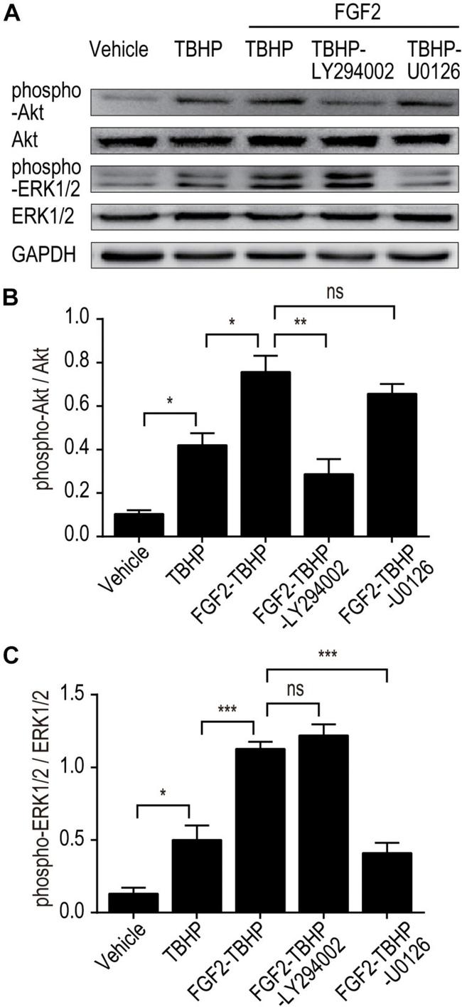
FIGURE 7 LY294002 and U0126 abolish the effect of FGF2 on the phosphorylation of Akt and ERK1/2 in vitro . (A) The phosphorylation of Akt and ERK1/2 was determined by Western blotting analysis. GAPDH was used as a loading control. Images are representative of five animals in each group. (B,C) The histograms show the normalized optical density analysis. LY294002 inhibited the effect of FGF2 treatment on the phosphorylation of Akt, whereas U0126 strikingly blocked the effect of FGF2 on the phosphorylation of ERK1/2. Representative data of five individual samples in each group. * P < 0.05, ** P < 0.01, *** P < 0.001; ns indicates not statistically significant.
Figure 5 The regulation effect of FGF10 on ER stress and ERK1/2 signaling pathway. (A) The expression levels of GRP-78, CHOP, XBP-1, ATF-4, ATF-6, PDI, ERK1/2, and phospho-ERK1/2 in kidney tissues of Sham group, I/R group and I/R-FGF10 group were determined by immunoblot analysis. FGF10 significantly increased the phosphorylation of ERK1/2. (B-I) The histograms show the normalized optical density analysis. Results are representative of five rats in each group. * P < 0.05, ** P < 0.01, *** P < 0.001, ns represents no significant difference.
Published figure using ERK1/ERK2 polyclonal antibody (Product # 82380) in Western Blot
Antibody specificity was demonstrated by siRNA mediated knockdown of the target protein. A549 cells were transfected with MAPK1 siRNA and decrease in signal intensity was observed in western blot application using Anti-ERK1/ERK2 Rabbit Polyclonal Antibody (Product # 82380). {KD}
Antibody specificity was demonstrated by siRNA mediated knockdown of target protein. MCF7 cells were transfected with ERK1 and ERK1/ERK2 siRNA and decrease in signal was observed in Western Blot using Anti-ERK1/ERK2 Polyclonal Antibody (Product # 82380). {KD}
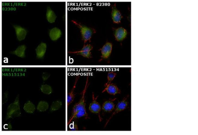
Antibody specificity was demonstrated by showing that antibodies raised against the same target protein perform similarly. Immunofluorescence of ERK1/ERK2 using ERK1/ERK2 polyclonal antibody (Product # 82380) along with another ERK1/ERK2 monoclonal antibody (Product # MA5-15134) shows similar expression of ERK1/ERK2 in RSC96 cells. {IAV}
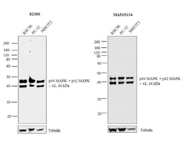
Antibody specificity was demonstrated by showing that antibodies raised against the same target protein perform similarly. Western blot of ERK1/ERK2 using ERK1/ERK2 Polyclonal Antibody (Product # 82380), tested in parallel with ERK1/ERK2 Monoclonal Antibody (Product # MA5-15134), shows similar expression of ERK1/ERK2 in the cell lines tested. {IAV}