



 全部商品分类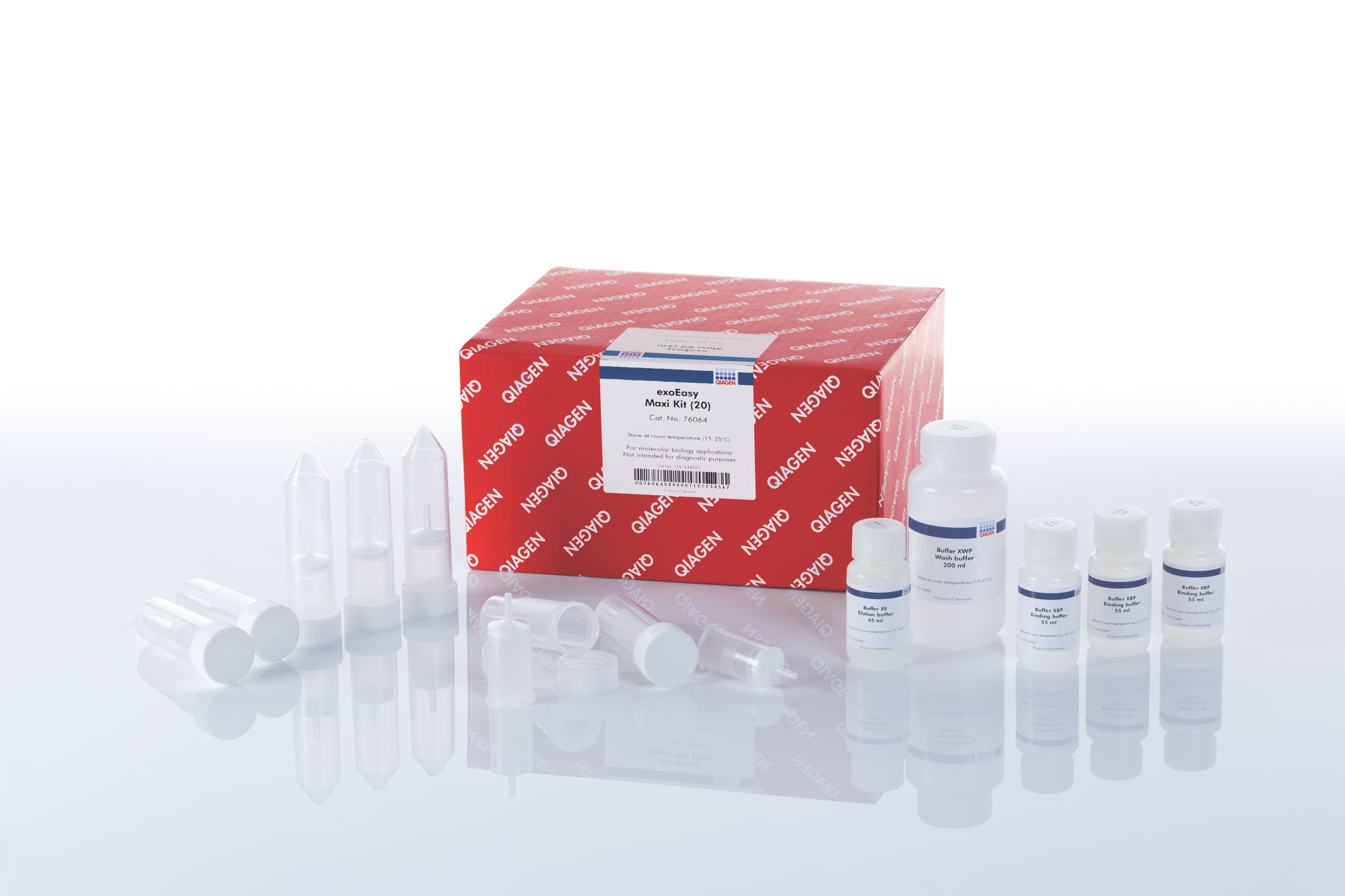


exoEasy Maxi Kit (20)


 用小程序,查商品更便捷



 收藏
收藏
 对比
对比 咨询
咨询
Features
- Isolate intact exosomes and other extracellular vesicles
- Suitable for functional testing and physical and biochemical analysis
- Purify exosomes and other extracellular vesicles in 25 minutes
- Process multiple samples in parallel using a simple spin-column procedure
- Use with up to 4 ml of plasma or serum or 32 ml of cell culture supernatant
- Suitable for MISEV-compliant protein characterization
Product Details
Performance
The exoEasy protocol for purification of exosomes and other extracellular vesicles is not only faster than ultracentrifugation, but also yields a cleaner preparation. Scanning electron microscopy shows that while both techniques deliver intact vesicles of the expected size, the preparation from ultracentrifugation also contains many smaller structures that do not match the expected size or shape for EVs (see figure “ Intact vesicles are eluted from the exoEasy membrane with higher purity compared with ultracentrifugation”).
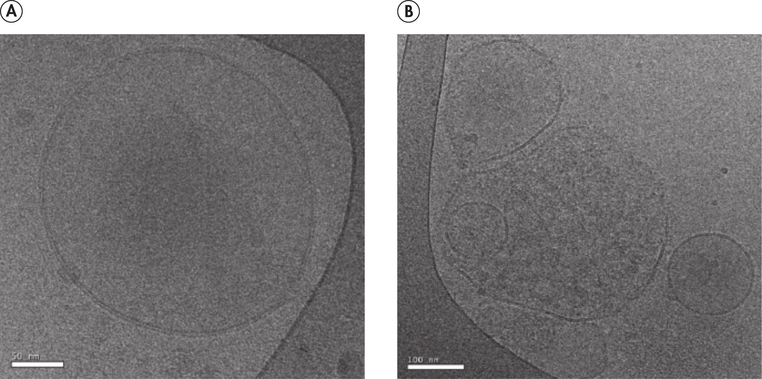
Fully intact, spherical vesicles of different sizes between approximately 50-300 nm diameter can also be observed using cryo-electron microscopy. (see figure " Visualization of EVs isolated using the exoEasy Kit by cryo-electron microscopy").
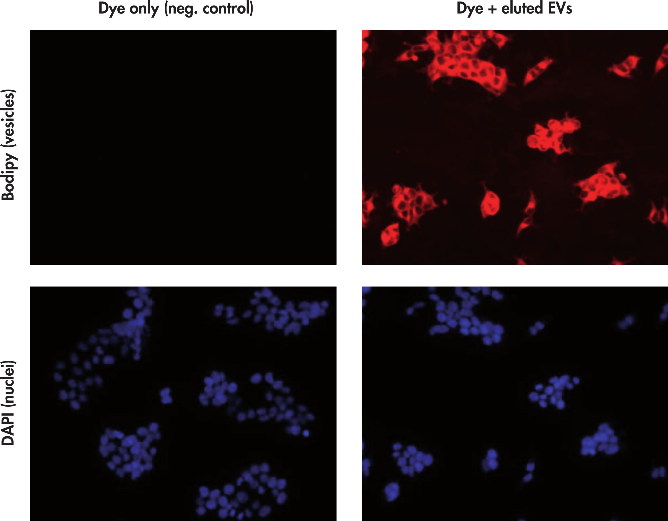
Isolated vesicles are fully functional and can be used for several downstream applications including cell culture work (see figure “ Extracellular vesicles isolated using the exoEasy Kit are taken up efficiently by target cells (customer data)”). Quality and quantity of eluted vesicles can be determined by a range of different methods. These include electron microscopy, physical characterization (e.g., Nanoparticle Tracking Analysis (NTA), or Tunable Resistive Pulse Sensing (TRPS)) and analysis of molecular constituents of vesicles, such as proteins, nucleic acids or lipids. Since particle counting techniques like NTA or TRPS do not distinguish vesicles from other particulate matter, particle counts performed in unprocessed biological fluids or crude preparations that still contain other particulate matter (e.g., protein complexes) may vastly overestimate the number of vesicles present.
To standardize protocols and reporting in the EV field, the International Society of Exosomes has developed the Minimal Information for Studies of Extracellular Vesicles (MISEV) guidelines. These guidelines provide examples of EV-enriched protein markers that can be used to characterize EVs. However, analyzing protein expression in intact EVs can be challenging due to the limited amount of material and low expression levels of many EV proteins.
In partnership with ProteinSimple (a Bio-Techne brand) QIAGEN has developed a standardized workflow for EV protein characterization. This new workflow combines a simple method for isolating EVs with automated protein separation and immunodetection of MISEV-recommended proteins.
Following isolation with the exoEasy Maxi Kit, protein expression of intact EVs can be characterized on Simple Western by ProteinSimple. Simple Western is a western blotting platform for automated protein separation and immunodetection. Signals from potentially contaminating proteins such as APOA1 are efficiently depleted during isolation. (See figure “ EV protein analysis across plasma fractions using Simple Western automated western blot”).
See figures
 Visualization of EVs isolated using the exoEasy kit by cryo-electron microscopy.
 Extracellular vesicles isolated using the exoEasy Kit are taken up efficiently by target cells (customer data).
 EV protein analysis across plasma fractions using Simple Western automated western blot (ProteinSimple, a Bio-Techne Brand).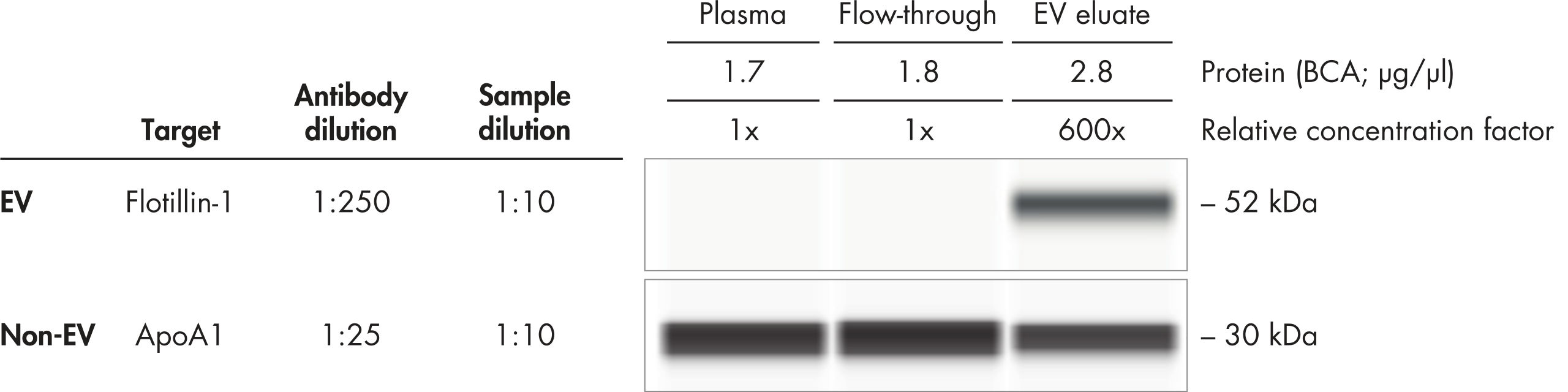
Principle
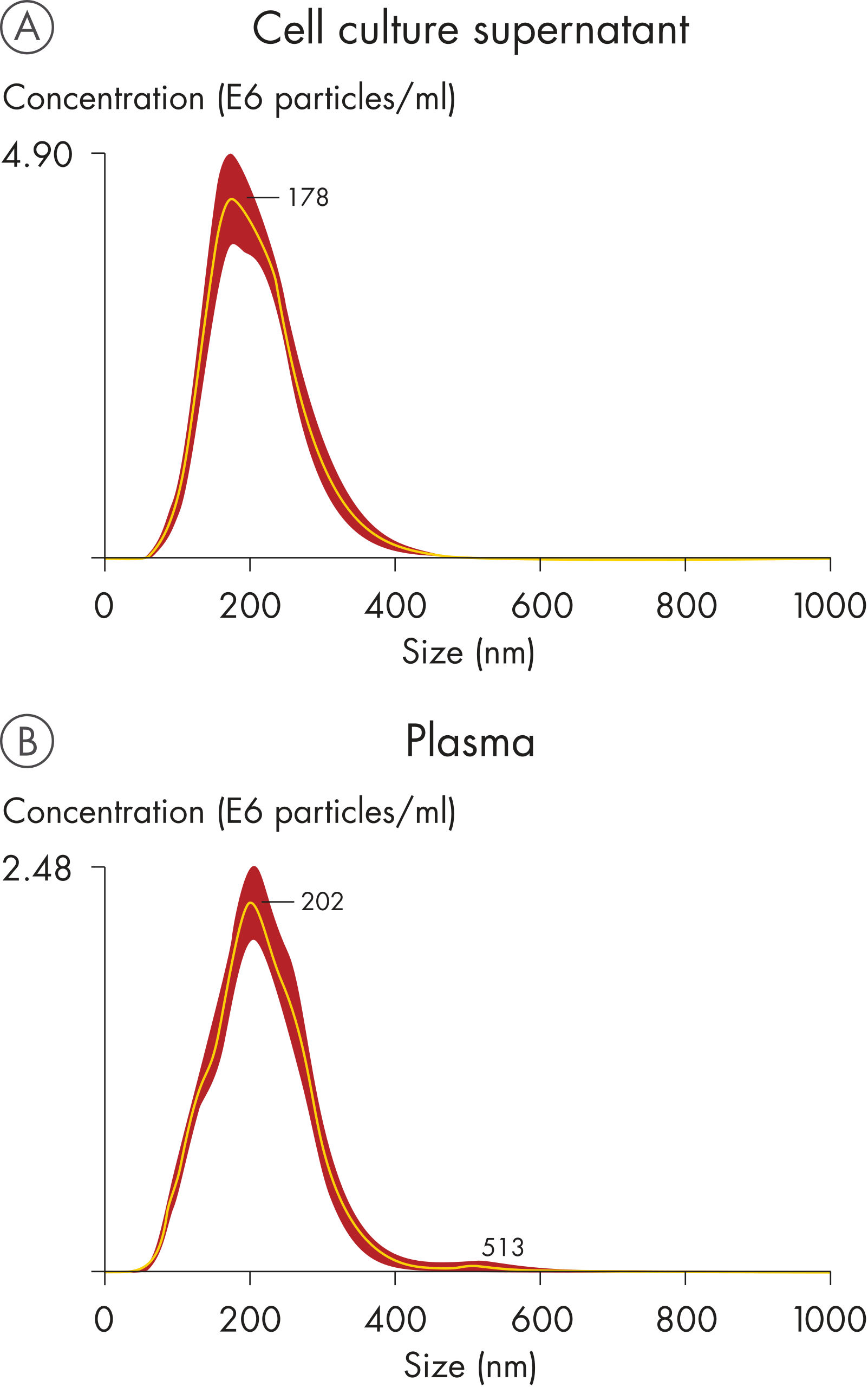
The exoEasy Maxi Kit uses a membrane-based affinity binding step to isolate exosomes and other EVs from serum and plasma or cell culture supernatant. The method does not distinguish EVs by size or cellular origin, and is not dependent on the presence of a particular epitope. Instead, it makes use of a generic, biochemical feature of vesicles to recover the entire spectrum of extracellular vesicles present in a sample (see figure “ Characterization of extracellular vesicles isolated using the exoEasy Maxi Kit by NTA”). It is therefore essential to completely remove cells, cell fragments, apoptotic bodies, etc., by centrifugation or filtration of samples before starting the protocol.
The technology developed with Exosome Diagnostics, Inc., uses a spin column format and specialized buffers to purify exosomes from pre-filtered biological fluids; up to 4 ml using plasma or serum. For cell culture supernatants, processing of up to 32 ml sample (4 column loading steps) has been successfully tested. However, the concentration of vesicles in supernatants depends strongly on the cell type and culture conditions; therefore, we recommend starting with no more than 16 ml of supernatant for sample material that has not been tested with the kit previously. Higher sample volumes may result in reduced recovery of vesicles. The procedure is very fast, consistent and highly suited for functional analysis of exosomes and other extracellular vesicles. Particulate matter other than vesicles, such as larger protein complexes that are especially abundant in plasma and serum, is largely removed during the binding and wash steps.
See figures
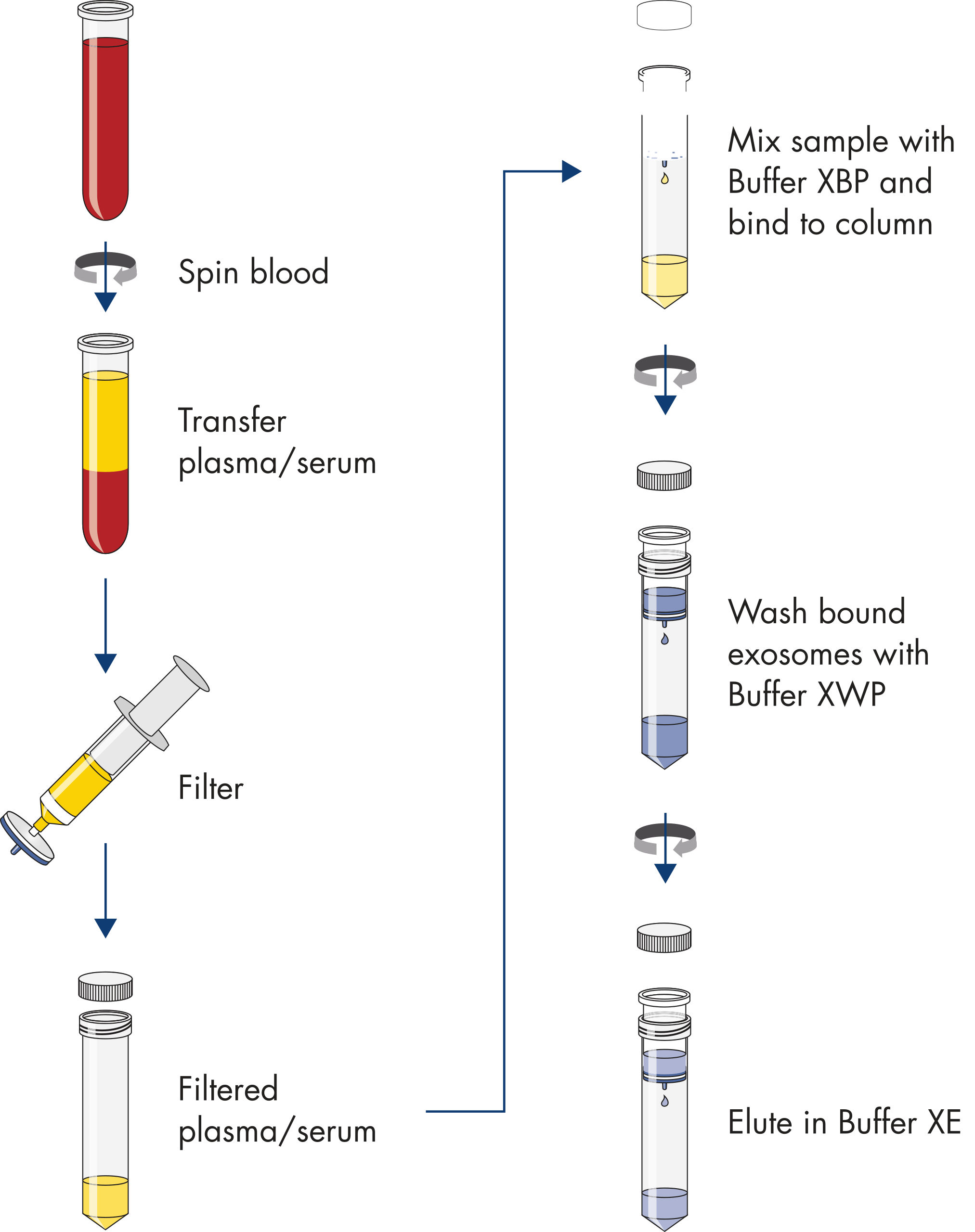
Procedure
Applications
- Physical characterization (e.g., NTA, TRPS)
- Electron microscopy
- Isolation and analysis of DNA, RNA*, proteins, lipids and other constituents
- Flow cytometry
- Antibody- or fluorescent dye-based labeling
- Uptake by recipient cells
Intact vesicles are eluted from the exoEasy membrane with higher purity compared with ultracentrifugation.
Scanning electron microscopy (SEM; 20,000x magnification) was performed on a solubilized pellet from ultracentrifugation of pre-filtered (0.8 µm) plasma compared to an exoEasy eluate. Both preparations contain vesicle-shaped structures with an expected size range from 50–200 nm (white arrows; scale bar 200 nm). The preparation from ultracentrifugation also includes many smaller, unidentified structures that do not match the expected shape and size of extracellular vesicles.