


 全部商品分类
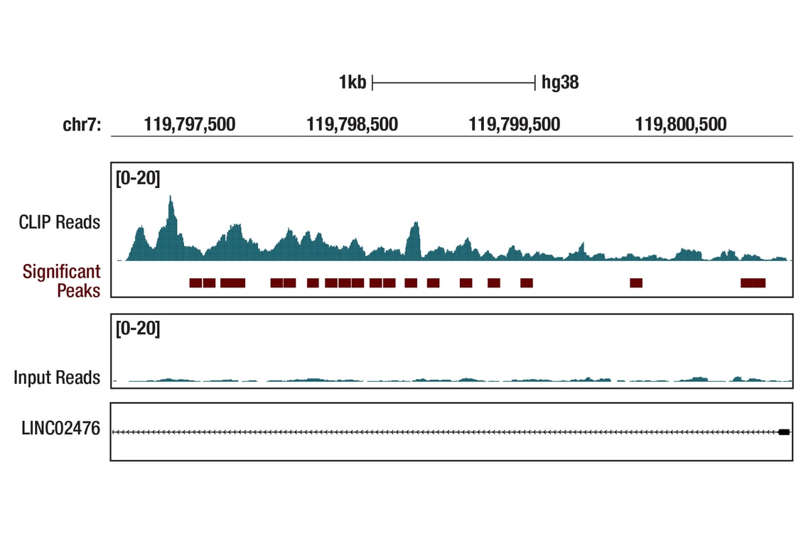


Ezh2 (D2C9) XP® Rabbit mAb

 下载产品说明书 下载COA 下载SDS
 用小程序,查商品更便捷


 收藏
收藏
 对比
对比 咨询
咨询
Monoclonal antibody is produced by immunizing animals with a synthetic peptide corresponding to residues surrounding Arg354 of human Ezh2 protein.
Product Usage Information
For optimal ChIP and ChIP-seq results, use 5 μl of antibody and 10 μg of chromatin (approximately 4 x 10⁶ cells) per IP. This antibody has been validated using SimpleChIP® Enzymatic Chromatin IP Kits.
The CUT&RUN dilution was determined using CUT&RUN Assay Kit #86652.
The CUT&Tag dilution was determined using CUT&Tag Assay Kit #77552.
| Application | Dilution |
|---|---|
| Western Blotting | 1:1000 |
| Simple Western™ | 1:10 - 1:50 |
| Immunoprecipitation | 1:300 |
| IHC Leica Bond | 1:50 - 1:200 |
| Immunohistochemistry (Paraffin) | 1:50 |
| Immunofluorescence (Frozen) | 1:100 - 1:400 |
| Immunofluorescence (Immunocytochemistry) | 1:100 - 1:400 |
| Flow Cytometry (Fixed/Permeabilized) | 1:100 - 1:400 |
| Chromatin IP | 1:100 |
| Chromatin IP-seq | 1:100 |
| CUT&RUN | 1:100 |
| CUT&Tag | 1:100 |
| eCLIP | 1:200 |
Specificity/Sensitivity
Species Reactivity:
Human, Mouse, Rat, Monkey
Supplied in 10 mM sodium HEPES (pH 7.5), 150 mM NaCl, 100 µg/ml BSA, 50% glycerol and less than 0.02% sodium azide. Store at –20°C. Do not aliquot the antibody.
For a carrier free (BSA and azide free) version of this product see product #16098.
参考图片
Enhanced cross-linking and immunoprecipitation (eCLIP) was performed with RNA from K-562 cells and Ezh2 (D2C9) XP® Rabbit mAb using a protocol based on the RBP-eCLIP Kit from EclipseBio. The figure shows binding across the LINC02476 transcript. Data is kindly provided by the laboratory of Dr. Gene Yeo and used with permission.
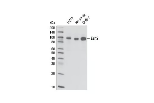
Western blot analysis of extracts from MCF7, Neuro-2a, and COS-7 cell lines using Ezh2 (D2C9) XP® Rabbit mAb.
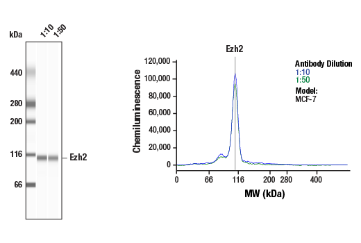
Simple Western™ analysis of lysates (0.1 mg/mL) from MCF-7 cells using Ezh2 (D2C9) XP® Rabbit mAb #5246. The virtual lane view (left) shows the target band (as indicated) at 1:10 and 1:50 dilutions of primary antibody. The corresponding electropherogram view (right) plots chemiluminescence by molecular weight along the capillary at 1:10 (blue line) and 1:50 (green line) dilutions of primary antibody. This experiment was performed under reducing conditions on the Jess™ Simple Western instrument from ProteinSimple, a BioTechne brand, using the 66 – 440 kDa separation module.
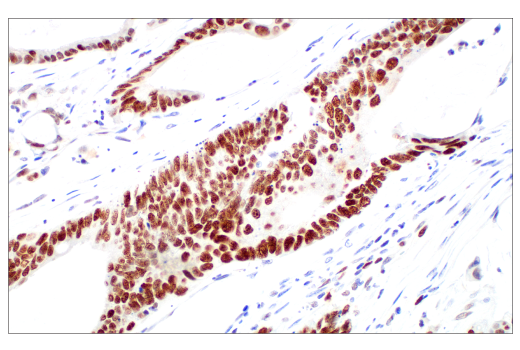
Immunohistochemical analysis of paraffin-embedded human colon adenocarcinoma using Ezh2 (D2C9) XP® Rabbit mAb performed on the Leica BOND Rx.
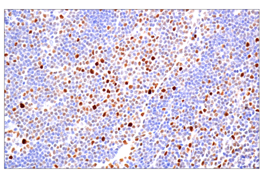
Immunohistochemical analysis of paraffin-embedded human B-cell non-Hodgkin lymphoma using Ezh2 (D2C9) XP® Rabbit mAb performed on the Leica BOND Rx.
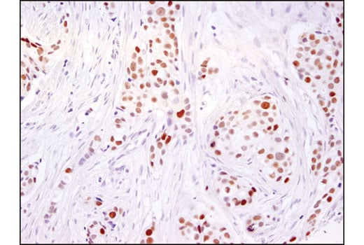
Immunohistochemical analysis of paraffin-embedded human breast carcinoma using Ezh2 (D2C9) XP® Rabbit mAb.
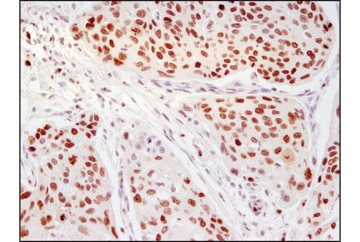
Immunohistochemical analysis of paraffin-embedded human cervical carcinoma using Ezh2 (D2C9) XP® Rabbit mAb.
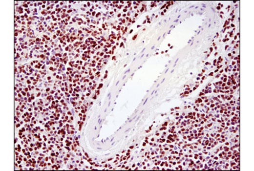
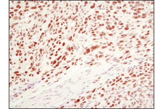
Immunohistochemical analysis of paraffin-embedded human lymphoma using Ezh2 (D2C9) XP® Rabbit mAb.
Immunohistochemical analysis of paraffin-embedded 4T1 mouse syngeneic tumor using Ezh2 (D2C9) XP® Rabbit mAb.
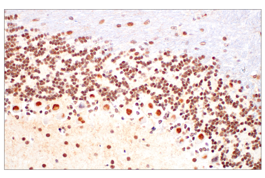
Immunohistochemical analysis of paraffin-embedded mouse brain using Ezh2 (D2C9) XP® Rabbit mAb.
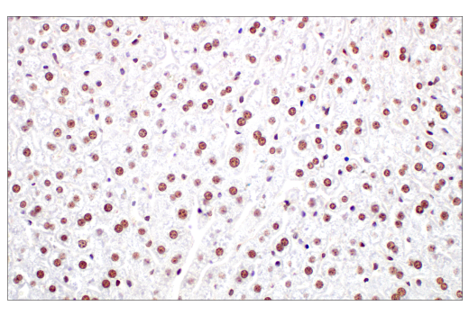
Immunohistochemical analysis of paraffin-embedded mouse liver using Ezh2 (D2C9) XP® Rabbit mAb.
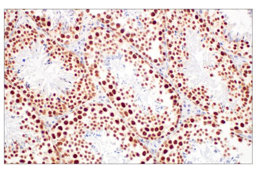
Immunohistochemical analysis of paraffin-embedded mouse testis using Ezh2 (D2C9) XP® Rabbit mAb.
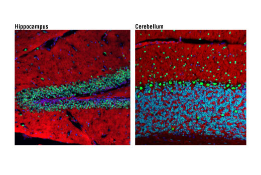
Confocal immunofluorescent analysis of mouse hippocampus (left) and cerebellum (right) using Ezh2 (D2C9) XP® Rabbit mAb (green). Actin filaments were labeled with DyLight™ 554 Phalloidin #13054 (red). Blue pseudocolor = DRAQ5® #4084 (fluorescent DNA dye).
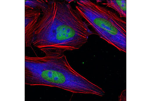
Confocal immunofluorescent analysis of HeLa cells using Ezh2 (D2C9) XP® Rabbit mAb (green) and S6 Ribosomal Protein (54D2) Mouse mAb #2317 (blue). Actin filaments were labeled with DY-554 phalloidin (red).
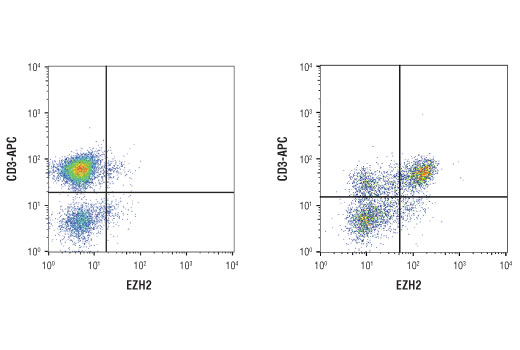
Flow cytometric analysis of human peripheral blood mononuclear cells untreated (left) and treated (right) with anti-human CD3 (10ug/ml, coated plates) and anti-human CD28 (5ug/ml) for 3 days at 37ºC using EZH2 (D2C9) XP® Rabbit mAb and co-stained with an anti-human CD3 antibody. Anti-rabbit IgG (H+L), F(ab')2 Fragment (Alexa Fluor 488 Conjugate) #4412 was used as a secondary antibody.
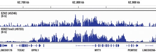
Chromatin immunoprecipitations were performed with cross-linked chromatin from Hela cells and either Ezh2 (D2C9) XP® Rabbit mAb or Tri-Methyl-Histone H3 (Lys27) (C36B11) Rabbit mAb, using SimpleChIP® Enzymatic Chromatin IP Kit (Magnetic Beads) #9003. DNA Libraries were prepared using DNA Library Prep Kit for Illumina® (ChIP-seq, CUT&RUN) #56795. EZH2 and H3K27me3 are known to associate with each other on chromatin. The figure shows binding of both EZH2 and H3K27me3 across the MYT1 gene. For additional ChIP-seq tracks, please download the product datasheet.
Chromatin immunoprecipitations were performed with cross-linked chromatin from Hela cells and either Ezh2 (D2C9) XP® Rabbit mAb or Tri-Methyl-Histone H3 (Lys27) (C36B11) Rabbit mAb, using SimpleChIP® Enzymatic Chromatin IP Kit (Magnetic Beads) #9003. DNA Libraries were prepared using DNA Library Prep Kit for Illumina® (ChIP-seq, CUT&RUN) #56795. EZH2 and H3K27me3 are known to associate with each other on chromatin. The figure shows binding of both EZH2 and H3K27me3 across chromosome 20 (upper), including MYT1 gene (lower).
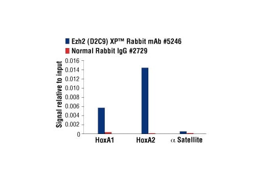
Chromatin immunoprecipitations were performed with cross-linked chromatin from NCCIT cells and either Ezh2 (D2C9) XP® Rabbit mAb or Normal Rabbit IgG #2729 using SimpleChIP® Enzymatic Chromatin IP Kit (Magnetic Beads) #9003. The enriched DNA was quantified by real-time PCR using SimpleChIP® Human HoxA1 Intron 1 Primers #7707, SimpleChIP® Human HoxA2 Promoter Primers #5517, and SimpleChIP® Human α Satellite Repeat Primers #4486. The amount of immunoprecipitated DNA in each sample is represented as signal relative to the total amount of input chromatin, which is equivalent to one.
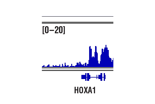
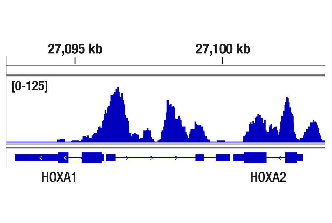
CUT&RUN was performed with NCCIT cells and Ezh2 (D2C9) XP® Rabbit mAb, using CUT&RUN Assay Kit #86652. DNA Libraries were prepared using DNA Library Prep Kit for Illumina® (ChIP-seq, CUT&RUN) #56795. The figures show binding across HoxA1, a known target gene of Ezh2 (see additional figure containing CUT&RUN-qPCR data).
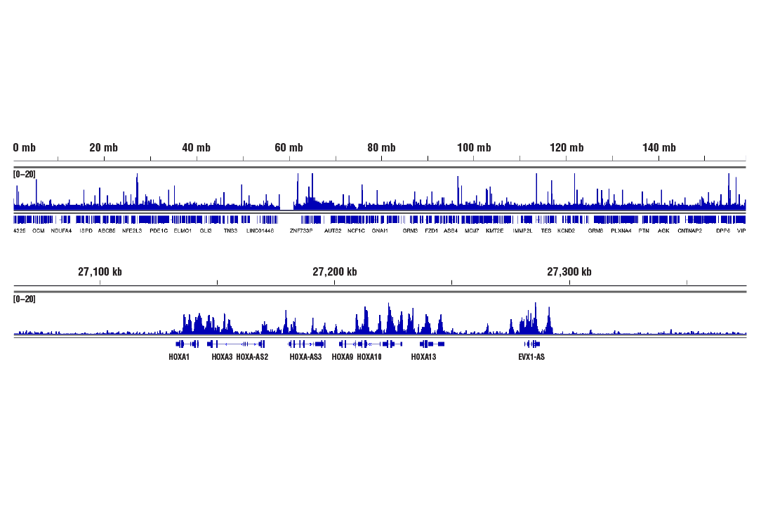
CUT&RUN was performed with NCCIT cells and Ezh2 (D2C9) XP® Rabbit mAb, using CUT&RUN Assay Kit #86652. DNA Libraries were prepared using DNA Library Prep Kit for Illumina® (ChIP-seq, CUT&RUN) #56795. The figures show binding across chromosome 7 (upper), including HoxA genes (lower), a known cluster of EZH2 target genes (see additional figure containing CUT&RUN-qPCR data).
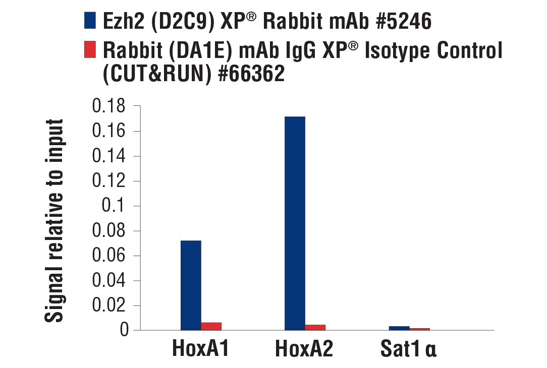
CUT&RUN was performed with NCCIT cells and either Ezh2 (D2C9) XP® Rabbit mAb or Rabbit (DA1E) mAb IgG XP® Isotype Control (CUT&RUN) #66362, using CUT&RUN Assay Kit #86652. The enriched DNA was quantified by real-time PCR using SimpleChIP® Human HoxA1 Intron 1 Primers #7707, SimpleChIP® Human HoxA2 Promoter Primers #5517, and SimpleChIP® Human α Satellite Repeat Primers #4486. The amount of immunoprecipitated DNA in each sample is represented as signal relative to the total amount of input chromatin, which is equivalent to one.
CUT&Tag was performed with NCCIT cells and Ezh2 (D2C9) XP® Rabbit mAb, using CUT&Tag Assay Kit #77552. DNA library was prepared using CUT&Tag Dual Index Primers and PCR Master Mix for Illumina Systems #47415. The figure shows binding across HOXA1, a known target gene of Ezh2 (see our ChIP-qPCR figure).
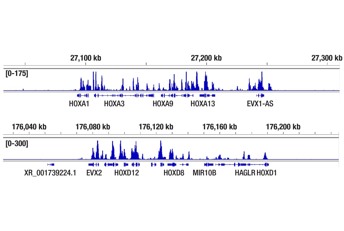
CUT&Tag was performed with NCCIT cells and Ezh2 (D2C9) XP® Rabbit mAb, using CUT&Tag Assay Kit #77552. DNA library was prepared using CUT&Tag Dual Index Primers and PCR Master Mix for Illumina Systems #47415. The figures show binding across the HOXA gene cluster (upper), and the HOXD gene cluster (lower), known target genes of Ezh2 (see our ChIP-qPCR figure).