The T45-2342 monoclonal antibody recognizes the mouse F4/80 antigen which is also known as EGF-like module-containing mucin-like hormone receptor-like 1 (EMR1). F4/80 is a 160 kDa glycoprotein that belongs to the EGF-TM7 family of seven-transmembrane spanning cell surface molecules. It is expressed on the surface of granulocytes and a wide range of mature tissue macrophages including, Kupffer cells, splenic red pulp macrophages, microglia, gut lamina propria macrophages, and Langerhans cells. F4/80 expression has also been reported on subpopulations of dendritic cells. F4/80 expression is heterogeneous and may be increased during inflammatory responses as observed in various mouse models of colitis, diabetes and brain injury.
商品描述
T45-2342
The T45-2342 monoclonal antibody recognizes the mouse F4/80 antigen which is also known as EGF-like module-containing mucin-like hormone receptor-like 1 (EMR1). F4/80 is a 160 kDa glycoprotein that belongs to the EGF-TM7 family of seven-transmembrane spanning cell surface molecules. It is expressed on the surface of granulocytes and a wide range of mature tissue macrophages including, Kupffer cells, splenic red pulp macrophages, microglia, gut lamina propria macrophages, and Langerhans cells. F4/80 expression has also been reported on subpopulations of dendritic cells. F4/80 expression is heterogeneous and may be increased during inflammatory responses as observed in various mouse models of colitis, diabetes and brain injury.
同种型
Rat WI, also known as Wistar (outbred) IgG2a, κ
克隆号
克隆 T45-2342 (RUO)
浓度
0.2 mg/ml
产品详情
PE
R-Phycoerythrin (PE), is part of the BD family of Phycobiliprotein dyes. This fluorochrome is a multimeric fluorescent phycobiliprotein with excitation maximum (Ex Max) of 496 nm and 566 nm and an emission maximum (Em Max) at 576 nm. PE is designed to be excited by the Blue (488 nm), Green (532 nm) and Yellow-Green (561 nm) lasers and detected using an optical filter centered near 575 nm (e.g., a 575/26-nm bandpass filter). As PE is excited by multiple lasers, this can result in cross-laser excitation and fluorescence spillover on instruments with various combinations of Blue, Green, and Yellow-Green lasers. Please ensure that your instrument’s configurations (lasers and optical filters) are appropriate for this dye.
研发参考(5)
1. Austyn JM., and Gordon S. F4/80, a monoclonal antibody directed specifically against the mouse macrophage. Eur J Immunol. 1981; 10:805-815. (Biology).
2. Gordon S, Hamann J, Lin HH, Stacey M. F4/80 and the related adhesion-GPCRs. Eur J Immunol. 2011; 41(9):2472-2476. (Biology).
3. Krüger T, Benke D, Eitner F, et al. Identification and functional characterization of dendritic cells in the healthy murine kidney and in experimental glomerulonephritis. J Am Soc Nephrol. 2004; 15(3):613-621. (Biology).
4. Leenen PJ, Radosević K, Voerman JS, et al. Heterogeneity of mouse spleen dendritic cells: in vivo phagocytic activity, expression of macrophage markers, and subpopulation turnover.. J Immunol. 1998; 160(5):2166-73. (Biology).
5. McKnight AJ, Macfarlane AJ, Dri P, Turley L, Willis AC, Gordon S. Molecular cloning of F4/80, a murine macrophage-restricted cell surface glycoprotein with Homology to the G-protein-linked transmembrane & hormone receptor family. J Biol Chem. 1996; 271:486. (Biology).
数据库链接
Entrez-Gene ID
13733
参考图片
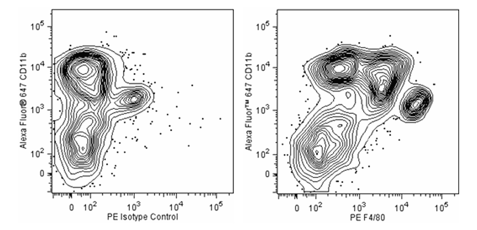
Two-color flow cytometric analysis of F4/80 expression on mouse splenic leucocytes. C57BL/6 mouse splenic leucocytes were preincubated with Purified Rat Anti-Mouse CD16/CD32 antibody (Mouse BD Fc Block™) (Cat. No. 553141/553142). The cells were then stained with Alexa Fluor® 647 Rat Anti-Mouse CD11b (Cat. No. 557686) and either PE Rat IgG2a Isotype Control (Cat. No. 553930; Left Panel) or PE Rat Anti-Mouse F4/80 antibody (Cat. No. 565410; Right Panel). The two-color flow cytometric contour plot showing the correlated expression of F4/80 (or Ig Isotype control staining) versus CD11b was derived from gated events with the forward and side light-scatter characteristics of viable monocytes. Flow cytometric analysis was performed using a BD LSRFortessa™ Cell Analyzer System.
Two-color flow cytometric analysis of F4/80 expression on mouse splenic leucocytes. C57BL/6 mouse splenic leucocytes were preincubated with Purified Rat Anti-Mouse CD16/CD32 antibody (Mouse BD Fc Block™) (Cat. No. 553141/553142). The cells were then stained with Alexa Fluor® 647 Rat Anti-Mouse CD11b (Cat. No. 557686) and either PE Rat IgG2a Isotype Control (Cat. No. 553930; Left Panel) or PE Rat Anti-Mouse F4/80 antibody (Cat. No. 565410; Right Panel). The two-color flow cytometric contour plot showing the correlated expression of F4/80 (or Ig Isotype control staining) versus CD11b was derived from gated events with the forward and side light-scatter characteristics of viable monocytes. Flow cytometric analysis was performed using a BD LSRFortessa™ Cell Analyzer System.


 全部商品分类


 下载产品说明书
 用小程序,查商品更便捷


 收藏
收藏
 对比
对比 咨询
咨询
