The T45-2342 monoclonal antibody recognizes the mouse F4/80 antigen which is also known as EGF-like module-containing mucin-like hormone receptor-like 1 (EMR1). F4/80 is a 160 kDa glycoprotein that belongs to the EGF-TM7 family of seven-transmembrane spanning cell surface molecules. It is expressed on the surface of granulocytes and a wide range of mature tissue macrophages including, Kupffer cells, splenic red pulp macrophages, microglia, gut lamina propria macrophages, and Langerhans cells. F4/80 expression has also been reported on subpopulations of dendritic cells. F4/80 expression is heterogeneous and may be increased during inflammatory responses as observed in various mouse models of colitis, diabetes and brain injury.
商品描述
T45-2342
The T45-2342 monoclonal antibody recognizes the mouse F4/80 antigen which is also known as EGF-like module-containing mucin-like hormone receptor-like 1 (EMR1). F4/80 is a 160 kDa glycoprotein that belongs to the EGF-TM7 family of seven-transmembrane spanning cell surface molecules. It is expressed on the surface of granulocytes and a wide range of mature tissue macrophages including, Kupffer cells, splenic red pulp macrophages, microglia, gut lamina propria macrophages, and Langerhans cells. F4/80 expression has also been reported on subpopulations of dendritic cells. F4/80 expression is heterogeneous and may be increased during inflammatory responses as observed in various mouse models of colitis, diabetes and brain injury.
同种型
Rat WI, also known as Wistar (outbred) IgG2a, κ
克隆号
克隆 T45-2342 (RUO)
浓度
0.2 mg/ml
产品详情
PerCP-Cy5.5
PerCP-Cy5.5 dye is part of the BD blue family of dyes. This tandem fluorochrome is comprised of a fluorescent protein complex (PerCP) with an excitation maximum (Ex Max) of 482 nm and an acceptor dye with an emission maximum (Em Max) at 676 nm. PerCP-Cy5 is designed to be excited by the blue laser (488-nm) and detected using an optical filter centered near 680 nm (e.g., a 695/40 nm bandpass filter). The donor dye can be partially excited by the Violet (405-nm) laser resulting in cross-laser excitation and fluorescence spillover. Please ensure that your instrument’s configurations (lasers and optical filters) are appropriate for this dye.
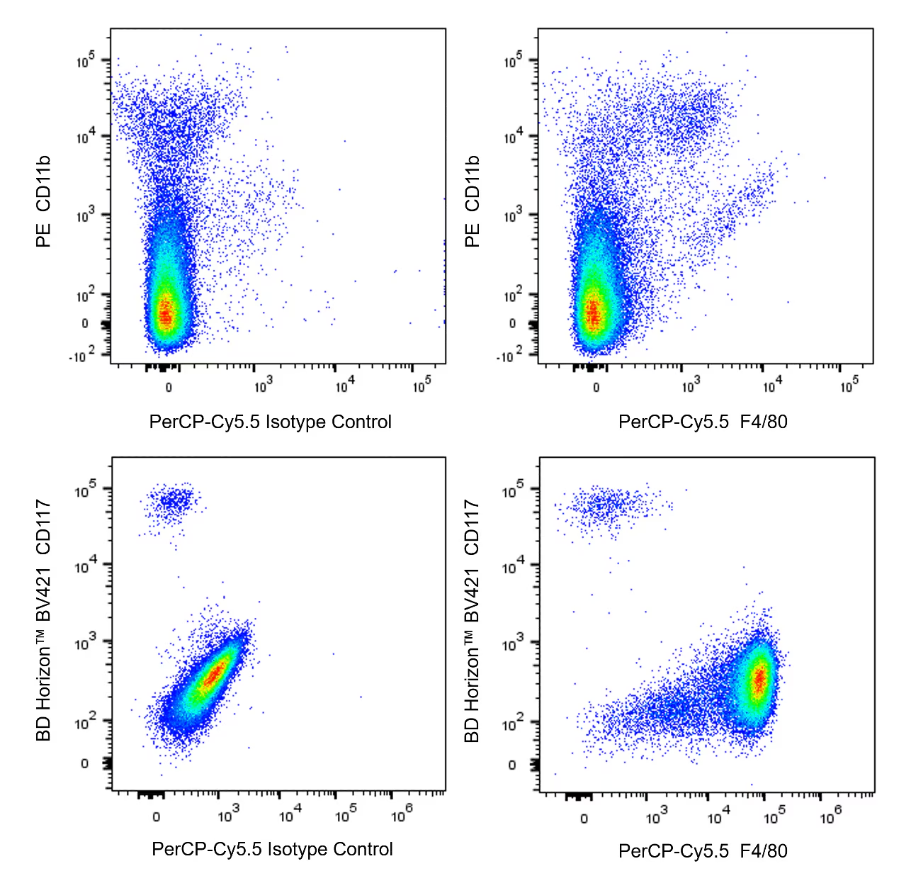
Multicolor flow cytometric analyses of F4/80 expression. The bivariate pseudocolor density plots showing the correlated expression of F4/80 (or Ig Isotype control staining) versus CD11b (Mouse Splenocytes; Top Plots) or CD117 (Mouse Peritoneal Exudate Cells; Bottom Plots) were derived from gated events with the forward and side light-scatter characteristics of viable (DAPI-negative for splenocytes or DRAQ7™-negative for PEC) monocytes. Flow cytometry and data analysis were performed using a BD LSRFortessa™ Cell Analyzer System and FlowJo™ software. Data shown on this Technical Data Sheet are not lot specific. Top Plots - Mouse splenocytes. C57BL/6 mouse splenic leucocytes were preincubated with Purified Rat Anti-Mouse CD16/CD32 antibody (Mouse BD Fc Block™) (Cat. No. 553141/553142) and stained with PE Rat Anti-CD11b antibody (Cat. No. 553311/557397/561689) and with either PerCP-Cy5.5 Rat IgG2a Isotype Control (Cat. No. 550765; Left Plot) or PerCP-Cy5.5 Rat Anti-Mouse F4/80 antibody (Cat. No. 567202; Right Plot) at 0.5 µg/test. DAPI Solution (Cat. No. 564907) was added to cells right before analysis. Bottom Plots - Mouse peritoneal exudate cells (PEC). C57BL/6 mouse PEC were similarly pretreated with Mouse BD Fc Block™, stained with BD Horizon™ BV421 Rat Anti-Mouse CD117 antibody (Cat. No. 566290/562609) and either PerCP-Cy5.5 Rat IgG2a Isotype Control (Left Plot) or PerCP-Cy5.5 Rat Anti-Mouse F4/80 antibody (Right Plot) at 0.5 µg/test. A solution containing DRAQ7™ (Cat. No. 564904) was added to cells right before analysis.


 全部商品分类


 下载产品说明书
 用小程序,查商品更便捷


 收藏
收藏
 对比
对比 咨询
咨询
