


 全部商品分类


 下载产品说明书 下载COA 下载SDS
 用小程序,查商品更便捷


 收藏
收藏
 对比
对比 咨询
咨询种属反应
已发表种属
宿主/亚型
Expression System
分类
类型
克隆号
抗原
偶联物
形式
浓度
纯化类型
保存液
内含物
保存条件
运输条件
RRID
产品详细信息
This antibody is predicted to react with mouse based on sequence homology.
Intact IgG appears on a non-reducing gel as ~150 kDa band and upon reduction generating a ~25 kDa light chain band and a ~50 kDa heavy chain.
Recombinant rabbit monoclonal antibodies are produced using in vitro expression systems. The expression systems are developed by cloning in the specific antibody DNA sequences from immunoreactive rabbits. Then, individual clones are screened to select the best candidates for production. The advantages of using recombinant rabbit monoclonal antibodies include: better specificity and sensitivity, lot-to-lot consistency, animal origin-free formulations, and broader immunoreactivity to diverse targets due to larger rabbit immune repertoire.
靶标信息
Focal Adhesion Kinase (FAK) is a 125 kDa non-receptor protein tyrosine kinase that acts as a substrate for Src and is a key element of integrin signaling. FAK plays an important role in cell spreading, differentiation, migration, cell death, and acceleration of the G1 to S phase transition of the cell cycle. FAK has a central catalytic domain and a C-terminal tail that localizes it to focal adhesions, which are sites where cells attach to the extracellular matrix via surface integrin receptors. Increased FAK tyrosine phosphorylation occurs upon integrin engagement with fibronectin. Adhesion of murine NIH3T3 fibroblasts to fibronectin promotes association of the Grb2 adapter protein and c-Src PTK with FAK in vivo, and also results in activation of the ERK2 MAP kinase. In v-Src-transformed NIH3T3, the association of v-Src, Grb2, and Sos with FAK is independent of cell adhesion to fibronectin. In vitro the Grb2 SH2 domain binds directly to tyrosine-phosphorylated FAK, and the binding site has been identified as Tyr925 by site directed mutagenesis. Several transcript variants encoding different isoforms have been found for the FAK gene, but the full-length natures of only three of them have been determined.
仅用于科研。不用于诊断过程。未经明确授权不得转售。
生物信息学
蛋白别名: EC 2.7.10.2; FADK 1; FAK-related non-kinase polypeptide; Focal adhesion kinase 1; focal adhesion kinase isoform FAK-Del33; Focal adhesion kinase-related nonkinase; FRNK; p125FAK; pp125 PTK2; pp125FAK; PPP1R71; Protein phosphatase 1 regulatory subunit 71; protein phosphatase 1, regulatory subunit 71; Protein-tyrosine kinase 2; PTK2 protein tyrosine kinase 2
基因别名: FADK; FAK; FAK1; FRNK; p125FAK; pp125FAK; PPP1R71; PTK2
UniProt ID:(Human) Q05397
Entrez Gene ID:(Human) 5747
参考图片
Immunofluorescent analysis of FAK1 was done on 70% confluent log phase A549 cells. The cells were fixed with 4% paraformaldehyde for 15 minutes; permeabilized with 0.25% Triton X-100 for 10 minutes followed by blocking with 5% BSA for 1 hour at room temperature. The cells were incubated with FAK1 Recombinant Rabbit Monoclonal Antibody (Product # 701094) at 2 µg-4 µg in 1% BSA and incubated for 3 hours at room temperature and then labeled with Alexa Fluor® 488 Goat anti-Rabbit IgG Secondary Antibody (Product # A-11008) at a dilution of 1:400 for 30 minutes at room temperature (Panel a: green). Nuclei (Panel b: blue) were stained with SlowFade® Gold Antifade Mountant with DAPI (Product # S36938). F-actin (Panel c: red) was stained with Alexa Fluor® 594 Phalloidin (Product # A12381). Panel d is a merged image showing membrane and cytoplasmic localization of FAK1. Panel e shows no primary antibody control. The images were captured at 20X magnification.
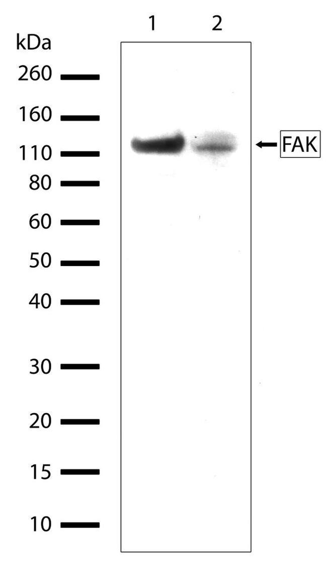
Western blot analysis of FAK in whole cell extracts of Jurkat (lane 1) and K562 (lane 2) cell lines using a FAK recombinant rabbit monoclonal antibody (Product # 701094) at a dilution of 1 µg/mL.
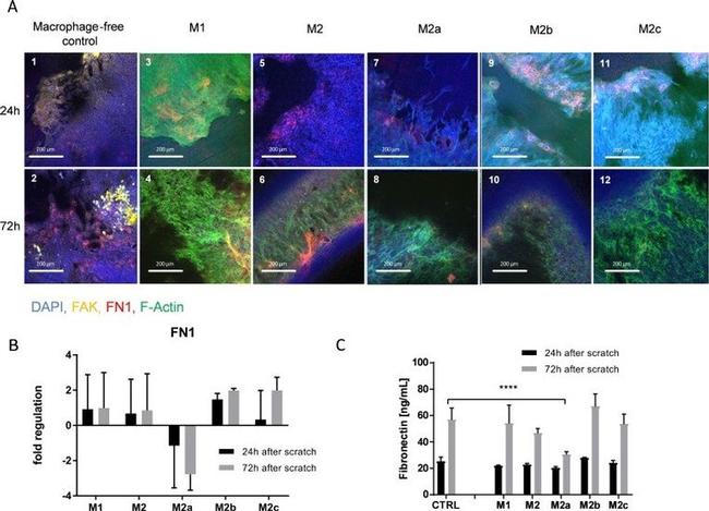
Fig 3 IHC of scratched cells and FN1 regulation/expression. A scratched epithelial cells incubated with different macrophage subtypes after 24h and 72h; DAPI stained in blue, FAK stained in yellow, FN1 stained in red, F-Actin stained in green. B FN1 mRNA regulation in the co-culture cell lysates measured after 24h and 72h Data are displayed as fold regulation compared to control medium containing macrophage maturation factors; Data is expressed as mean +- SD; n = 3 C FN1 expression measured in the supernatants via ELISA after 24h and 72h; Data is expressed as mean +- SD; n = 3 (**** = p>0.0001)
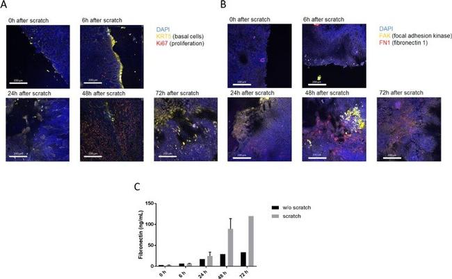
Fig 2 IHC of scratched cells and fibronectin expression. A IHC microscopic images of scratched cells at t = 0h, 6h, 24h, 48h and 72h; DAPI in blue, KRT5 in yellow, Ki67 in red. B IHC microscopic images of scratched cells at t = 0h, 6h, 24h, 48h and 72h; DAPI in blue, FAK in yellow, FN1 in red. C ELISA of FN1 in the supernatant after 0h, 6h, 24h, 48h and 72h with and without scratch.