


 全部商品分类


BD Pharmingen™ Purified Mouse Anti-Rat CD32

 下载产品说明书 下载SDS
 用小程序,查商品更便捷


 收藏
收藏
 对比
对比 咨询
咨询
参考图片
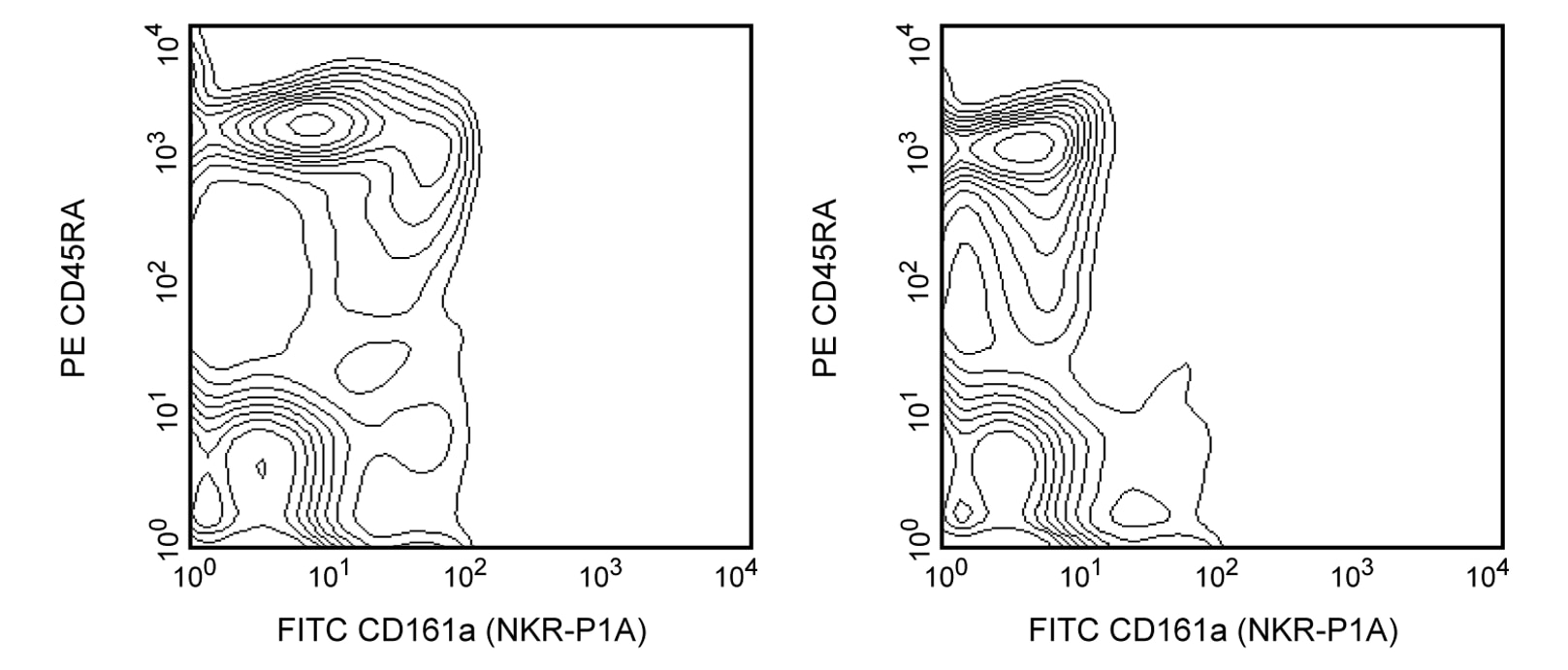
Blocking of Fc-mediated binding to FcγII receptors (CD32) on rat splenocytes. Lewis rat splenocytes were pre-incubated with purified isotype control mAb A112-2 (Cat. No. 553487, left panel) or Rat BD Fc Block™ purified anti-rat CD32 mAb D34-485 (right panel). Two-color staining was performed with FITC-conjugated anti-rat NKR-P1A mAb 10/78 (Cat. No. 555008) and PE-conjugated anti-rat CD45RA mAb OX-33 (Cat. No. 551402/554884). Note how the dim staining of B lymphocytes (OX-33+ cells) by anti-CD161a (NKR-P1A) (left panel) is reduced when Rat BD Fc Block™ is used before staining (right panel). Flow cytometry was performed on a BD FACScan™ flow cytometry system.
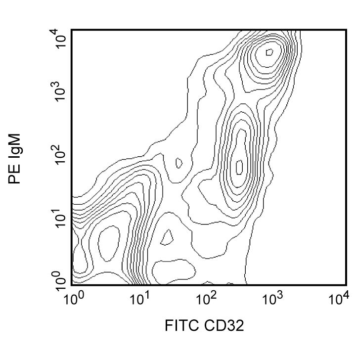
Two-color analysis of the expression of Cd32 on rat splenocytes. Lewis rat splenocytes were stained with purified D34-485 mAb, followed by FITC-conjugated polyclonal goat anti-mouse Ig (Cat. No. 554001), then PE-conjugated anti-rat IgM G35-238 (Cat. No. 553888). Double-positive cells are B lymphocytes (IgM+ cells), which express CD32 on the cell surface. Flow cytometry was performed on a BD FACScan™ flow cytometry system.
Blocking of Fc-mediated binding to FcγII receptors (CD32) on rat splenocytes. Lewis rat splenocytes were pre-incubated with purified isotype control mAb A112-2 (Cat. No. 553487, left panel) or Rat BD Fc Block™ purified anti-rat CD32 mAb D34-485 (right panel). Two-color staining was performed with FITC-conjugated anti-rat NKR-P1A mAb 10/78 (Cat. No. 555008) and PE-conjugated anti-rat CD45RA mAb OX-33 (Cat. No. 551402/554884). Note how the dim staining of B lymphocytes (OX-33+ cells) by anti-CD161a (NKR-P1A) (left panel) is reduced when Rat BD Fc Block™ is used before staining (right panel). Flow cytometry was performed on a BD FACScan™ flow cytometry system.