


 全部商品分类
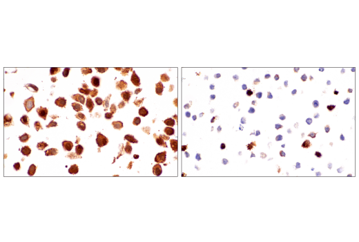


 下载产品说明书 下载SDS
 用小程序,查商品更便捷


 收藏
收藏
 对比
对比 咨询
咨询
The Ferroptosis Antibody Sampler Kit provides an economical means of detecting proteins involved in ferroptosis. The kit includes enough antibodies to perform two western blot experiments with each primary antibody.
参考图片
Immunohistochemical analysis of paraffin-embedded HeLa cell pellet (left, high-expressing) or HL-60 cell pellet (right, low-expressing) using FTH1 (D1D4) Rabbit mAb.
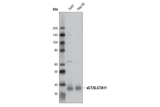
Western blot analysis of extracts from Huh7 and Hep G2 cells using xCT/SLC7A11 (D2M7A) Rabbit mAb.
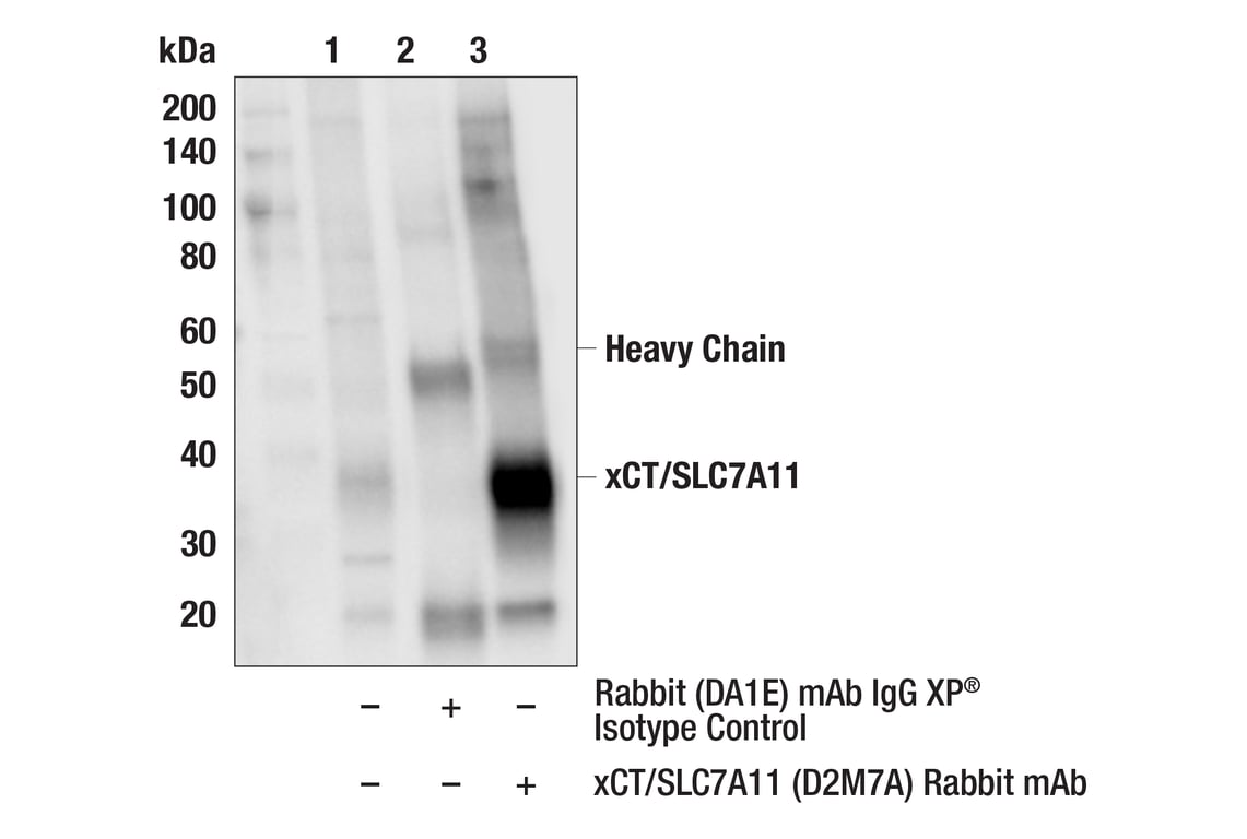
Immunoprecipitation of xCT/SLC7A11 protein from A549 cell extracts. Lane 1 is 10% input, lane 2 is Rabbit (DA1E) mAb IgG XP® Isotype Control #3900, and lane 3 is xCT/SLC7A11 (D2M7A) Rabbit mAb. Western blot analysis was performed using xCT/SLC7A11 (D2M7A) Rabbit mAb. Anti-rabbit IgG, HRP-linked Antibody #7074 was used as a secondary antibody.
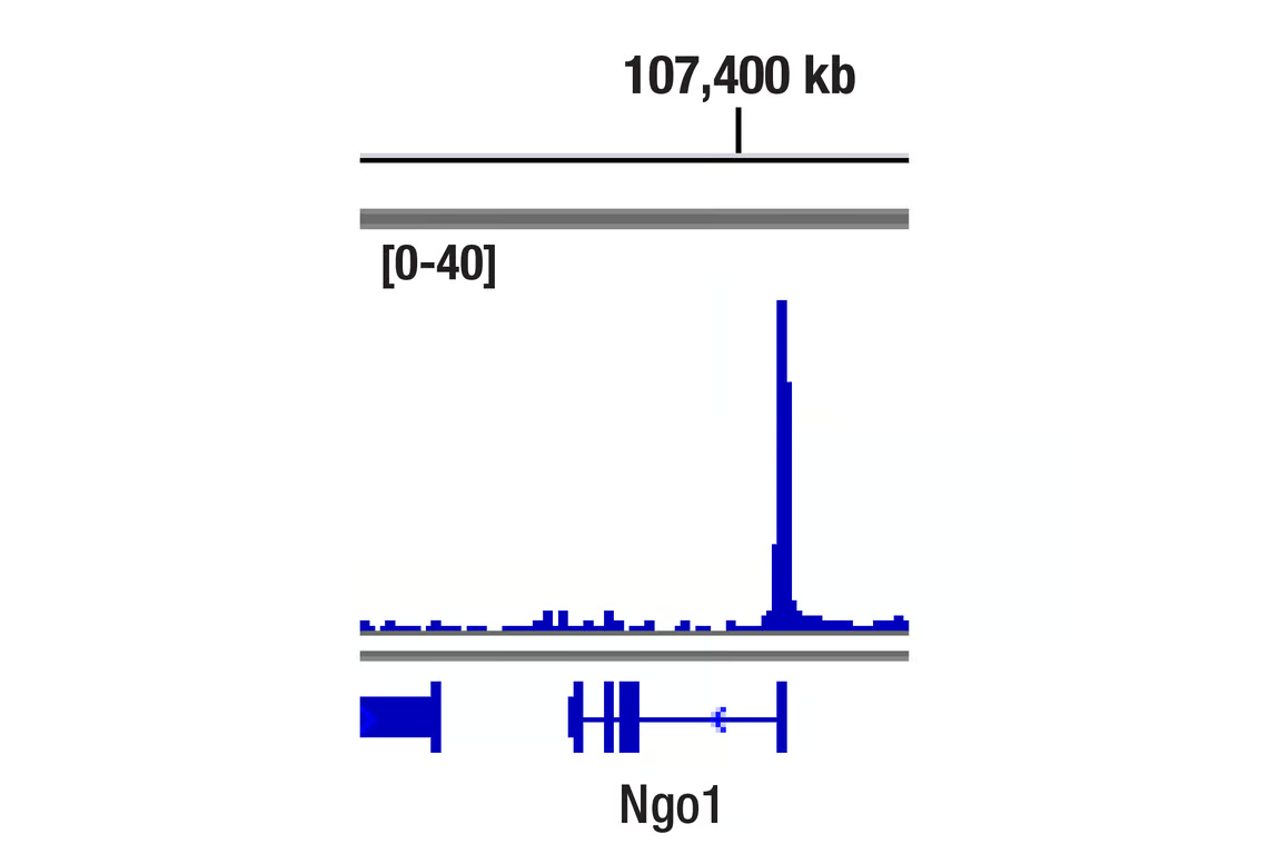
Chromatin immunoprecipitations were performed with cross-linked chromatin from MEF cells treated with DEM (50 μM, 3 hr) and NRF2 (D1Z9C) XP® Rabbit mAb, using SimpleChIP® Plus Enzymatic Chromatin IP Kit (Magnetic Beads) #9005. DNA Libraries were prepared using DNA Library Prep Kit for Illumina Systems (ChIP-seq, CUT&RUN) #56795. The figure shows binding across NQO1, a known target gene of NRF2 (see additional figure containing ChIP-qPCR data).
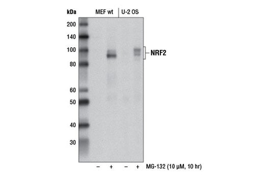
Western blot analysis of extracts from MEF wt and U-2 OS cells, untreated (-) or treated with MG-132 #2194 (10 μM, 10 hr; +), using NRF2 (D1Z9C) XP® Rabbit mAb.
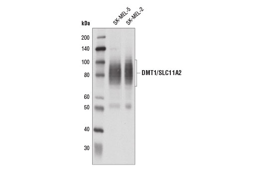
Western blot analysis of extracts from SK-MEL-5 and SK-MEL-2 cells using DMT1/SLC11A2 (D3V8G) Rabbit mAb.
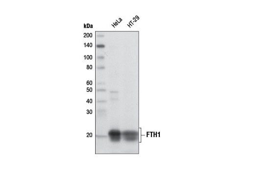
Western blot analysis of extracts from HeLa and HT-29 cells using FTH1 (D1D4) Rabbit mAb.
Simple Western™ analysis of lysates (1.0 mg/mL) from HeLa cells using FTH1 (D1D4) Rabbit mAb #4393. The virtual lane view (left) shows the target (as indicated) at 1:10 and 1:50 dilutions of primary antibody. The corresponding electropherogram view (right) plots chemiluminescence by molecular weight along the capillary at 1:10 (blue line) and 1:50 (green line) dilutions of primary antibody. This experiment was performed under reducing conditions on the Jess™ Simple Western instrument from ProteinSimple, a BioTechne brand, using the 12-230 kDa separation module.
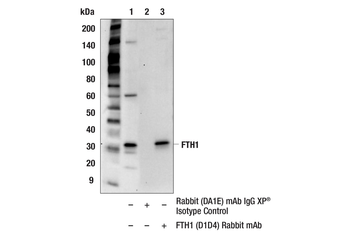
Immunoprecipitation of FTH1 from HeLa cell extracts. Lane 1 is 10% input, lane 2 is precipitated with Rabbit (DA1E) mAb IgG XP® Isotype Control #3900, and lane 3 is FTH1 (D1D4) Rabbit mAb. Western blot was performed using FTH1 (D1D4) Rabbit mAb.
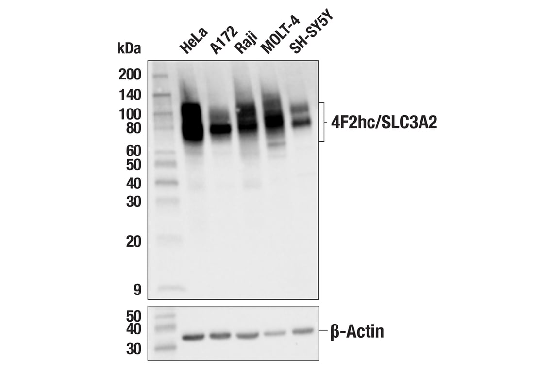
Western blot analysis of extracts from various cell lines using 4F2hc/SLC3A2 (D3F9D) XP® Rabbit mAb (upper) and β-actin (D6A8) Rabbit mAb #8457 (lower).
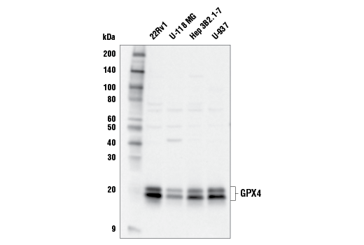
Western blot analysis of extracts from various cell lines using GPX4 Antibody.
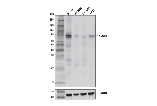
Western blot analysis of extracts from various cell lines using NCOA4 (E8H8Z) Rabbit mAb (upper) or β-Actin (D6A8) Rabbit mAb #8457 (lower). As expected, HT-1080 and OVCAR-4 cells are low for NCOA4 expression.
After the primary antibody is bound to the target protein, a complex with HRP-linked secondary antibody is formed. The LumiGLO® is added and emits light during enzyme catalyzed decomposition.
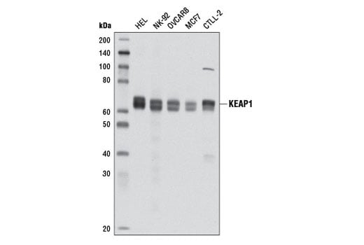
Western blot analysis of extracts from various cell lines using KEAP1 (D6B12) Rabbit mAb.
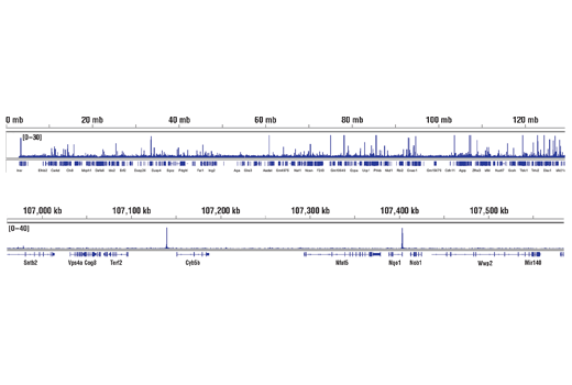
Chromatin immunoprecipitations were performed with cross-linked chromatin from MEF cells treated with DEM (50 μM, 3 hr) and NRF2 (D1Z9C) XP® Rabbit mAb, using SimpleChIP® Plus Enzymatic Chromatin IP Kit (Magnetic Beads) #9005. DNA Libraries were prepared using DNA Library Prep Kit for Illumina® (ChIP-seq, CUT&RUN) #56795. The figure shows binding across chromosome 8 (upper), including NQO1 (lower), a known target gene of NRF2 (see additional figure containing ChIP-qPCR data).
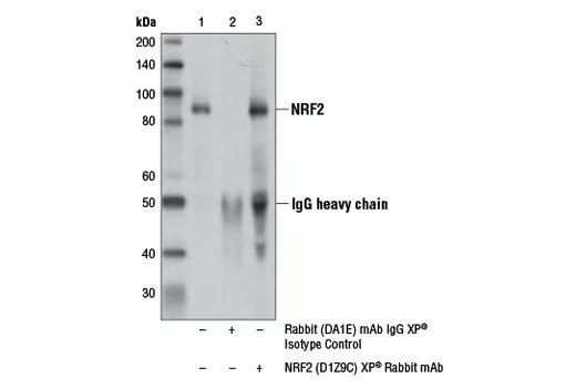
Immunoprecipitation of NRF2 from MEF wt cell extracts treated with MG-132 #2194 (10 μM, 10 hr) using Rabbit (DA1E) mAb IgG XP® Isotype Control #3900 (lane 2) or NRF2 (D1Z9C) XP® Rabbit mAb (lane 3). Lane 1 is 10% input. Western blot analysis was performed using NRF2 (D1Z9C) XP® Rabbit mAb (lane 3).
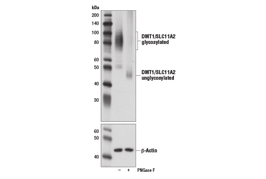
Western blot analysis of extracts from human SK-MEL-5 cells, untreated (-) or treated with PNGase F (+), using DMT1/SLC11A2 (D3V8G) Rabbit mAb (upper) and β-Actin (13E5) Rabbit mAb #4970 (lower).
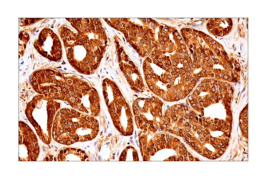
Immunohistochemical analysis of paraffin-embedded human ductal breast carcinoma using FTH1 (D1D4) Rabbit mAb.
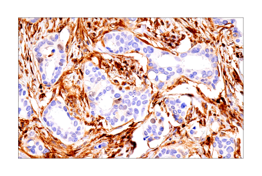
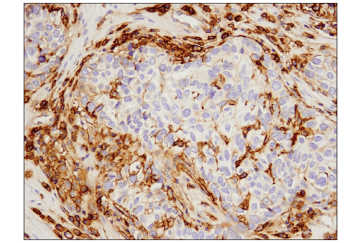
Immunohistochemical analysis of paraffin-embedded human breast carcinoma using 4F2hc/SLC3A2 (D3F9D) XP® Rabbit mAb.
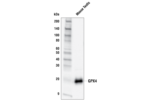
Western blot analysis of extracts from mouse testis using GPX4 Antibody.
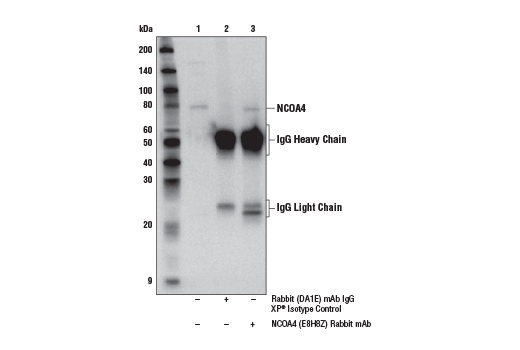
Immunoprecipitation of NCOA4 protein from HeLa cell extracts. Lane 1 is 10% input, lane 2 is Rabbit (DA1E) mAb IgG XP® Isotype Control #3900, and lane 3 is NCOA4 (E8H8Z) Rabbit mAb. Western blot analysis was performed using NCOA4 (E8H8Z) Rabbit mAb. Anti-rabbit IgG, HRP-linked Antibody #7074 was used as a secondary antibody.
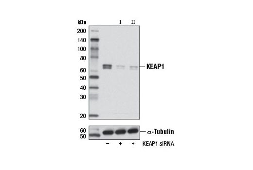
Western blot analysis of extracts from OVCAR8 cells, transfected with 100 nM SignalSilence® Control siRNA (Unconjugated) #6568 (-), SignalSilence® KEAP1 siRNA I #5285 (+) or SignalSilence® KEAP1 siRNA II #5289 (+), using KEAP1 (D6B12) Rabbit mAb (upper) or α-Tubulin (11H10) Rabbit mAb #2125 (lower). The KEAP1 (D6B12) Rabbit mAb confirms silencing of KEAP1 expression, while the α-Tubulin (11H10) Rabbit mAb is used as a loading control.
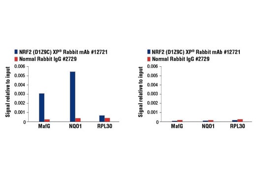
Chromatin immunoprecipitations were performed with cross-linked chromatin from MEF NRF2 wild-type (left) and NRF2 knock-out (right) cells, both treated with DEM (50 μM, 3 hr), and NRF2 (D1Z9C) XP® Rabbit mAb or Normal Rabbit IgG #2729 using SimpleChIP® Enzymatic Chromatin IP Kit (Magnetic Beads) #9003. The enriched DNA was quantified by real-time PCR using mouse MafG intron 1 primers, SimpleChIP® Mouse NQO1 Promoter Primers #12635, and SimpleChIP® Mouse RPL30 Intron 2 Primers #7015. The amount of immunoprecipitated DNA in each sample is represented as signal relative to the total amount of input chromatin, which is equivalent to one.
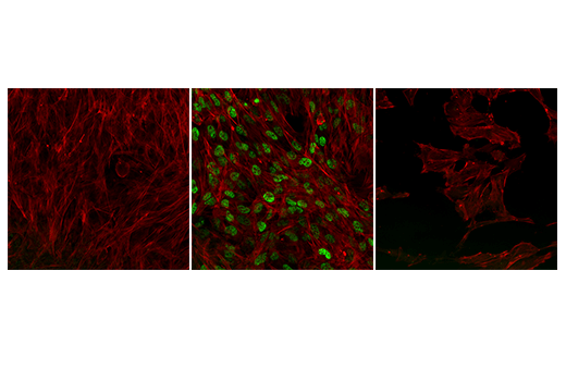
Confocal immunofluorescent analysis of wild-type NRF2 MEF cells, untreated (left) or treated with MG-132 #2194 (10 μM, 8 hr; center), and NRF2 knock-out MEF cells treated with MG-132 #2194 (10 μM, 8 hr; right), using NRF2 (D1Z9C) XP® Rabbit mAb (green pseudocolor). Actin filaments were labeled with Alexa Fluor® 488 Phalloidin #8878 (red pseudocolor).
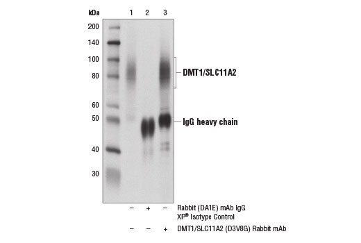
Immunoprecipitation of DMT1 from SK-MEL-5 cell extracts using Rabbit (DA1E) mAb IgG XP® Isotype Control #3900 (lane 2) or DMT1/SLC11A2 (D3V8G) Rabbit mAb (lane 3). Lane 1 is 10% input. Western blot analysis was performed using DMT1/SLC11A2 (D3V8G) Rabbit mAb.
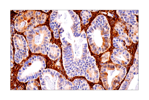
Immunohistochemical analysis of paraffin-embedded human prostate adenocarcinoma using FTH1 (D1D4) Rabbit mAb.
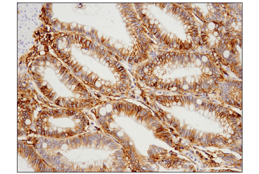
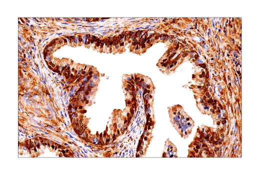
Immunohistochemical analysis of paraffin-embedded human colon carcinoma using 4F2hc/SLC3A2 (D3F9D) XP® Rabbit mAb.
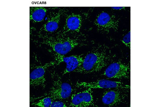
Confocal immunofluorescent analysis of OVCAR8 cells using KEAP1 (D6B12) Rabbit mAb (green). Blue pseudocolor = DRAQ5® #4084 (fluorescent DNA dye).
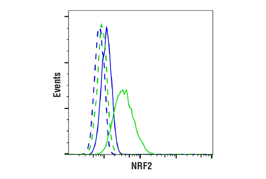
Flow cytometric analysis of MEF wt cells, untreated (blue) or treated with MG-132 #2194 (10uM, 4 hrs; green) using NRF2 (D1Z9C) XP® Rabbit mAb (solid line) compared to concentration-matched Rabbit (DA1E) mAb IgG XP® Isotype Control #3900 (dashed line). Anti-rabbit IgG (H+L), F(ab')2 Fragment (Alexa Fluor® 488 Conjugate) #4412 was used as a secondary antibody.
Immunohistochemical analysis of paraffin-embedded human endometrioid adenocarcinoma using FTH1 (D1D4) Rabbit mAb.
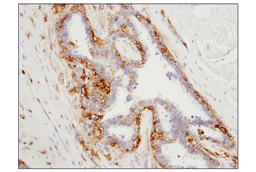
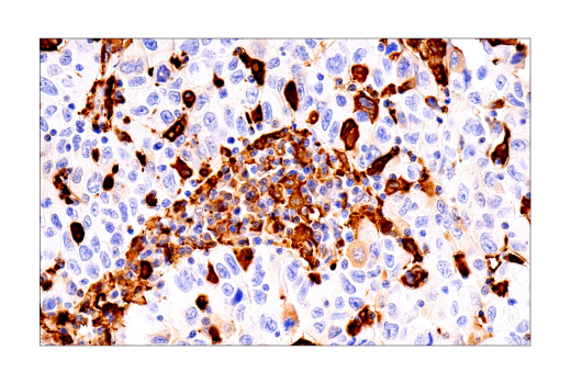
Immunohistochemical analysis of paraffin-embedded human ovarian carcinoma using 4F2hc/SLC3A2 (D3F9D) XP® Rabbit mAb.
Immunohistochemical analysis of paraffin-embedded human prostate adenocarcinoma using FTH1 (D1D4) Rabbit mAb.
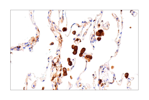
Immunohistochemical analysis of paraffin-embedded human non-small cell lung carcinoma using FTH1 (D1D4) Rabbit mAb.
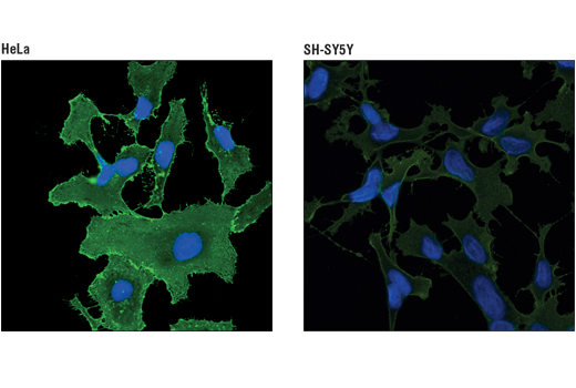
Confocal immunofluorescent analysis of HeLa (left, high expressing) or SH-SY5Y (right, low expressing) cells using 4F2hc/SLC3A2 (D3F9D) XP® Rabbit mAb (green). Blue pseudocolor = DRAQ5® #4084 (fluorescent DNA dye).
Immunohistochemical analysis of paraffin-embedded normal human lung using FTH1 (D1D4) Rabbit mAb.
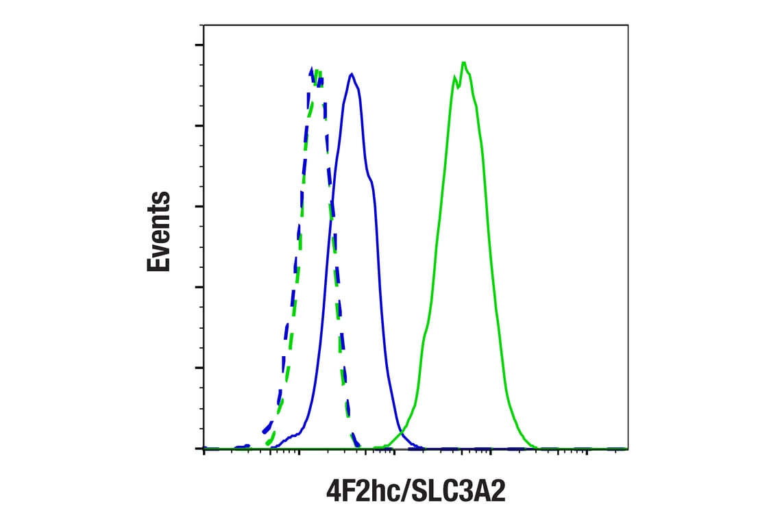
Flow cytometric analysis of SH-SY5Y cells (blue) and HeLa cells (green) using 4F2hc/SLC3A2 (D3F9D) XP® Rabbit mAb (solid lines) or a concentration-matched Rabbit (DA1E) mAb IgG XP® Isotype Control #3900 (dashed lines). Anti-rabbit IgG (H+L), F(ab')2 Fragment (Alexa Fluor® 488 Conjugate) #4412 was used as a secondary antibody.
Immunohistochemical analysis of paraffin-embedded normal mouse colon using FTH1 (D1D4) Rabbit mAb.
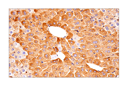
Immunohistochemical analysis of paraffin-embedded normal mouse liver using FTH1 (D1D4) Rabbit mAb.
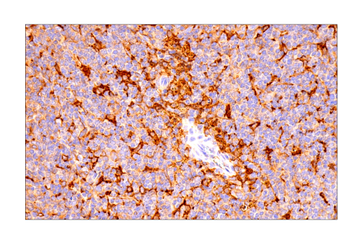
Immunohistochemical analysis of paraffin-embedded normal mouse spleen using FTH1 (D1D4) Rabbit mAb.
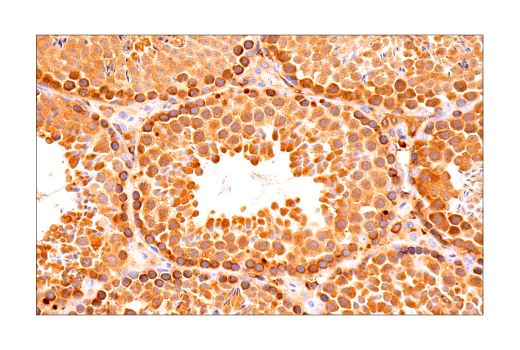
Immunohistochemical analysis of paraffin-embedded normal mouse testis using FTH1 (D1D4) Rabbit mAb.
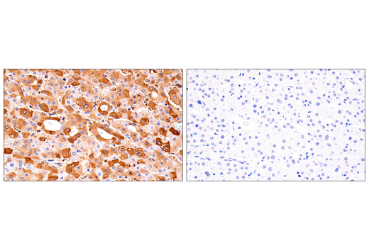
Immunohistochemical analysis of paraffin-embedded human hepatocellular carcinoma using FTH1 (D1D4) Rabbit mAb (left) compared to concentration-matched Rabbit (DA1E) mAb IgG XP® Isotype Control #3900 (right).