


 全部商品分类
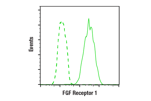


 用小程序,查商品更便捷


Monoclonal antibody is produced by immunizing animals with a recombinant protein specific to the carboxy terminus of human FGF receptor 1 protein.
Product Usage Information
| Application | Dilution |
|---|---|
| Western Blotting | 1:1000 |
| Immunoprecipitation | 1:50 |
| Immunohistochemistry (Paraffin) | 1:100 - 1:400 |
| Immunofluorescence (Immunocytochemistry) | 1:200 - 1:800 |
| Flow Cytometry (Fixed/Permeabilized) | 1:200 - 1:800 |
Specificity/Sensitivity
Species Reactivity:
Human, Mouse, Rat, Monkey
Supplied in 10 mM sodium HEPES (pH 7.5), 150 mM NaCl, 100 µg/ml BSA, 50% glycerol and less than 0.02% sodium azide. Store at –20°C. Do not aliquot the antibody.
For a carrier free (BSA and azide free) version of this product see product #76123.
参考图片
Flow cytometric analysis of A-204 cells using FGF Receptor 1 (D8E4) XP® Rabbit mAb (solid line) compared to concentration-matched Rabbit (DA1E) mAb IgG XP® Isotype control #3900 (dashed line). Anti-rabbit IgG (H+L), F(ab')2 Fragment (Alexa Fluor® 488 Conjugate) #4412 was used as a secondary antibody.
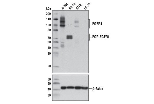
Western blot analysis of extracts from A-204 (FGFR1 positive), KG-1a (FGFR1 oncogenic partner-FGFR1 fusion), A172 (FGFR1 low), and HT-29 (FGFR1 negative) cells using FGF Receptor 1 (D8E4) XP® Rabbit mAb (upper) and β-Actin (D6A8) Rabbit mAb #8457 (lower).
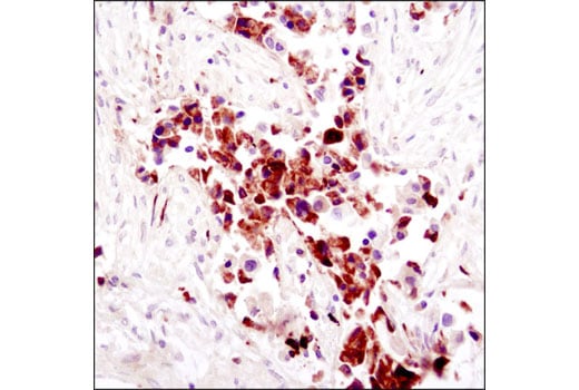
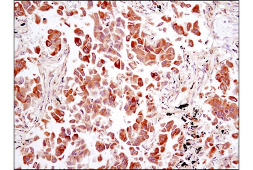
Immunohistochemical analysis of paraffin-embedded human breast carcinoma using FGF Receptor 1 (D8E4) XP® Rabbit mAb.
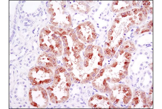
Immunohistochemical analysis of paraffin-embedded human kidney using FGF Receptor 1 (D8E4) XP® Rabbit mAb.
Immunohistochemical analysis of paraffin-embedded human lung carcinoma using FGF Receptor 1 (D8E4) XP® Rabbit mAb.
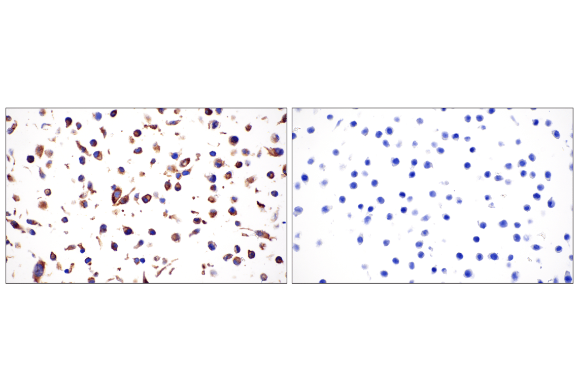
Immunohistochemical analysis of paraffin-embedded A-204 cell pellet (left, positive) or HT-29 cell pellet (right, negative) using FGF Receptor 1 (D8E4) XP® Rabbit mAb.
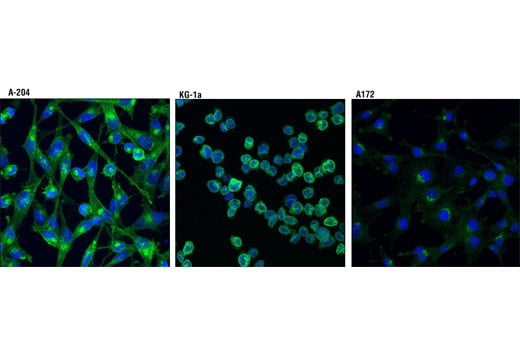
Confocal immunofluorescent analysis of A204 cells (positive, left), KG-1 cells (positive, middle) and A172 cells (weak expression, right) using FGF Receptor 1 (D8E4) XP® Rabbit mAb (green). Blue pseudocolor= DRAQ5® #4084 (fluorescent DNA dye).