Flow cytometry, Intracellular staining (flow cytometry) (Tested During Development)
产品介绍
产品介绍
产品信息
简单描述
BD Horizon™ Fixable Viability Stain 780 (FVS780) is useful for discrimination of viable from non-viable mammalian cells in multicolor flow cytometric applications. This dye reacts with and covalently binds to cell-surface and intracellular amines. Permeable plasma cell membranes, such as those present in necrotic cells, allow for the intracellular diffusion of the dye and covalent binding to higher overall concentrations of amines than in non-permeable live cells. Therefore, necrotic cells present in a typical
in vitro
assay label with higher levels of dye increasing their fluorescence intensity 10-20 fold over that of viable cells. The labeled cells can be fixed with formaldehyde for downstream decontamination, freezing and/or permeabilization and subsequent intracellular staining while maintaining stable viability stain fluorescence.
BD Horizon™ Fixable Viability Stain 780 is excited by the Red laser (with an excitation maximum of 759 nm), and has a fluorescence emission maximum of 780 nm.
商品描述
BD Horizon™ Fixable Viability Stain 780 (FVS780) is useful for discrimination of viable from non-viable mammalian cells in multicolor flow cytometric applications. This dye reacts with and covalently binds to cell-surface and intracellular amines. Permeable plasma cell membranes, such as those present in necrotic cells, allow for the intracellular diffusion of the dye and covalent binding to higher overall concentrations of amines than in non-permeable live cells. Therefore, necrotic cells present in a typical
in vitro
assay label with higher levels of dye increasing their fluorescence intensity 10-20 fold over that of viable cells. The labeled cells can be fixed with formaldehyde for downstream decontamination, freezing and/or permeabilization and subsequent intracellular staining while maintaining stable viability stain fluorescence.
BD Horizon™ Fixable Viability Stain 780 is excited by the Red laser (with an excitation maximum of 759 nm), and has a fluorescence emission maximum of 780 nm.
克隆号
(RUO)
应用
实验应用
Flow cytometry, Intracellular staining (flow cytometry) (Tested During Development)
目标/特异性
Live/Dead Discriminator Dyes
文献
文献
研发参考(2)
1. Perfetto SP, Chattopadhyay PK, Lamoreaux L, et al. Amine reactive dyes: an effective tool to discriminate live and dead cells in polychromatic flow cytometry. J Immunol Methods. 2006; 313(1–2):199-208. (Methodology).
2. Perfetto SP, Chattopadhyay PK, Lamoreaux L, et al. Amine-reactive dyes for dead cell discrimination in fixed samples. Curr Protoc Cytom. 9(9.34)(Methodology).
参考图片
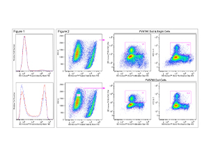
Figure 1. Fluorescent staining of Jurkat cells with BD Horizon™ Fixable Viability Stain 780. Human Jurkat cells were treated (16 hr) with 0.025% DMSO (Top Plot) or 5 μM camptothecin (Bottom Plot) and stained with BD Horizon™ Fixable Viability Stain 780 (Cat. No. 565388). Cells were either not fixed (solid line histograms), or fixed in BD Cytofix™ Fixation Buffer (Cat. No. 554655) and permeabilized in Perm/Wash Buffer I (Cat. No. 557885) (dashed line histograms). Histograms were derived from gated events with the light scattering characteristics of Jurkat cells. Flow cytometry was performed using a BD™ LSRII Cell Analyzer System. Figure 2. Analysis of proliferating mouse splenocytes for surface and intracellular markers. BALB/c splenocytes were stained (10 min, 37°C) with BD Horizon™ Violet Proliferation Dye 450 (Cat. No. 562158), washed twice, and then cultured (3 days) with Purified NA/LE Hamster Anti-Mouse CD3e (Cat. No. 553057) and Hamster Anti-Mouse CD28 (Cat. No. 553294) antibodies. The cells were restimulated (4 hr) with PMA, Ionomycin, and BD GolgiStop™ Protein Transport Inhibitor (Monensin) (Cat. No. 554724). Cells were harvested, stained with BD Horizon™ Fixable Viability Stain 780, fixed and permeabilized using a BD Cytofix/Cytoperm™ Fixation/Permeabilization Solution Kit (Cat. No. 554714), and then stained with BD Horizon™ BUV395 Anti-Mouse CD4 (Cat. No. 563790) and FITC Anti-Mouse IL-2 (Cat. No. 554427) antibodies. Two-color dot plots showing VPD450 fluorescence versus CD4 expression (Middle Plots) or IL-2 expression (Right Plots) were derived from either total cells (Top; FVS780 Dull and Bright Cells) or previously viable cells (Bottom; FVS780 Dull Cells) with the light scatter characteristics of intact cells. CD4+ and IL-2+ cell gates were based on an FMO or unstimulated cell control, respectively. Flow cytometry was performed on a BD LSRFortessa™ Cell Analyzer system.


 全部商品分类


 下载产品说明书
 用小程序,查商品更便捷


 收藏
收藏
 对比
对比 咨询
咨询