


 全部商品分类
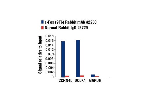


c-Fos (9F6) Rabbit mAb


 下载产品说明书 下载COA 下载SDS
 用小程序,查商品更便捷


 收藏
收藏
 对比
对比 咨询
咨询
Monoclonal antibody is produced by immunizing animals with a synthetic peptide corresponding to residues near the amino terminus of human c-Fos protein.
Product Usage Information
For optimal ChIP results, use 10 μl of antibody and 10 μg of chromatin (approximately 4 x 10⁶ cells) per IP. This antibody has been validated using SimpleChIP® Enzymatic Chromatin IP Kits.
| Application | Dilution |
|---|---|
| Western Blotting | 1:1000 |
| Simple Western™ | 1:10 - 1:50 |
| Immunofluorescence (Frozen) | 1:1600 - 1:3200 |
| Immunofluorescence (Immunocytochemistry) | 1:3200 - 1:12800 |
| Flow Cytometry (Fixed/Permeabilized) | 1:1600 - 1:6400 |
| Chromatin IP | 1:50 |
Specificity/Sensitivity
Species Reactivity:
Human, Mouse, Rat
Supplied in 10 mM sodium HEPES (pH 7.5), 150 mM NaCl, 100 µg/ml BSA, 50% glycerol and less than 0.02% sodium azide. Store at –20°C. Do not aliquot the antibody.
For a carrier free (BSA and azide free) version of this product see product #53345.
参考图片
Chromatin immunoprecipitations were performed with cross-linked chromatin from PC-12 cells starved overnight and treated with Human β-Nerve Growth Factor (h-βNGF) #5221 (50ng/ml) for 2h, and either c-Fos (9F6) Rabbit mAb or Normal Rabbit IgG #2729 using SimpleChIP® Enzymatic Chromatin IP Kit (Magnetic Beads) #9003. The enriched DNA was quantified by real-time PCR using SimpleChIP® Rat CCRN4L Promoter Primers #7983, rat DCLK1 promoter primers, and SimpleChIP® Rat GAPDH Promoter Primers #7964. The amount of immunoprecipitated DNA in each sample is represented as signal relative to the total amount of input chromatin, which is equivalent to one.
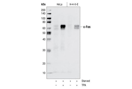
Western blot analysis of extracts from HeLa and H-4-II-E cells serum-starved overnight and TPA-stimulated for 4 hours, using c-Fos (9F6) Rabbit mAb.
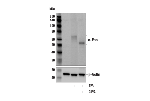
Western blot analysis of extracts from NIH/3T3 cells, serum-starved overnight and then either untreated (lane 1), stimulated for 4 hours with TPA (12-O-Tetradecanoylphorbol-13-Acetate) #4174 (lane 2), or stimulated with TPA for 4 hours then treated with λ-phosphatase (lane 3). c-Fos (9F6) Rabbit mAb #2250 (upper) and β-Actin (13E5) Rabbit mAb (HRP Conjugate) #5125 (lower).
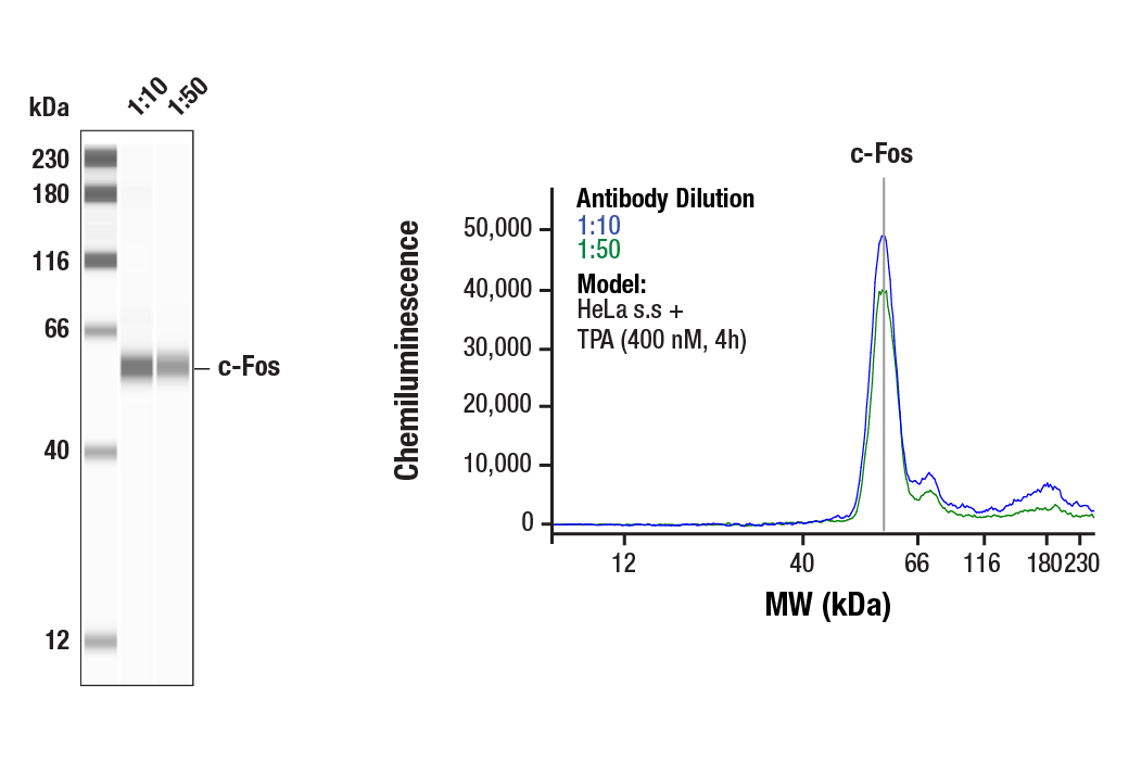
Simple Western™ analysis of lysates (1.0 mg/mL) from serum-starved HeLa cells treated with TPA (400 nM, 4h) using c-Fos (9F6) Rabbit mAb #2250. The virtual lane view (left) shows the target band (as indicated) at 1:10 and 1:50 dilutions of primary antibody. The corresponding electropherogram view (right) plots chemiluminescence by molecular weight along the capillary at 1:10 (blue line) and 1:50 (green line) dilutions of primary antibody. This experiment was performed under reducing conditions on the Jess™ Simple Western instrument from ProteinSimple, a BioTechne brand, using the 12-230 kDa separation module.
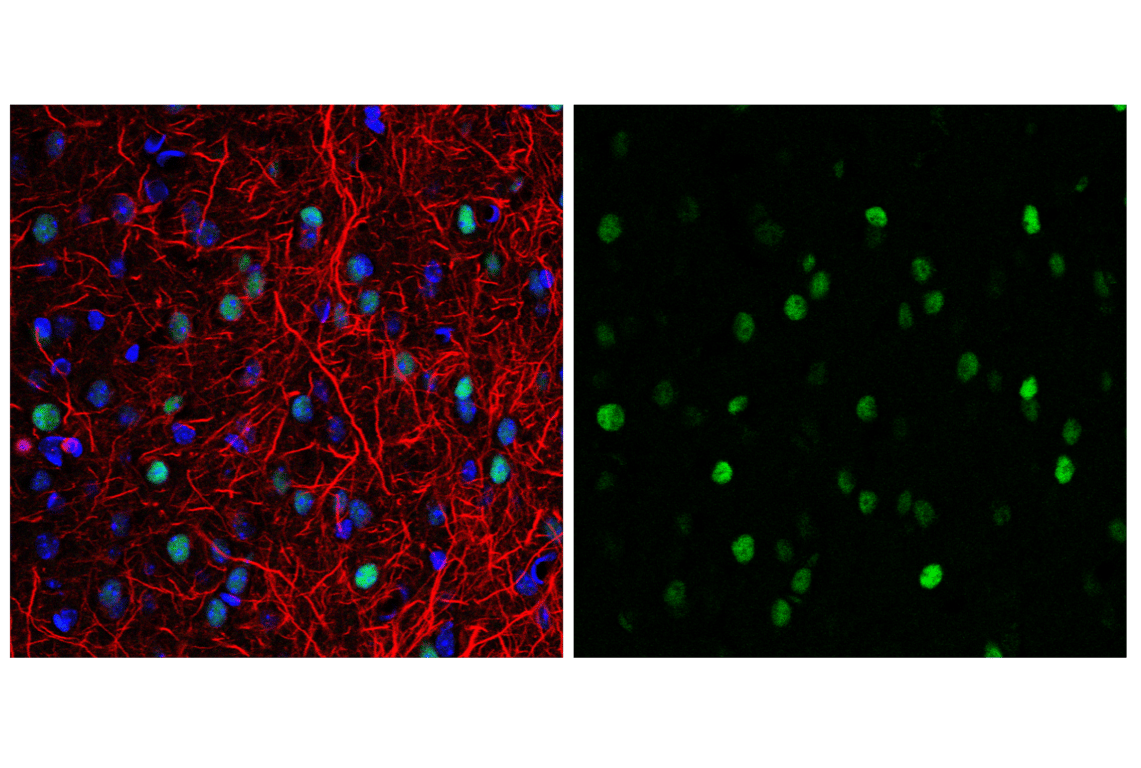
Confocal immunofluorescent analysis of mouse cerebral cortex using c-Fos (9F6) Rabbit mAb #2250 (green) and Neurofilament-L (DA2) Mouse mAb #2835 (red). Samples were mounted in ProLong® Gold Antifade Reagent with DAPI #8961 (blue).
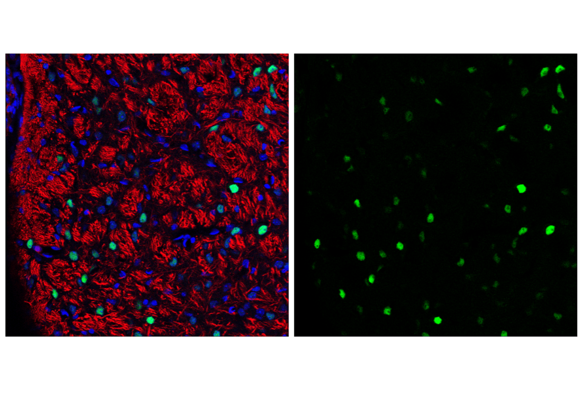
Confocal immunofluorescent analysis of mouse pons using c-Fos (9F6) Rabbit mAb #2250 (green) and Neurofilament-L (DA2) Mouse mAb #2835 (red). Samples were mounted in ProLong® Gold Antifade Reagent with DAPI #8961 (blue).
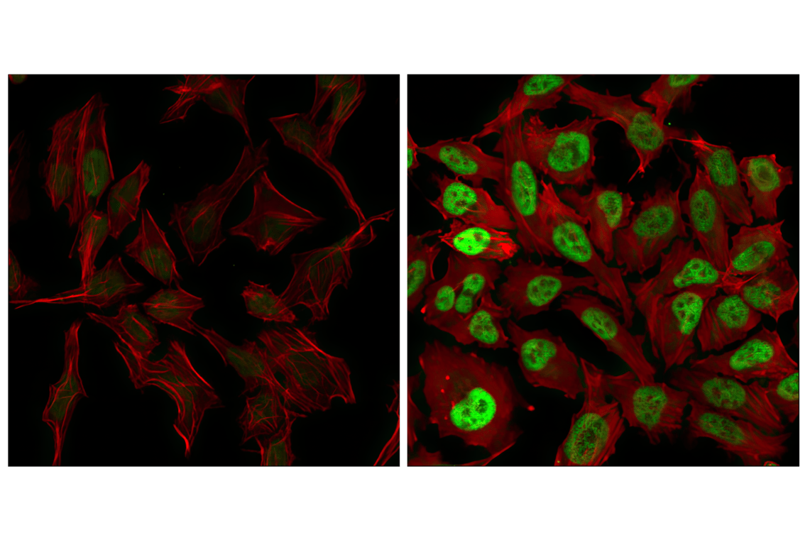
Confocal immunofluorescent analysis of HeLa cells serum-starved (left) or treated with TPA (#9905) for 4 hours (right) and labeled with c-Fos (9F6) Rabbit mAb (green). Actin filaments have been labeled with Alexa Fluor® 555 phalloidin (red).
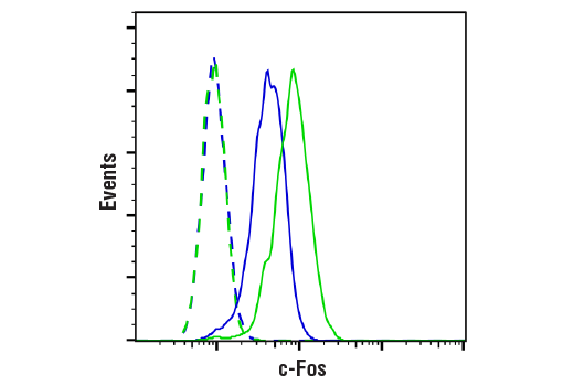
Flow cytometric analysis of HeLa cells serum starved overnight, untreated (blue, moderate expression) or treated with TPA #4174 (200 nM, 4 hr; green, high expression) using c-Fos (9F6) Rabbit mAb (solid lines) or concentration-matched Rabbit (DA1E) mAb IgG XP® Isotype Control #3900 (dashed lines). Anti-rabbit IgG (H+L), F(ab')2 Fragment (Alexa Fluor® 488 Conjugate) #4412 was used as a secondary antibody.