


 全部商品分类


 下载产品说明书 下载SDS
 用小程序,查商品更便捷


 收藏
收藏
 对比
对比 咨询
咨询

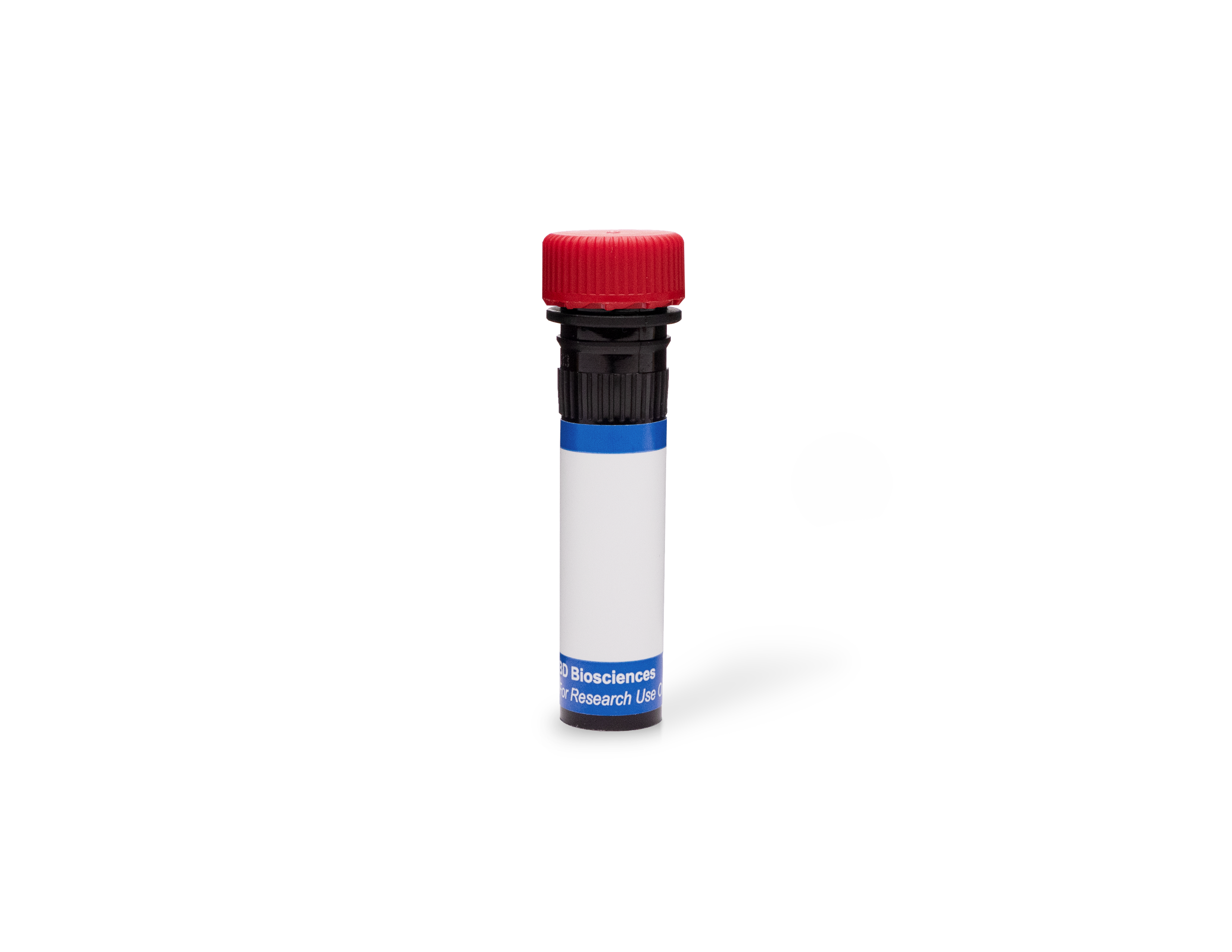
参考图片
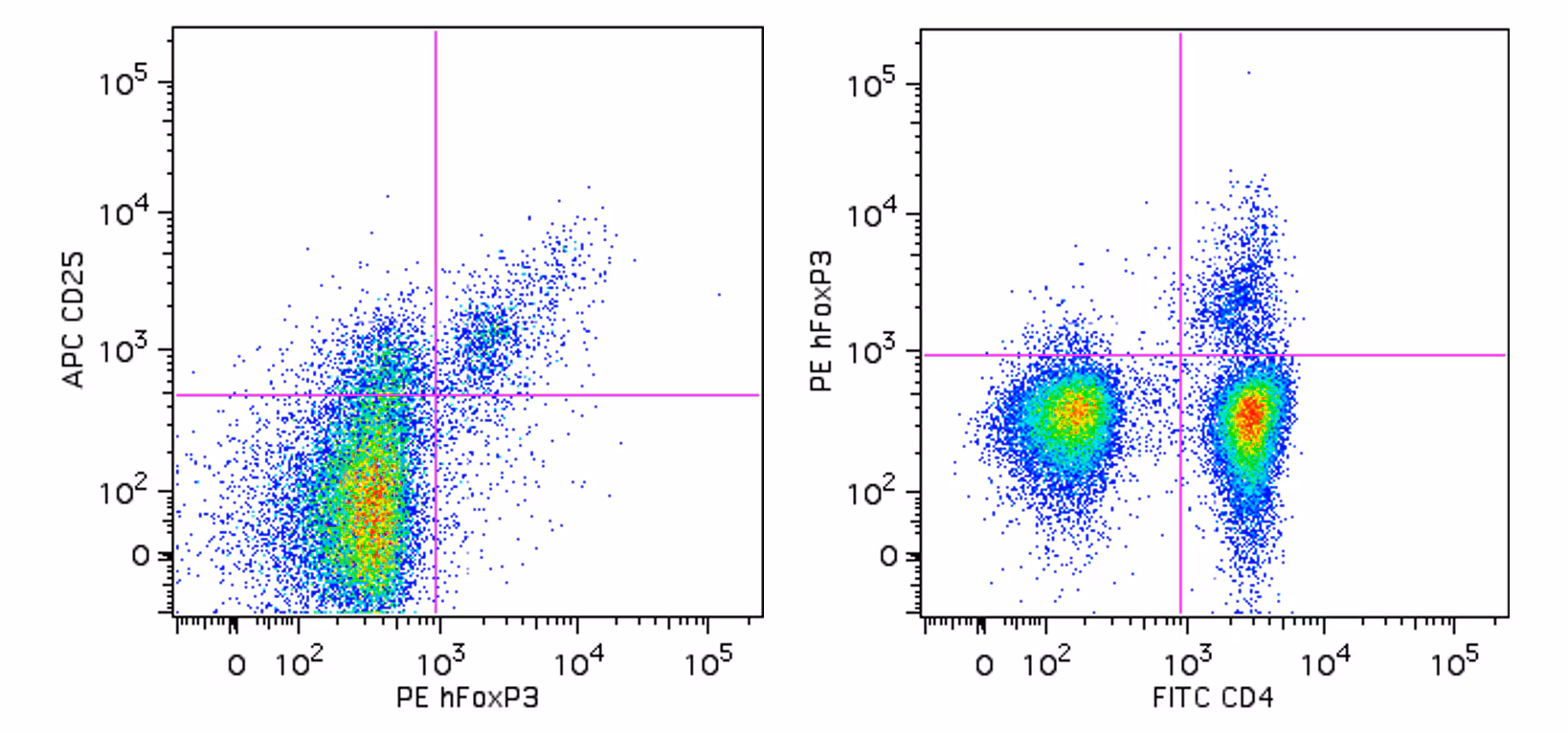
Flow cytometric analysis of FoxP3 expressed in human lymphocytes. Human peripheral blood mononuclear cells (PBMC) were stained with FITC anti-human CD4 (clone RPA-T4, Cat. No. 555346) and APC anti-human CD25 (clone 2A3, Cat. 340938) simultaneously. The PBMC were fixed and permeabilized (see Recommended Assay Procedure) followed by intracellular staining with PE anti-human FoxP3 (clone 236A; Cat No. 560852). Two-color flow cytometric dot plots were derived from gated events based on the light scattering characteristics of lymphocytes and fluorescence characteristics of CD4+ or CD25+, respectively, shown as either FoxP3 versus CD25 (left panel) or CD4 versus FoxP3 (right panel). Flow cytometry was performed on a BD LSRII™ flow cytometry system.
Flow cytometric analysis of FoxP3 expressed in human lymphocytes. Human peripheral blood mononuclear cells (PBMC) were stained with FITC anti-human CD4 (clone RPA-T4, Cat. No. 555346) and APC anti-human CD25 (clone 2A3, Cat. 340938) simultaneously. The PBMC were fixed and permeabilized (see Recommended Assay Procedure) followed by intracellular staining with PE anti-human FoxP3 (clone 236A; Cat No. 560852). Two-color flow cytometric dot plots were derived from gated events based on the light scattering characteristics of lymphocytes and fluorescence characteristics of CD4+ or CD25+, respectively, shown as either FoxP3 versus CD25 (left panel) or CD4 versus FoxP3 (right panel). Flow cytometry was performed on a BD LSRII™ flow cytometry system.