


 全部商品分类
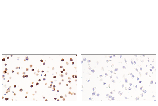


Exosomal Marker Antibody Sampler Kit

 下载产品说明书 下载SDS
 用小程序,查商品更便捷


 收藏
收藏
 对比
对比 咨询
咨询
The Exosomal Marker Antibody Sampler Kit provides an economical means to evaluate the presence of exosomal markers. The kit includes enough primary antibody to perform two western blot experiments for each target.
参考图片
Immunohistochemical analysis of paraffin-embedded Ramos cell pellet (left, positive) or A549 cell pellet (right, negative) using CD54/ICAM-1 (E3Q9N) XP® Rabbit mAb.
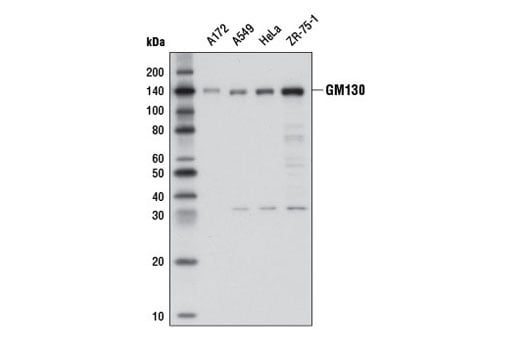
Western blot analysis of extracts from various cell lines using GM130 (D6B1) XP® Rabbit mAb.
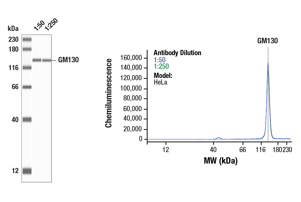
Simple Western™ analysis of lysates (0.1 mg/mL) from HeLa cells using GM130 (D6B1) XP® Rabbit mAb #12480. The virtual lane view (left) shows the target band (as indicated) at 1:50 and 1:250 dilutions of primary antibody. The corresponding electropherogram view (right) plots chemiluminescence by molecular weight along the capillary at 1:50 (blue line) and 1:250 (green line) dilutions of primary antibody. This experiment was performed under reducing conditions on the Jess™ Simple Western instrument from ProteinSimple, a BioTechne brand, using the 12-230 kDa separation module.
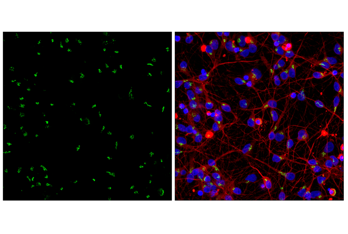
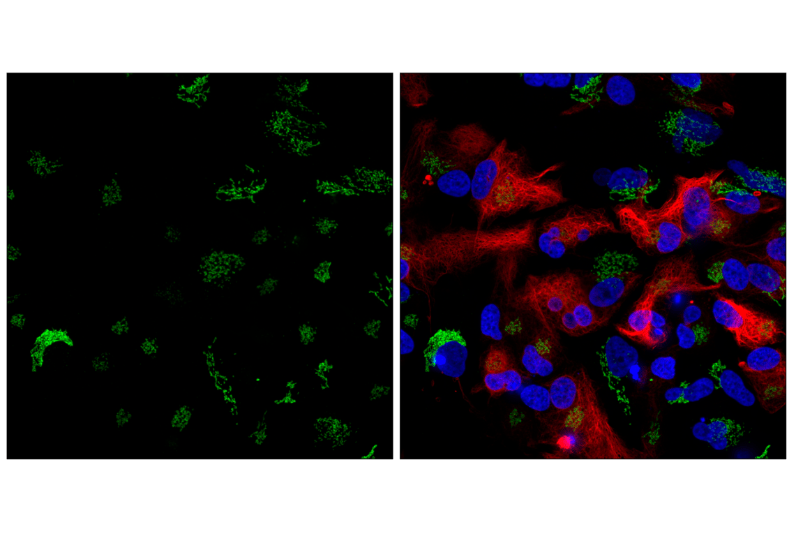
Confocal immunofluorescent analysis of human iPSC-derived cortical glutamatergic neurons at 7 days in vitro using GM130 (D6B1) XP® Rabbit mAb (green), β3-Tubulin (E9F3E) Mouse mAb #45058 (red), and DAPI #4083 (blue). iCell GlutaNeurons were kindly provided by FUJIFILM Cellular Dynamics, Inc.
Confocal immunofluorescent analysis of human iPSC-derived astrocyte cells at 7 days in vitro using GM130 (D6B1) XP® Rabbit mAb (green), GFAP (GA5) Mouse mAb #3670 (red), and DAPI #4083 (blue). iCell Astrocytes 2.0 were kindly provided by FUJIFILM Cellular Dynamics, Inc.
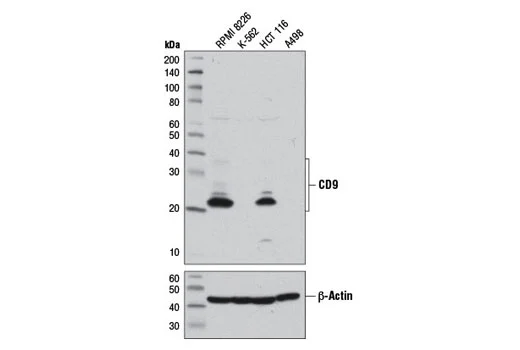
Western blot analysis of extracts from various cell lines using CD9 (D8O1A) Rabbit mAb (upper) or β-Actin (D6A8) Rabbit mAb #8457 (lower).
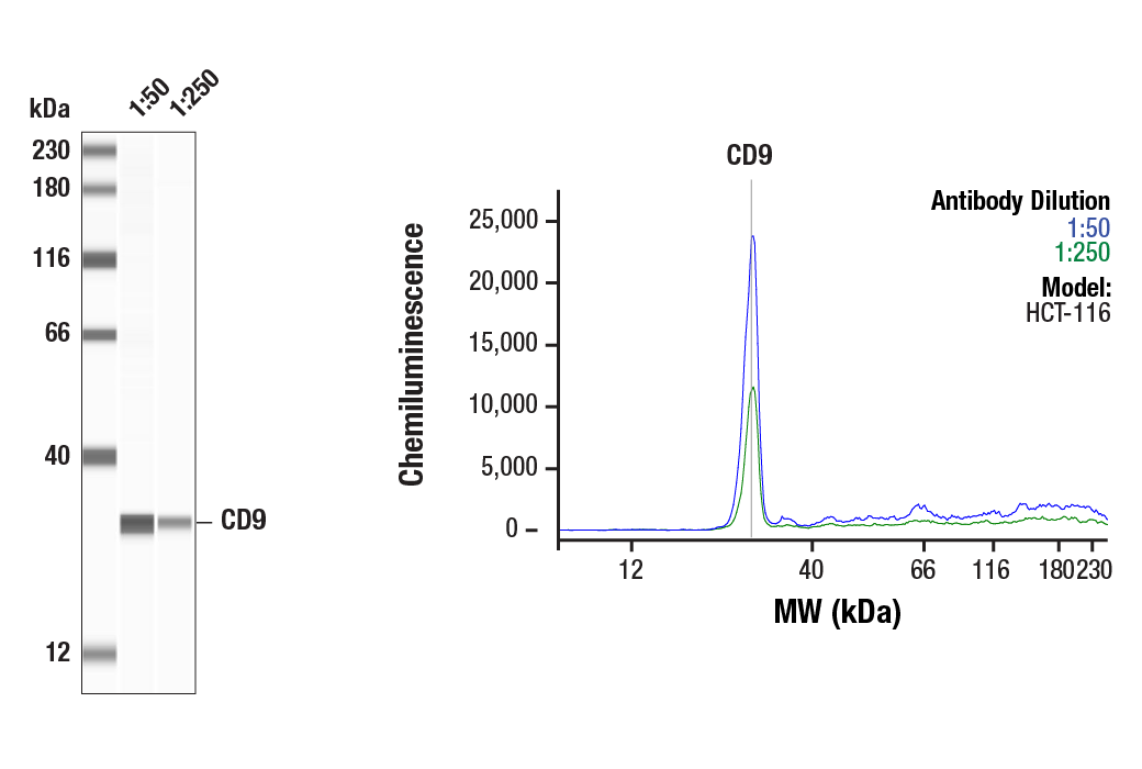
Simple Western™ analysis of lysates (1.0 mg/mL) from HCT-116 cells using CD9 (D8O1A) Rabbit mAb #13174. The virtual lane view (left) shows the target band (as indicated) at 1:50 and 1:250 dilutions of primary antibody. The corresponding electropherogram view (right) plots chemiluminescence by molecular weight along the capillary at 1:50 (blue line) and 1:250 (green line) dilutions of primary antibody. This experiment was performed under reducing conditions on the Jess™ Simple Western instrument from ProteinSimple, a BioTechne brand, using the 12-230 kDa separation module.
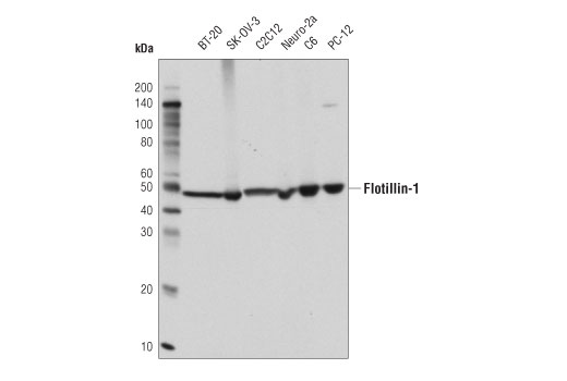
Western blot analysis of extracts from various cell lines using Flotillin-1 (D2V7J) XP® Rabbit mAb.
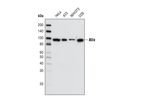
Western blot analysis of cell extracts from various cell types using Alix (3A9) Mouse mAb.
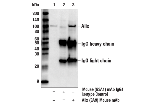
Immunoprecipitation of Alix from HeLa cell extracts. Lane 1 is 10% input, lane 2 is precipitated with Mouse (G3A1) mAb IgG1 Isotype Control #5415, and lane 3 is Alix (3A9) Mouse mAb, #2171. Western blot was performed using Alix (3A9) Mouse mAb.
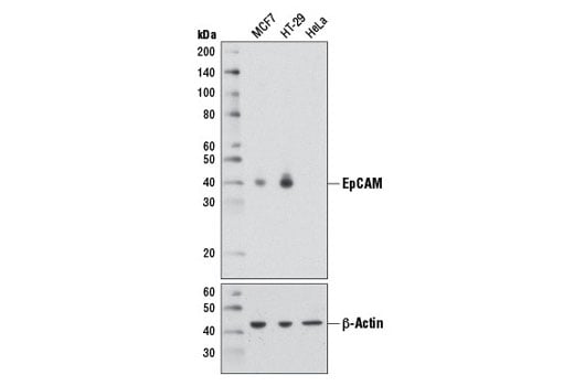
Western blot analysis of extracts from MCF7 (EpCAM positive), HT-29 (EpCAM positive), and HeLa (EpCAM negative) cells using EpCAM (D1B3) Rabbit mAb (upper) or β-Actin (D6A8) Rabbit mAb #8457 (lower).
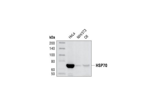
Western blot analysis of extracts from HeLa, NIH/3T3 and C6 cells, using HSP70 (D69) Antibody.
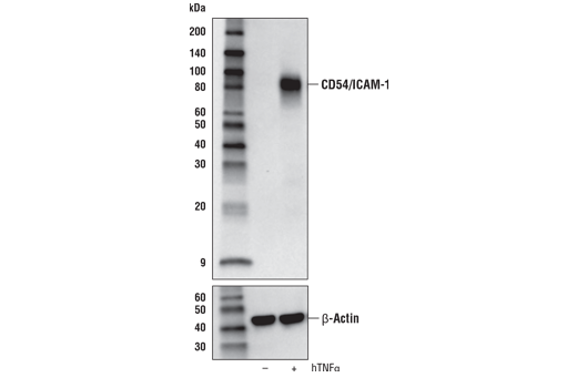
Western blot analysis of extracts from A549 cells, untreated (-) or treated with hTNF-α #8902 (10 ng/mL, 6 hr; +), using CD54/ICAM-1 (E3Q9N) XP® Rabbit mAb (upper) or β-Actin (D6A8) Rabbit mAb #8457 (lower).
After the primary antibody is bound to the target protein, a complex with HRP-linked secondary antibody is formed. The LumiGLO® is added and emits light during enzyme catalyzed decomposition.
After the primary antibody is bound to the target protein, a complex with HRP-linked secondary antibody is formed. The LumiGLO* is added and emits light during enzyme catalyzed decomposition.
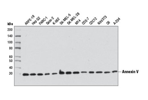
Western blot analysis of extracts from various cell lines using Annexin V Antibody.
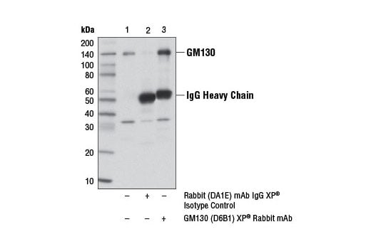
Immunoprecipitation of GM130 protein from ZR-75-1 cell extracts, using Rabbit (DA1E) mAb IgG XP® Isotype Control #3900 (lane 2) or GM130 (D6B1) XP® Rabbit mAb (lane 3). Lane 1 is 10% input. Western blot analysis was performed using GM130 (D6B1) XP® Rabbit mAb.
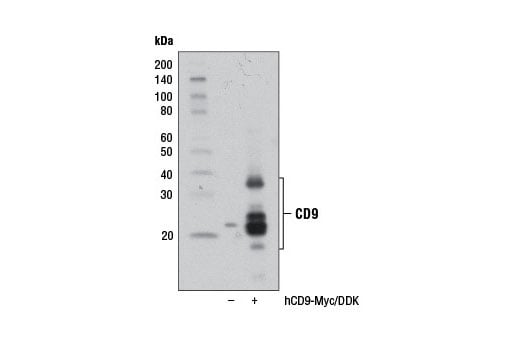
Western blot analysis of extracts from 293T cells, mock transfected (-) or transfected with a construct expressing Myc/DDK-tagged full-length human CD9 protein (hCD9-Myc/DDK; +), using CD9 (D8O1A) Rabbit mAb.
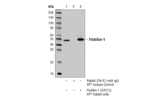
Immunoprecipitation of flotillin-1 protein from BT-20 cell extracts. Lane 1 is 10% input, lane 2 is Rabbit (DA1E) mAb IgG XP® Isotype Control #3900, and lane 3 is Flotillin-1 (D2V7J) XP® Rabbit mAb. Western blot analysis was performed using Flotillin-1 (D2V7J) XP® Rabbit mAb.
Immunohistochemical analysis of paraffin-embedded human urothelial carcinoma using CD54/ICAM-1 (E3Q9N) XP® Rabbit mAb performed on the Leica® BOND™ Rx.
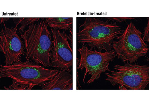
Confocal immunofluorescent analysis of HeLa cells, untreated (left) or treated with Brefeldin A #9972 (200 μM, 30 min; right), using GM130 (D6B1) XP® Rabbit mAb (green). Actin filaments were labeled with DY-554 phalloidin (red). Blue pseudocolor = DRAQ5® #4084 (fluorescent DNA dye).
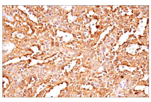
Immunohistochemical analysis of paraffin-embedded human squamous cell lung carcinoma using CD54/ICAM-1 (E3Q9N) XP® Rabbit mAb performed on the Leica® BOND™ Rx.
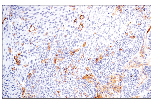
Immunohistochemical analysis of paraffin-embedded human hepatocellular carcinoma using Flotillin-1 (D2V7J) XP® Rabbit mAb in the presence of control peptide (left) or antigen-specific peptide (right).
Immunohistochemical analysis of paraffin-embedded human prostate adenocarcinoma using CD54/ICAM-1 (E3Q9N) XP® Rabbit mAb performed on the Leica® BOND™ Rx.
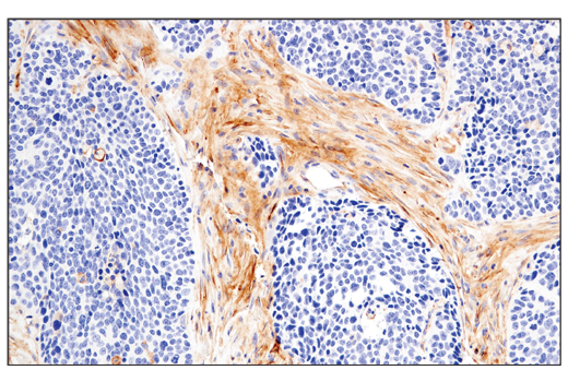
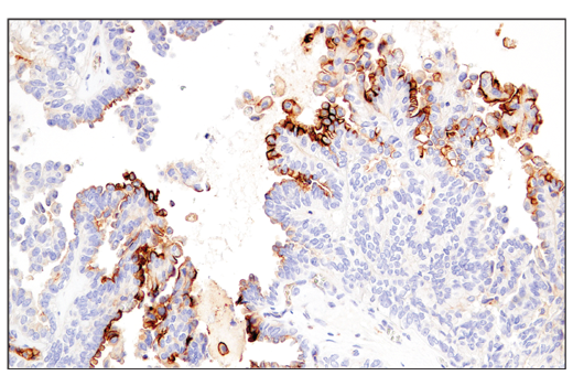
Immunohistochemical analysis of paraffin-embedded human colon carcinoma using CD54/ICAM-1 (E3Q9N) XP® Rabbit mAb.
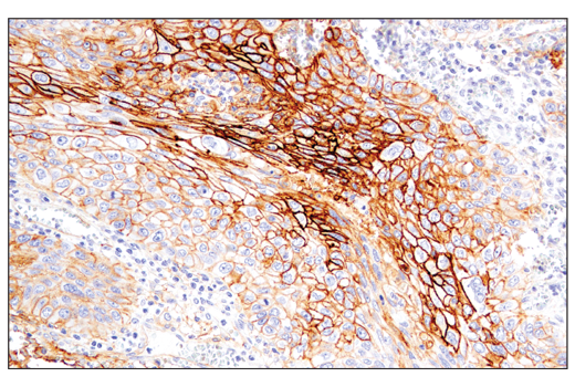
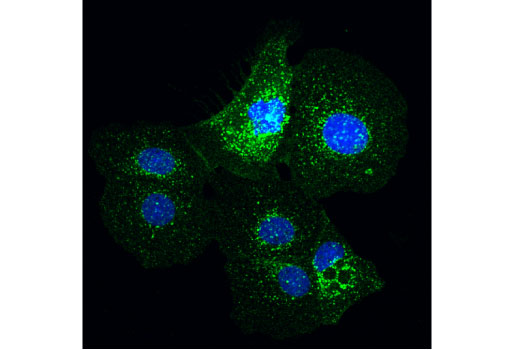
Confocal immunofluorescent analysis of BT-20 cells using Flotillin-1 (D2V7J) XP® Rabbit mAb (green). Blue pseudocolor = DRAQ5® #4084 (fluorescent DNA dye).
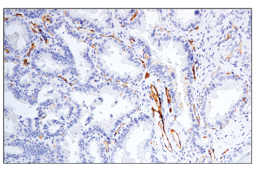
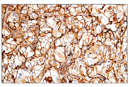
Immunohistochemical analysis of paraffin-embedded human renal cell carcinoma using CD54/ICAM-1 (E3Q9N) XP® Rabbit mAb.
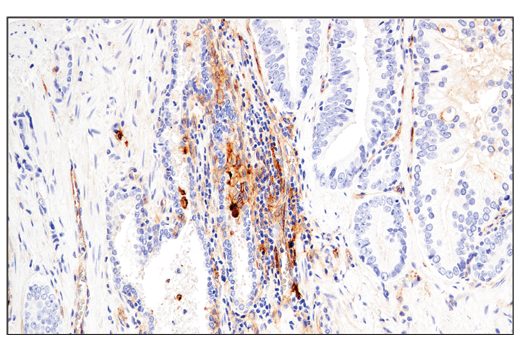
Immunohistochemical analysis of paraffin-embedded human endometrioid adenocarcinoma using CD54/ICAM-1 (E3Q9N) XP® Rabbit mAb.
Immunohistochemical analysis of paraffin-embedded human prostate carcinoma using CD54/ICAM-1 (E3Q9N) XP® Rabbit mAb.
Immunohistochemical analysis of paraffin-embedded human tonsil using CD54/ICAM-1 (E3Q9N) XP® Rabbit mAb.
Immunohistochemical analysis of paraffin-embedded normal rhesus monkey spleen using CD54/ICAM-1 (E3Q9N) XP® Rabbit mAb.
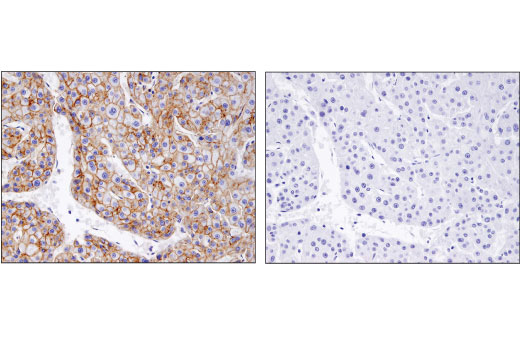
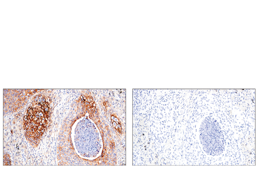
Immunohistochemical analysis of paraffin-embedded human squamous cell lung carcinoma using CD54/ICAM-1 (E3Q9N) XP® Rabbit mAb (left) compared to concentration matched Rabbit (DA1E) mAb IgG XP® Isotype Control #3900 (right).