


 全部商品分类
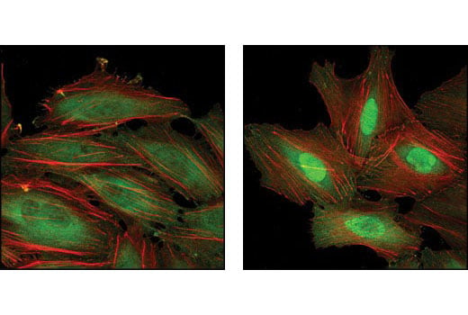


Anti-rat IgG (H+L), (Alexa Fluor ® 488 Conjugate)

 下载产品说明书 下载COA 下载SDS
 用小程序,查商品更便捷


 收藏
收藏
 对比
对比 咨询
咨询
Anti-rat IgG (H+L) antibody was conjugated to Alexa Fluor® 488 fluorescent dye under optimal conditions and formulated at 2 mg/ml. This product has been optimized for use as a secondary antibody in immunofluorescent applications. Cell Signaling Technology’s strict quality control procedures assure that each conjugate provides optimal specificity and fluorescence.
Product Usage Information
The optimal dilution of the anti-species antibody should be determined for each primary antibody by titration. However, a final dilution shown below should yield acceptable results for immunofluorescent assays:Immunofluorescent: 1:500 - 1:2000
Specificity/Sensitivity
Species Reactivity:
Rat
Supplied in 0.1 M sodium phosphate, 0.1 M sodium chloride, pH 7.5, 5 mM sodium azide. Store at 4°C. Do not aliquot the antibody. Protect from light. Do not freeze.
参考图片
Confocal immunofluorescent analysis of HeLa cells, untreated (left) or heat shocked (right), using HSP70 (6B3) Rat mAb #4873 detected with Anti-rat IgG (H+L), (Alexa Fluor® 488 Conjugate) (green). Actin filaments have been labeled with Alexa Fluor® 555 phalloidin (red).
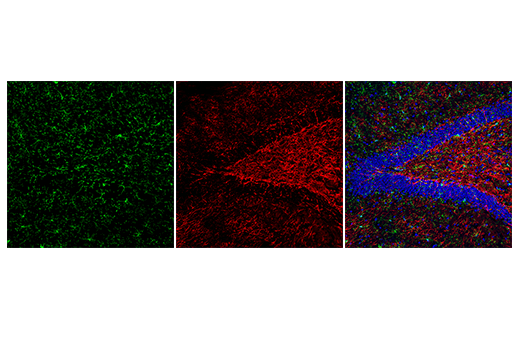
Confocal immunofluorescent analysis of mouse hippocampus using F4/80 (BM8.1) Rat mAb #71299 detected with Anti-rat IgG (H+L), (Alexa Fluor® 488 Conjugate) #4416 (green) and Myelin Basic Protein (D8X4Q) XP® Rabbit mAb #78896 detected with Anti-rabbit IgG (H+L), F(ab')2 Fragment (Alexa Fluor® 555 Conjugate) #4413 (red). Sections were mounted in ProLong® Gold Antifade Reagent with DAPI #8961 (blue).