


 全部商品分类


Anti-rabbit IgG (H+L), F(ab‘) 2 Fragment (Alexa Fluor ® 555 Conjugate)

 下载产品说明书 下载COA 下载SDS
 用小程序,查商品更便捷


 收藏
收藏
 对比
对比 咨询
咨询
Anti-Rabbit IgG (H+L) F(ab')2 Fragment was conjugated to Alexa Fluor® 555 fluorescent dye under optimal conditions and formulated at 2 mg/ml. This F(ab')2 fragment product results in less non-specific binding, as it lacks the Fc domain that can bind to the cells with Fc receptors.
Product Usage Information
The optimal dilution of the anti-species antibody should be determined for each primary antibody by titration. However, a final dilution of 1:500–1:2000 should yield acceptable results for immunofluorescent and flow cytometry assays.
Specificity/Sensitivity
Species Reactivity:
Rabbit
Supplied in 0.1 M sodium phosphate, 0.1 M sodium chloride, pH 7.5, 5 mM sodium azide. Store at 4°C. Do not aliquot the antibody. Protect from light. Do not freeze.
参考图片
High content analysis of A549 cells exposed to varying concentrations of LY294002 #9901 for 3 hrs, followed by 100 ng/mL EGF for 20 minutes. With increasing concentrations of LY294002, a significant decrease (~5 fold) in phospho-S6 Ribosomal Protein (Ser235/236) signal as compared to the uninhibited control was observed. When using phospho-S6 as a measurement, the IC50 of this compound was 3.06 μM. Data were generated on the Acumen HCS platform using Anti-Rabbit IgG (H+L), F(ab')2 Fragment (Alexa Fluor® 555 Conjugate).
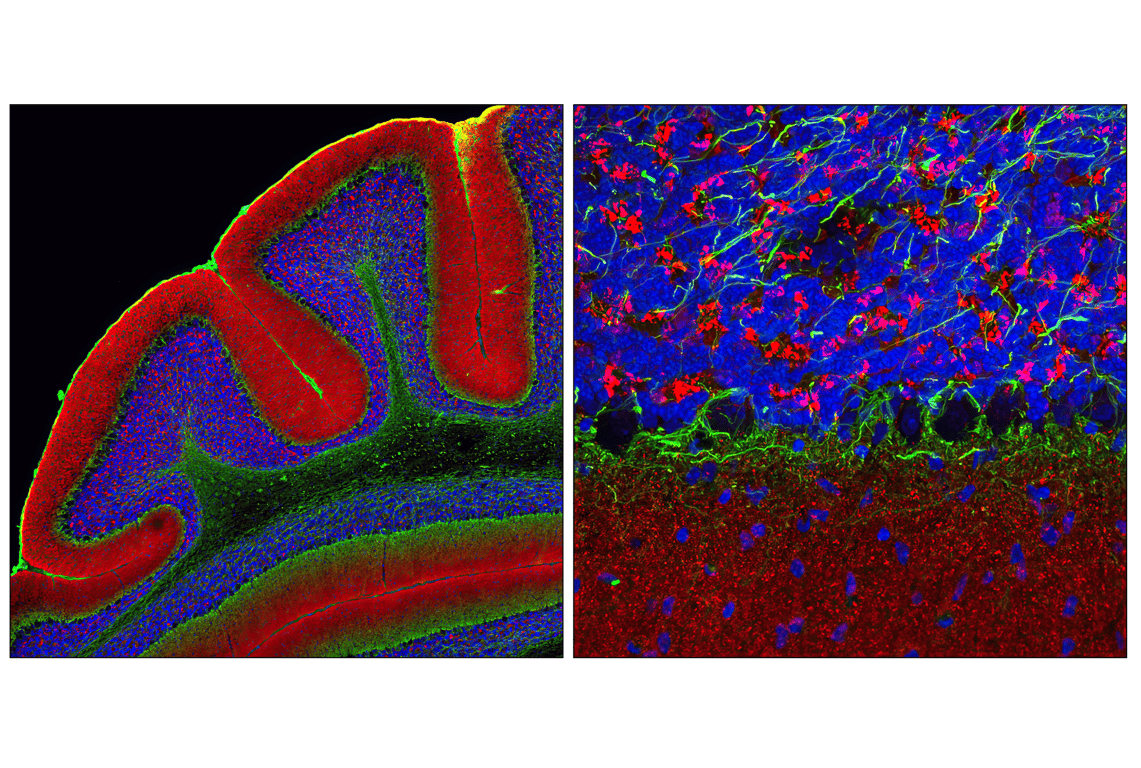
Confocal immunofluorescent analysis of mouse olfactory bulb using Calbindin (D1I4Q) XP® Rabbit mAb #13176 detected with Anti-rabbit IgG (H+L), F(ab')2 Fragment (Alexa Fluor® 555 Conjugate) #4413 (red) and Neurofilament-L (DA2) Mouse mAb #2835 detected with Anti-mouse IgG (H+L), F(ab')2 Fragment (Alexa Fluor® 647 Conjugate) #4410 (yellow pseudocolor). Sections were mounted in ProLong® Gold Antifade Reagent with DAPI #8961 (blue).
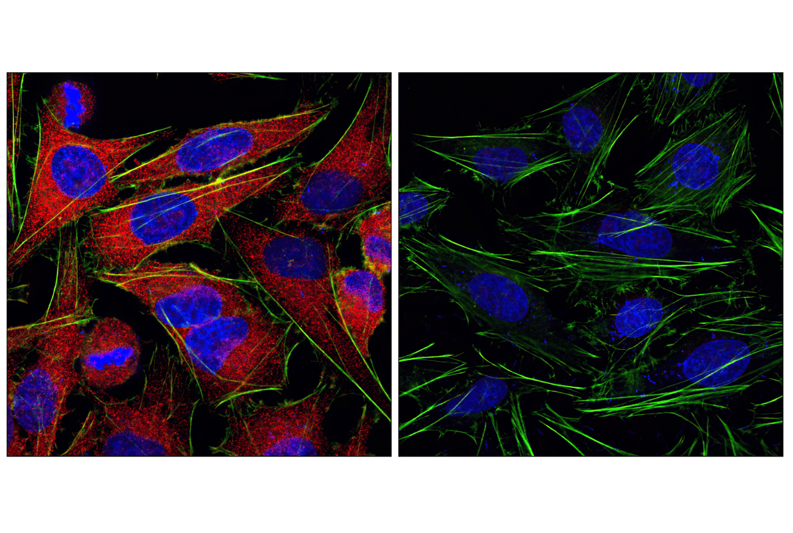
Confocal immunofluorescent analysis of HeLa cells labeled with MEK1/2 (47E6) Rabbit mAb #9126 detected with Anti-Rabbit IgG (H+L), F(ab')2 Fragment (Alexa Fluor® 555 Conjugate) (red, left) compared to an isotype control (right). Actin filaments have been labeled with fluorescein phalloidin (green). Blue pseudocolor = DRAQ5® #4084 (fluorescent DNA dye).
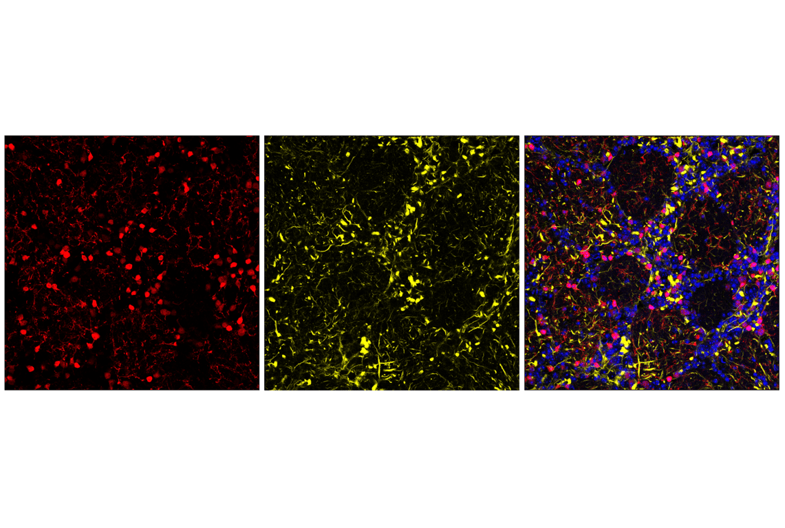
Confocal immunofluorescent analysis of mouse cerebellum using α-Synuclein Antibody (IF Preferred) #2628 detected with Anti-Rabbit IgG (H+L), F(ab')2 Fragment (Alexa Fluor® 555 Conjugate) (red) and Neurofilament-L (DA2) Mouse mAb #2835 detected with Anti-Mouse IgG (H+L), F(ab')2 Fragment (Alexa Fluor® 488 Conjugate) #4408 (green). Blue pseudocolor = DRAQ5® #4084 (fluorescent DNA dye).
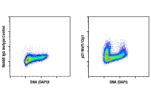
Flow cytometric analysis of Daudi cells using p21 Waf1/Cip1 (12D1) Rabbit mAb #2947 (right) and DAPI #4083, or concentration-matched Rabbit (DA1E) mAb IgG XP® Isotype Control #3900 (left). Anti-rabbit IgG (H+L), F(ab')2 Fragment (Alexa Fluor® 555 Conjugate) was used as a secondary antibody.