


 全部商品分类
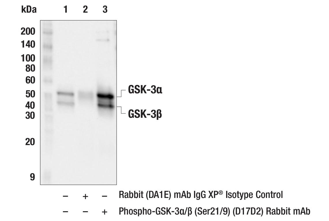


GSK-3 Antibody Sampler Kit

 下载产品说明书 下载SDS
 用小程序,查商品更便捷


 收藏
收藏
 对比
对比 咨询
咨询
The GSK-3 Antibody Sampler Kit contains primary and secondary antibodies to perform two Western blots with each antibody.
参考图片
Immunoprecipitation of phospho-Akt (Ser473) from HeLa cell extracts. Lane 1 is 10% input, lane 2 is Rabbit (DA1E) mAb IgG XP® Isotype Control #3900, and lane 3 is Phospho-GSK-3α/β (Ser21/9) (D17D2) Rabbit mAb. Western blot analysis was performed using Phospho-GSK-3α/β (Ser21/9) (D17D2) Rabbit mAb. Mouse Anti-rabbit IgG (Conformation Specific) (L27A9) mAb #3678 was used as a secondary antibody.
Western blot analysis of extracts from wild-type, GSK-3α (-/-), and GSK3β (-/-) mouse embryonic fibroblasts (MEFs) using GSK-3β (D5C5Z) XP® Rabbit mAb (upper) and GSK-3α/β (D75D3) XP® Rabbit mAb #5676 (lower). (MEF wild type, GSK-3α (-/-), and GSK-3β (-/-) cells were kindly provided by Dr. Jim Woodgett, University of Toronto, Canada).
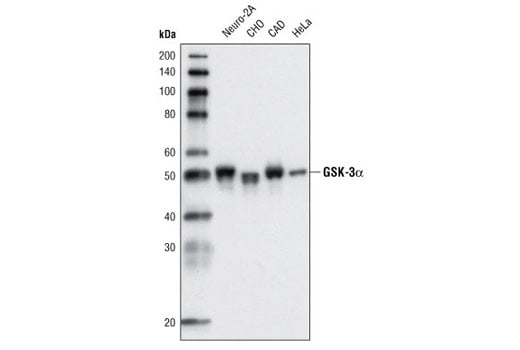
Western blot analysis of extracts from various cell types using GSK-3α (D80E6) Rabbit mAb.
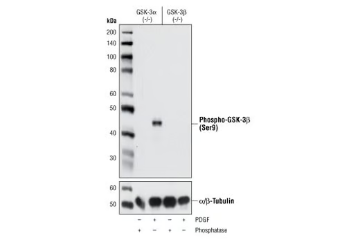
Western blot analysis of extracts from GSK-3α (-/-) (lanes 1,2) and GSK-3β (-/-) (lanes 3,4) mouse embryonic fibroblast (MEF) cells, λ phosphatase or PDGF-treated, using Phospho-GSK-3β (Ser9) (D85E12) XP® Rabbit mAb (upper) and α/β-Tubulin Antibody #2148 (lower). (MEF wild type, GSK-3α (-/-) and GSK-3β (-/-) cells were kindly provided by Dr. Jim Woodgett, University of Toronto, Canada).
After the primary antibody is bound to the target protein, a complex with HRP-linked secondary antibody is formed. The LumiGLO® is added and emits light during enzyme catalyzed decomposition.
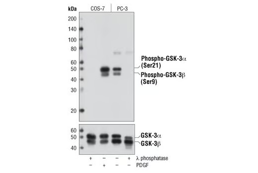
Western blot analysis of extracts from COS-7 cells, λ phosphatase- or PDGF-treated (100 μg/ml, 15 min), and PC-3 cells, untreated or λ phosphatase-treated, using Phospho-GSK-3α/β (Ser21/9) (D17D2) Rabbit mAb (upper) and GSK-3α/β (D75D3) XP® Rabbit mAb #5676 (lower).
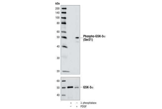
Western blot analysis of extracts from COS-7 cells, λ-phosphatase or PDGF-treated, using Phospho-GSK-3α (Ser21) (36E9) Rabbit mAb (upper) or GSK-3α Antibody #9338 (lower).
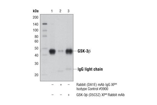
Immunoprecipitation of GSK-3β from PC-12 cell extracts, using Rabbit (DA1E) mAb IgG XP® Isotype Control #3900 (lane 2) or GSK-3β (D5C5Z) XP® Rabbit mAb (lane 3). Lane 1 is 10% input. Western blot analysis was performed using GSK-3β (D5C5Z) XP® Rabbit mAb.
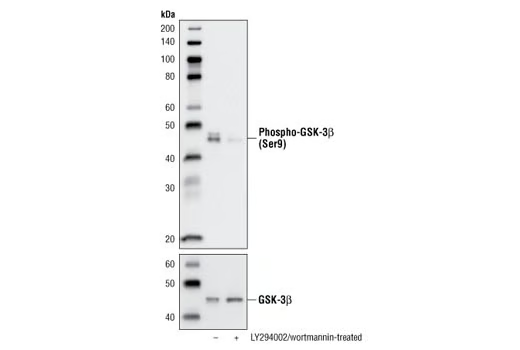
Western blot analysis of extracts from PC-3 cells, untreated or LY294002/wortmannin-treated, using Phospho-GSK-3β (Ser9) (D85E12) XP® Rabbit mAb (upper) or GSK-3β (27C10) Rabbit mAb #9315 (lower).
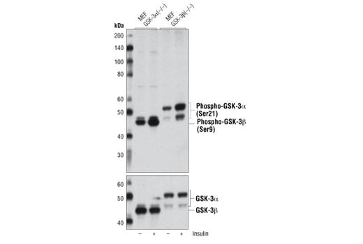
Western blot analysis of extracts from GSK-3α (-/-) and GSK-3β (-/-) mouse embryonic fibroblasts (MEF), untreated or insulin-treated (100 ng/ml, 20 min) , using Phospho-GSK-3α/β (Ser21/9) (D17D2) Rabbit mAb (upper) and GSK-3α/β (D75D3) XP® Rabbit mAb #5676 (lower). (MEF GSK-3α (-/-) and GSK-3β (-/-) cells were kindly provided by Dr. Jim Woodgett, University of Toronto, Canada).
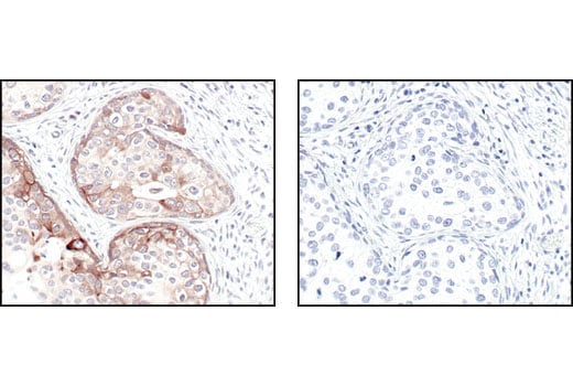
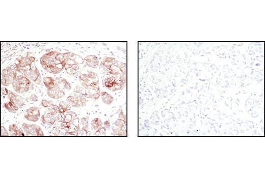
Immunohistochemical analysis of paraffin-embedded human breast carcinoma , untreated (left) or lambda phosphatase treated (right), using Phospho-GSK-3alpha (Ser 21) (36E9) Rabbit mAb.
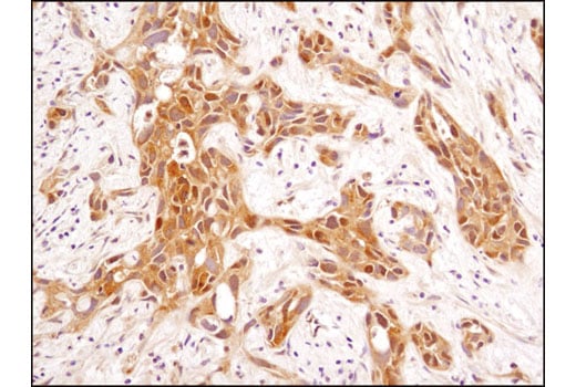
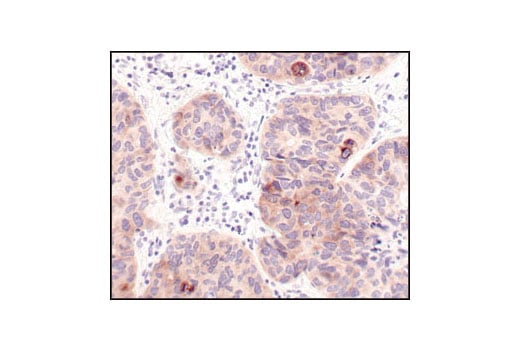
Immunohistochemical analysis of paraffin-embedded human breast carcinoma using GSK-3β (D5C5Z) XP® Rabbit mAb.
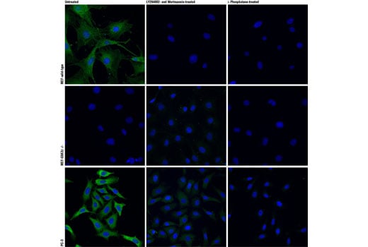
Confocal immunofluorescent analysis of wild type mouse embryonic fibroblasts (MEFs) (top row), GSK-3β (-/-) MEFs (middle row) , or PC-3 cells (bottom row), untreated (left), LY294002- and Wortmannin-treated (#9901 and #9951 respectively; center) or lambda phosphatase-treated (right), using Phospho-GSK-3β (Ser9) (D85E12) XP® Rabbit mAb (green). Actin filaments were labeled with DY-554 phalloidin (red). Blue pseudocolor = DRAQ5® #4084 (fluorescent DNA dye). (MEF wild type and GSK-3β (-/-) cells were kindly provided by Dr. Jim Woodgett, University of Toronto, Canada).
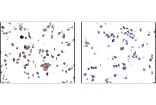
Immunohistochemical analysis of paraffin-embedded LNCaP cells, untreated (left) or LY294002-treated (right), using Phospho-GSK-3alpha (Ser 21) (36E9) Rabbit mAb.
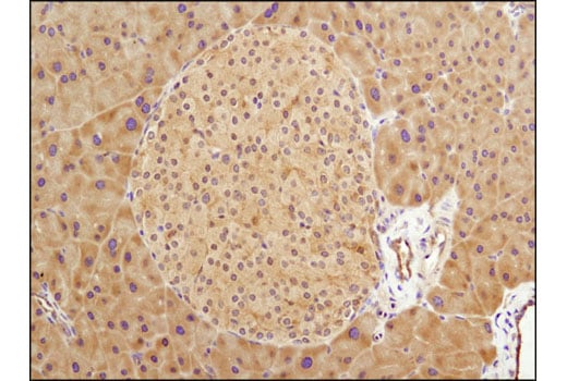
Immunohistochemical analysis of paraffin-embedded mouse pancreas using GSK-3β (D5C5Z) XP® Rabbit mAb.
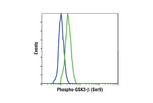
Flow cytometric analysis of NIH/3T3 cells, untreated (blue) or PDGF-treated (green), using Phospho-GSK-3β (Ser9) (D85E12) XP® Rabbit mAb.
Immunohistochemical analysis of paraffin-embedded human breast carcinoma using Phospho-GSK-3α (Ser21) (36E9) Rabbit mAb in the presence of control peptide (left) or Phospho-GSK-3α (Ser21) (36E9) Blocking Peptide #1027 (right).
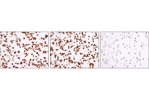
Immunohistochemical analysis of paraffin-embedded MEF cell pellets, wild type (left), GSK-3α (-/-) (middle) and GSK-3β (-/-) (right) using GSK-3β (D5C5Z) XP® Rabbit mAb. (MEF wild type, GSK-3β (-/-), and GSK-3α (-/-) cells were kindly provided by Dr. Jim Woodgett, University of Toronto, Canada).
Immunohistochemical analysis of paraffin-embedded human lung carcinoma using Phospho-GSK-3alpha (Ser 21) (36E9) Rabbit mAb.
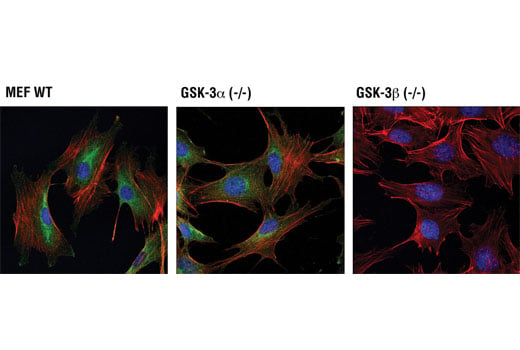
Confocal immunofluorescent analysis of wild-type mouse embryonic fibroblasts (MEFs) (left), GSK-3α (-/-) MEFs (center)and GSK-3β (-/-) MEFs (right) using GSK-3β (D5C5Z) XP® Rabbit mAb (green). Actin filaments were labeled with DY-554 phalloidin (red). Blue pseudocolor = DRAQ5® #4084 (fluorescent DNA dye). (MEF wild type, GSK-3α (-/-), and GSK-3β (-/-) cells were kindly provided by Dr. Jim Woodgett, University of Toronto, Canada).
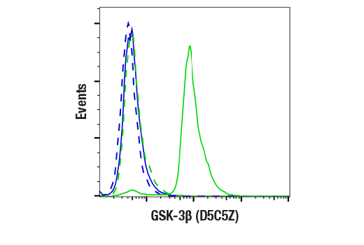
Flow cytometric analysis of GSK-3β (-/-) MEFs (blue, negative) and wild type mouse embryonic fibroblasts (MEFs) (green, positive) using GSK-3β (D5C5Z) XP® Rabbit mAb or a concentration-matched Rabbit (DA1E) mAb IgG XP® Isotype Control #3900 (dashed lines). Anti-rabbit IgG (H+L), F(ab')2 Fragment (Alexa Fluor® 488 Conjugate) #4412 was used as a secondary antibody. (MEF wild type and GSK-3β (-/-) cells were kindly provided by Dr. Jim Woodgett, University of Toronto, Canada).
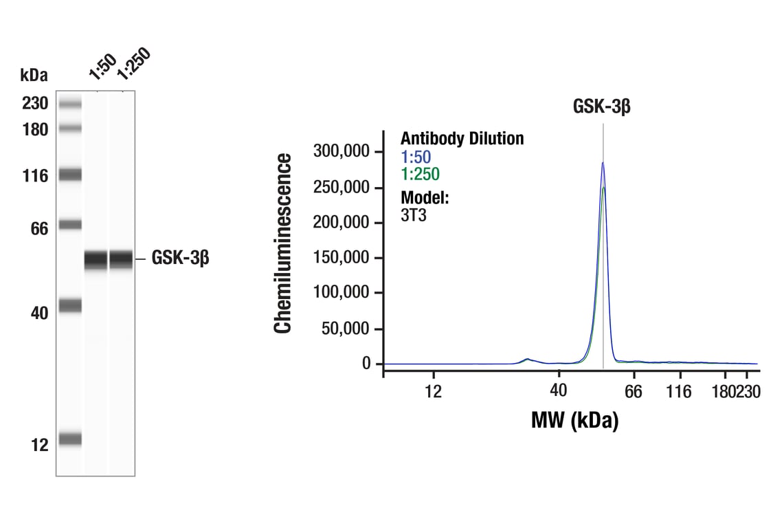
Simple Western™ analysis of lysates (1 mg/mL) from 3T3 cells using GSK-3β (D5C5Z) XP® Rabbit mAb #12456. The virtual lane view (left) shows a single target band (as indicated) at 1:50 and 1:250 dilutions of primary antibody. The corresponding electropherogram view (right) plots chemiluminescence by molecular weight along the capillary at 1:50 (blue line) and 1:250 (green line) dilutions of primary antibody. This experiment was performed under reducing conditions on the Jess™ Simple Western instrument from ProteinSimple, a BioTechne brand, using the 12-230 kDa separation module.
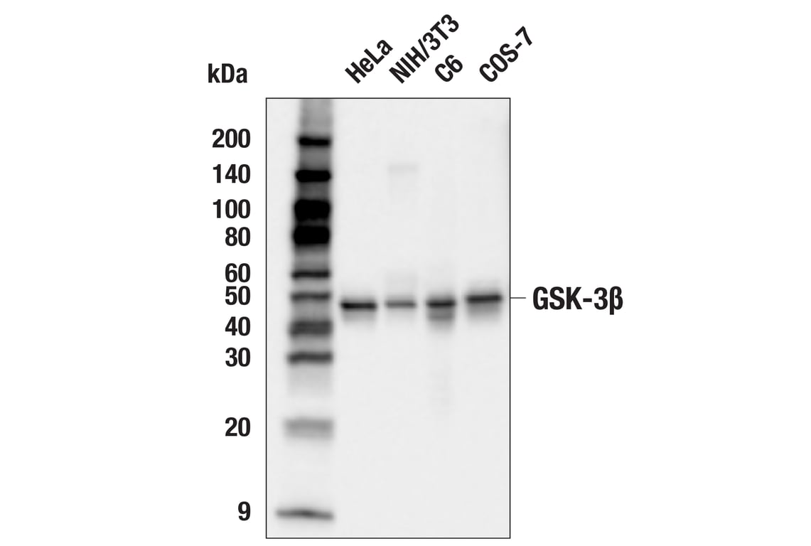
Western blot analysis of extracts from various cell lines using GSK-3β (D5C5Z) XP® Rabbit mAb.
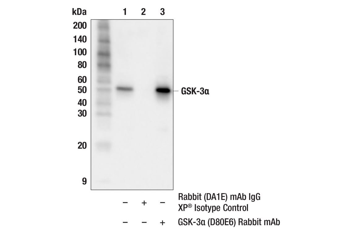
Immunoprecipitation of GSK-3α from HeLa cell extracts. Lane 1 is 10% input, lane 2 is Rabbit (DA1E) mAb IgG XP® Isotype Control #3900, and lane 3 is GSK-3α (D80E6) Rabbit mAb. Western blot analysis was performed using GSK-3α (D80D1) Rabbit mAb #4818. Mouse Anti-rabbit IgG (Conformation Specific) (L27A9) mAb (HRP Conjugate) #5127 was used as a secondary antibody.
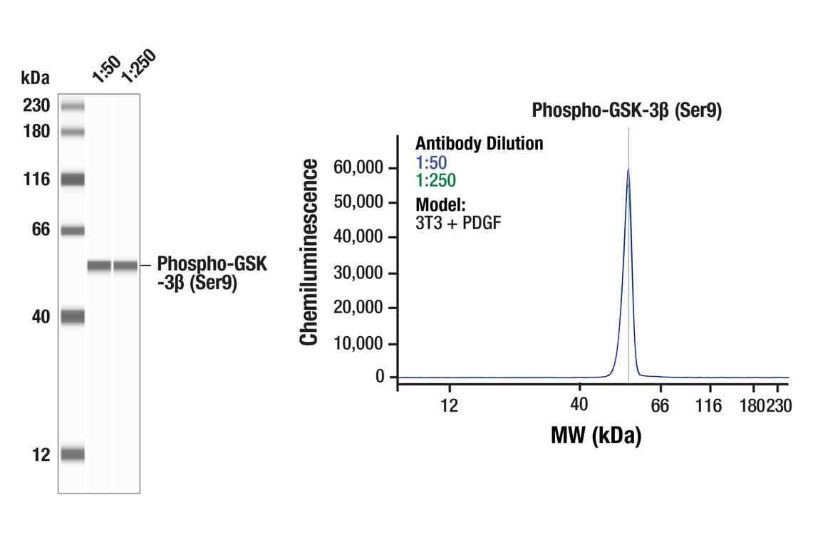
Simple Western™ analysis of lysates (0.1 mg/mL) from PDGF-treated 3T3 cells using Phospho-GSK-3β (Ser9) (D85E12) XP® Rabbit mAb #5558. The virtual lane view (left) shows the target band (as indicated) at 1:50 and 1:250 dilutions of primary antibody. The corresponding electropherogram view (right) plots chemiluminescence by molecular weight along the capillary at 1:50 (blue line) and 1:250 (green line) dilutions of primary antibody. This experiment was performed under reducing conditions on the Jess™ Simple Western instrument from ProteinSimple, a BioTechne brand, using the 12-230 kDa separation module.
Western blot analysis of extracts from various cell lines treated with Insulin (100nM, 20 min.) or PDGF (100ng/mL, 5 min.) using Phospho-GSK-3α/β (Ser21/9) (D17D2) Rabbit mAb (upper), GSK-3α/β (D75D3) Rabbit mAb #5676 (middle), or β-Actin (D6A8) Rabbit mAb #8457 (lower).